
Diphtheria micro with photos
Reading File
This is a Visual + Broad query. I'll run parallel searches across headings and paragraphs for comprehensive Diphtheria microbiology coverage with images.
Finding Sources
Finding Sources
The best microbiology source is Sherris & Ryan's Medical Microbiology. Let me read that block and Harrison's for comprehensive coverage, plus check for images.
Reading File
Reading File
Now let me get the detailed bacteriology section from Sherris and search for images:
Reading File
Finding Sources
Excellent content. Now let me read the rest of the Sherris diphtheria section and look for image references:
Reading File
Reading File
I found image URLs. Let me now fetch all images and continue reading the Sherris section for more content and images:
Loading Image
Reading File
Let me fetch the remaining key images and continue reading for more content:
Loading Image
Reading File
Excellent! Let me fetch the remaining 2 clinical images and read the lab diagnosis/treatment sections:
Loading Image
Loading Image
Reading File
I now have all the content and images needed. Let me also quickly look for the Gram stain / morphology image and metachromatic granules content from Jawetz:
Reading File
I now have comprehensive material from all major sources. Let me compose the full answer.
Diphtheria - Microbiology
The Organism: Corynebacterium diphtheriae
C. diphtheriae (from the Greek diphtheria, meaning "leather") is a small, pleomorphic, catalase-positive Gram-positive rod. Key morphological features:
- Club-shaped ends (Greek koryne = club), wider at one pole
- Snapping division creates characteristic arrangements: "Chinese letters," palisades, or V- and L-shaped pairs
- Non-spore-forming, non-motile, aerobic
- Metachromatic granules (Babes-Ernst granules/volutin) - polyphosphate-rich granules that stain reddish-purple with alkaline methylene blue or Loeffler's stain, while the cell body stains blue. These are visible as "beaded rods"
- Three biotypes: gravis, mitis, and intermedius (gravis tends to cause more severe disease)
Staining: Smears stained with alkaline methylene blue or Gram stain show beaded rods in typical Chinese-letter arrangements.
Toxin Genetics and Structure
The gene for diphtheria toxin (DT) is encoded in the genome of a lysogenic bacteriophage (corynephage beta). Only strains carrying this phage (tox+ strains) produce toxin. Toxin production is repressed by the DtxR protein, a repressor that responds to iron - low iron concentrations (as found in tissues) de-repress the gene and allow toxin production.
DT is a classic A-B toxin (535 amino acids):
- B subunit - binds to heparin-binding EGF-like precursor receptor on eukaryotic cell surfaces
- A subunit - the enzymatically active component
Mechanism of Action
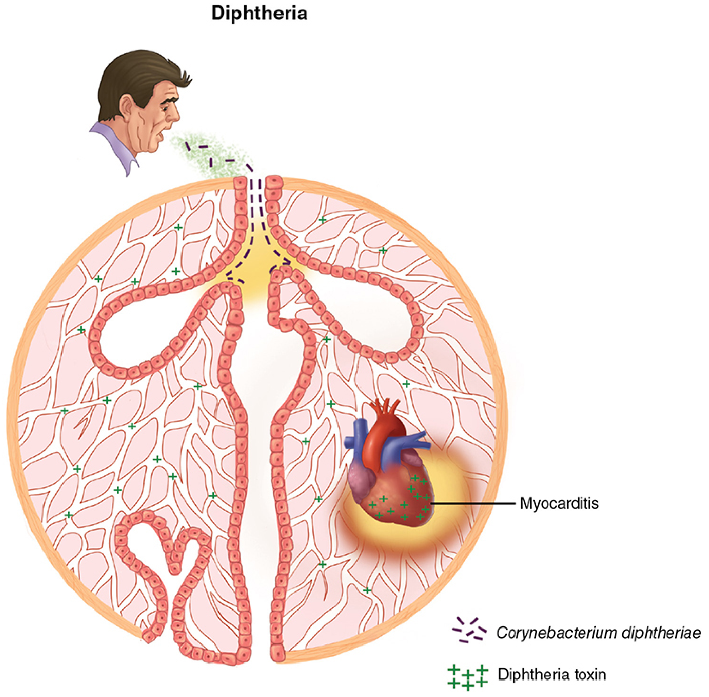
Fig. 1 - Diphtheria overview: C. diphtheriae spreads by respiratory droplets, infects the throat without invasion, and produces toxin that is absorbed into the bloodstream to cause myocarditis (Sherris & Ryan's Medical Microbiology)
Step-by-step:
- B subunit binds its receptor on host cell
- The entire A-B complex enters the cell via receptor-mediated endocytosis
- In the acidified endosomal vacuole, the toxin unfolds
- The A subunit translocates from the vacuole into the cytosol
- A subunit catalyzes NAD+-dependent ADP-ribosylation of elongation factor 2 (EF-2)
- EF-2 is required for translocation of polypeptidyl-tRNA from acceptor to donor ribosomal sites
- ADP-ribosylation of EF-2 irreversibly arrests protein synthesis → cell death
This mechanism is shared with Pseudomonas aeruginosa Exotoxin A (same target, same mechanism).
Potency: The 50% lethal dose is ~100 ng/kg body weight in sensitive species (guinea pigs, humans). Mice and rats are naturally resistant.
Pseudomembrane Formation and Pathology
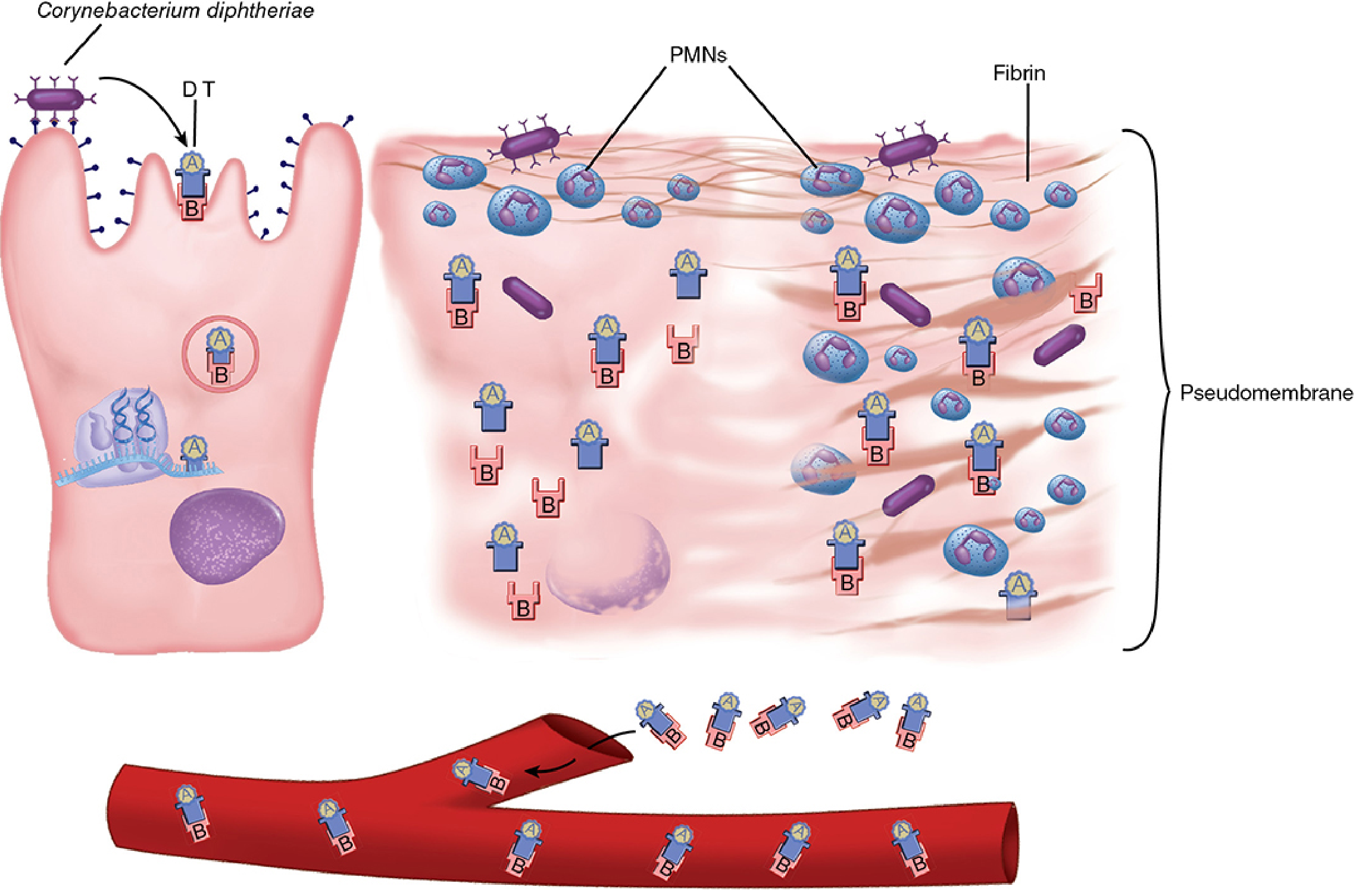
Fig. 2 - Cellular pathogenesis: (Left) C. diphtheriae binds epithelial cells and secretes DT, which enters the cell and ADP-ribosylates EF-2. (Middle) Dying cells recruit PMNs and fibrin. (Right) Destruction coalesces into a pseudomembrane; toxin enters the bloodstream (Sherris & Ryan's Medical Microbiology)
- C. diphtheriae has little invasive capacity - it does NOT invade deep tissues and rarely enters the bloodstream
- DT acts locally on epithelial cells → necrosis and superficial inflammation
- PMNs + fibrin + cellular debris form the pseudomembrane: an adherent, leathery, gray-white/gray membrane
- The pseudomembrane is tightly adherent - forcible removal causes bleeding (tears capillaries)
- It can extend from the tonsils/uvula/pharynx down through the larynx and into the tracheobroncial tree
Clinical Manifestations
Incubation: 2-4 days
Respiratory diphtheria
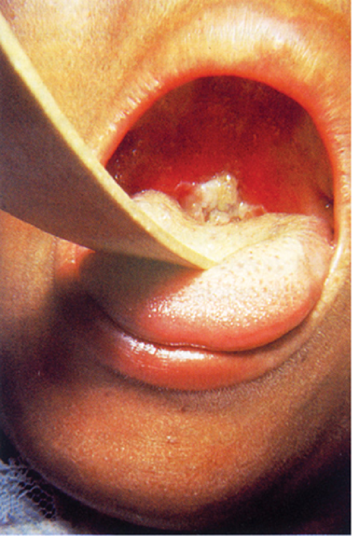
Fig. 3 - Typical diphtheritic pseudomembrane adherent to the oropharynx of a child (Sherris & Ryan's Medical Microbiology)
- Malaise, sore throat, low-grade fever
- Gray-white pseudomembrane on tonsils, uvula, soft palate, or pharyngeal wall
- "Bull neck" - cervical adenitis + marked neck edema from regional lymph node involvement
- Membrane can extend to larynx/trachea → respiratory obstruction and suffocation
Systemic (toxin-mediated) effects
Myocarditis (most serious):
- Detected by ECG in 2/3 of patients
- Clinically significant cardiac dysfunction in up to 25%
- Appears at week 2-3
- Manifests as cardiac enlargement, arrhythmias, and congestive heart failure
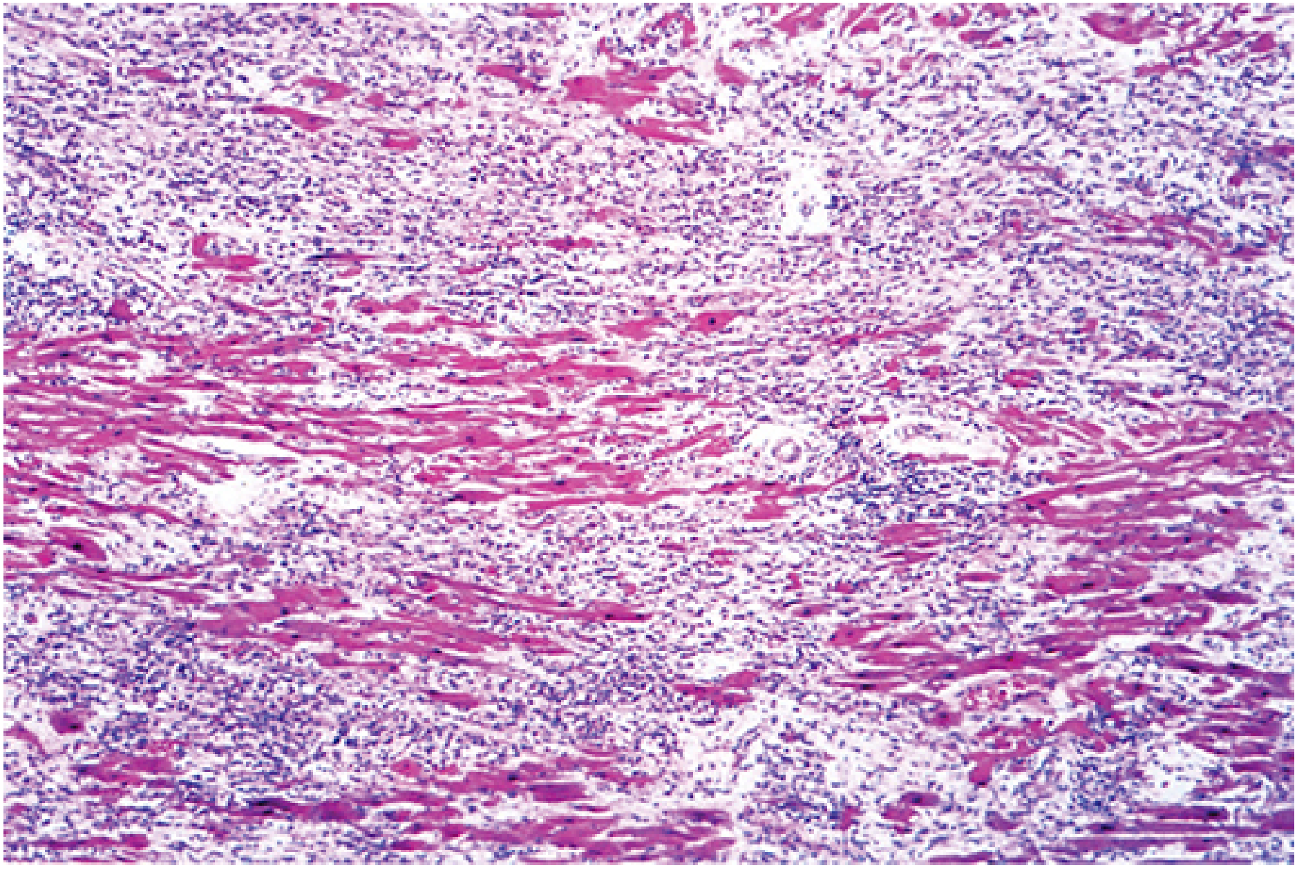
Fig. 4 - Diphtheritic myocarditis: H&E section showing myocardial necrosis and dense acute inflammatory infiltrate from a fatal case of diphtheria (Sherris & Ryan's Medical Microbiology)
Neuropathy (appears later):
- Demyelination of nerves
- Palatal palsy (most common), occulomotor palsy, peripheral neuropathy
- Diaphragm involvement can cause respiratory failure
- Generally reversible
Other organ toxicity: Fatty infiltration/necrosis of liver, kidney tubular necrosis, adrenal gland hemorrhage.
Cutaneous diphtheria
- Ulcerative skin lesions, often fail to heal, may form membrane
- More common in tropics and among homeless/alcoholic populations
- Toxin absorption is usually slight → less systemic disease
- Does promote development of antitoxin antibodies
Epidemiology
- Transmission: respiratory droplets, direct contact with cutaneous lesions, fomites
- Convalescent pharyngeal/nasal carriers can harbor the organism for weeks to months
- Rare where immunization is widely practiced (USA: last respiratory case 2003)
- Risk factors: unvaccinated, crowded living, low SES, alcoholism
- Notable epidemic: Former Soviet Union 1990-1995 (>47,000 cases, >1,700 deaths) after public health breakdown - controlled by mass vaccination
- Globally, 8,638 cases reported in 2021 (WHO), with many more unreported
- Recent uptick among unvaccinated asylum seekers in Europe (source: West Africa, Afghanistan)
Laboratory Diagnosis
| Step | Method |
|---|---|
| Initial diagnosis | Clinical - do NOT delay antitoxin while awaiting labs |
| Direct smear | Alkaline methylene blue → beaded rods with metachromatic granules (not definitive alone) |
| Culture medium | Loeffler's coagulated serum (rapid growth, prominent granules) and Tinsdale/Tellurite medium (black colonies due to tellurite reduction) |
| Identification | Biochemical reactions, colony morphology on tellurite agar |
| Toxigenicity testing | Elek test (immunodiffusion in agar), PCR for tox gene, or Vero cell cytotoxicity assay |
| MALDI-TOF | Increasingly used for species identification |
Important: The lab MUST be notified in advance of suspected diphtheria - routine throat culture procedures do not detect C. diphtheriae, and most labs don't stock the required media. Swabs should be taken from beneath or at the edge of the membrane and placed in Amies transport media.
Treatment
| Component | Details |
|---|---|
| Diphtheria antitoxin | Equine antiserum - must be given promptly as it only neutralizes free (circulating) toxin, not cell-bound toxin |
| Antibiotics | Erythromycin (drug of choice) or penicillin - eliminate the organism and stop further toxin production |
| Supportive care | Airway management (intubation/tracheostomy), cardiac monitoring |
Immunity and Prevention
- Natural infection stimulates antitoxin IgG (antibodies directed at toxin, not organism)
- Toxoid vaccine: formalin-treated DT - retains antigenicity, loses toxicity
- Schedule: 3-4 doses beginning in first year of life (as DTaP), boosters every 10 years (as Tdap)
- Fully immunized individuals can still be colonized with C. diphtheriae but disease is mild - serious disease only in unimmunized or incompletely immunized individuals
- Protective antitoxin titer: ≥0.01 IU/mL; ~30% of men aged 60-69 fall below this level
Key Microbiology Summary
| Property | Detail |
|---|---|
| Gram stain | Gram-positive pleomorphic rods (club-shaped) |
| Arrangement | Chinese letters, palisades, V/L shapes |
| Special stain | Metachromatic granules with methylene blue |
| Toxin gene | Carried by lysogenic corynephage (tox+ strains only) |
| Toxin regulation | Iron-regulated (DtxR repressor) - low iron → more toxin |
| Toxin mechanism | ADP-ribosylation of EF-2 → protein synthesis arrest |
| Culture | Tinsdale/Loeffler's media; black colonies on tellurite |
| Treatment | Antitoxin + erythromycin |
| Prevention | Toxoid vaccine (DTaP/Tdap) |
Sources: Sherris & Ryan's Medical Microbiology, 8th Ed. | Jawetz, Melnick & Adelberg's Medical Microbiology, 28th Ed. | Harrison's Principles of Internal Medicine, 22nd Ed. (2025)
This is a shared conversation. Sign in to Orris to start your own chat.