
Sweet syndrome
Sweet syndrome skin lesions neutrophilic dermatosis
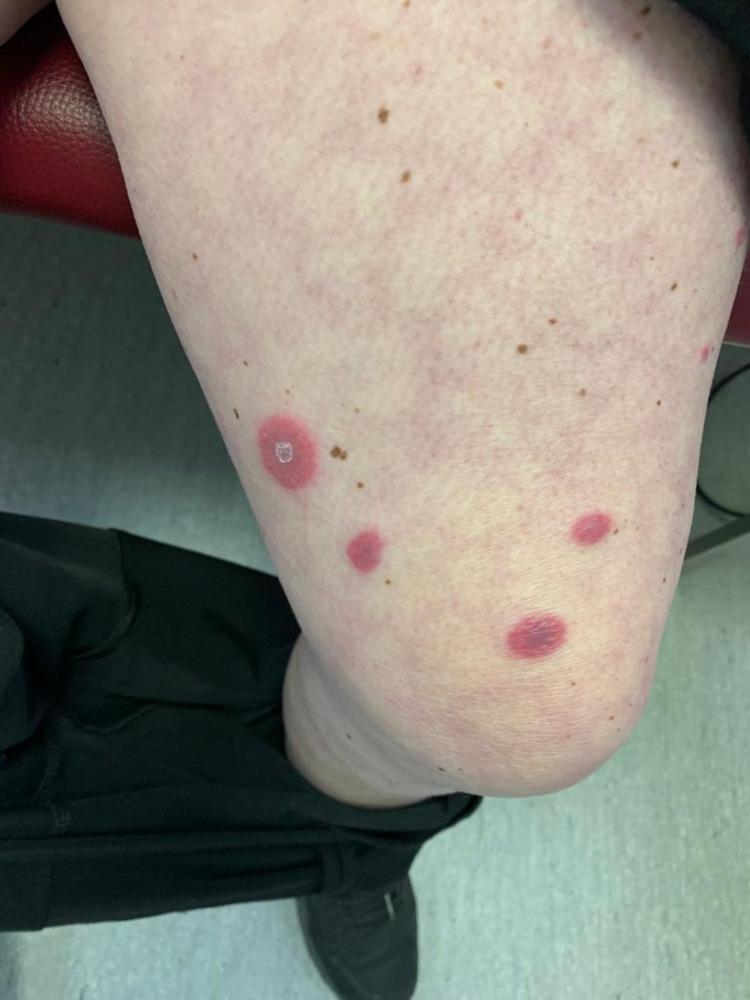
This clinical photograph shows the anterior thigh of a patient with several cutaneous lesions characteristic of Sweet syndrome (acute febrile neutrophilic dermatosis). The primary findings are multiple well-demarcated, erythematous to violaceous, edematous plaques and nodules. The lesions exhibit a scattered distribution across the fair-toned skin. One prominent lesion demonstrates a 'targetoid' or annular morphology with a lighter central zone, while other smaller lesions appear as solid, deep-red papulonodular infiltrates. The surrounding skin shows mild inflammatory erythema but is otherwise intact with visible benign hyperpigmented macules (freckles). These visual features represent classical neutrophilic infiltration of the dermis, often associated with systemic conditions such as inflammatory bowel disease (IBD). This image serves as a teaching tool for recognizing the dermatologic manifestations of reactive neutrophilic dermatoses in a clinical setting.
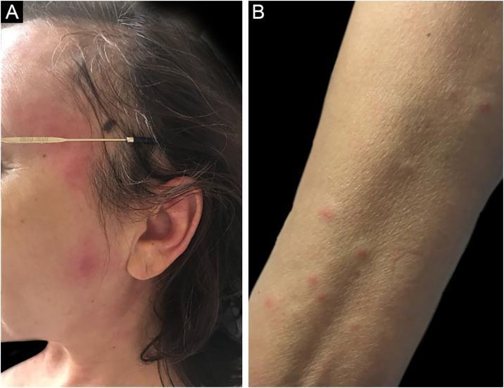
Two-panel clinical photograph illustrating the dermatological manifestations of Sweet syndrome (acute febrile neutrophilic dermatosis) in a patient with acute myeloid leukemia (AML). Panel A shows the lateral aspect of the face featuring multiple erythematous-edematous plaques and papules. The lesions are red-to-violaceous, raised, and relatively well-defined, concentrated on the cheek and periorbital region. Panel B displays the anterior forearm with smaller, discrete erythematous papules scattered across the skin surface. The lesions in both sites lack secondary features such as scaling, crusting, or ulceration. This visual demonstrates the classic 'succulent' or edematous appearance of neutrophilic dermatoses. The educational focus is on recognizing malignancy-associated skin signs, specifically the abrupt onset of painful, inflammatory lesions in a patient with an underlying hematologic disorder like MDS or AML.
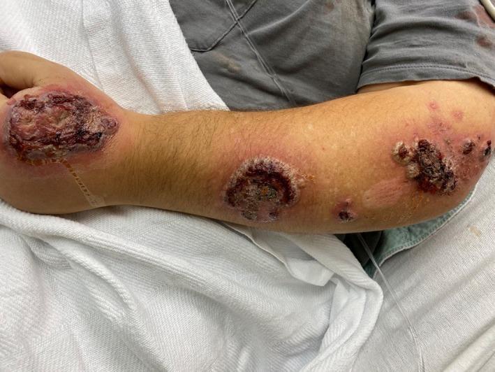
This clinical photograph displays severe dermatological manifestations on the left forearm and hand of a patient, consistent with an acute febrile neutrophilic dermatosis (Sweet syndrome). The primary findings include multiple large, well-demarcated, erythematous plaques and nodules. Significant features of these lesions include central ulceration with dark hemorrhagic crusting and focal areas of necrosis. The borders of the plaques are notably edematous, raised, and intensely inflamed, creating a pseudo-vesicular or succulent appearance characteristic of neutrophilic infiltration. Smaller, discrete erythematous papules and patches are scattered proximally along the forearm. Widespread diffuse erythema and apparent soft tissue edema are visible in the surrounding skin, with some adjacent areas showing post-inflammatory hypopigmentation. The distribution involves the dorsal hand, wrist, and extensors of the forearm, representing a common clinical presentation for this condition. This image serves as an educational example of the bullous or ulcerative variant of Sweet syndrome, highlighting the importance of distinguishing these lesions from infectious processes like cellulitis or necrotizing fasciitis.
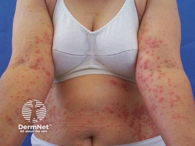
Clinical photograph of extensive neutrophilic dermatosis consistent with Sweet syndrome. Modality: color clinical photography of exposed and covered surfaces under standard white-light illumination. Findings: widespread, symmetric eruption involving the trunk and both proximal arms with numerous erythematous papules and plaques, some with subtle edema and superficial crusting. Lesions are tender on palpation and may coalesce into larger plaques, creating a figurate, arcuate, or annular morphology in places. The color is bright to violaceous red, with sharp borders against surrounding normal skin. Distribution is truncal and limb-centric, no mucosal involvement evident in the supplied views. Interpretation: the dermal eruption is characteristic for an acute febrile neutrophilic dermatosis (Sweet syndrome) and should prompt evaluation for systemic associations including hematologic malignancy, infection, inflammatory disorders, or drug exposure. Correlation with fever, leukocytosis, and elevated inflammatory markers strengthens the clinical diagnosis. If biopsy is performed, histopathology typically shows dense neutrophilic infiltrate in the upper dermis with edema and absence of vasculitis. Differential diagnoses include erythema nodosum, erythema multiforme, leukocytoclastic vasculitis, and drug eruption. The image supports dermatologic assessment, guides workup for underlying disease, and monitors treatment response to systemic corticosteroids or immunomodulatory therapy. This phenotype is essential for education, differential diagnosis practice, and research on neutrophilic dermatoses.
"Sweet syndrome" OR "acute febrile neutrophilic dermatosis"
Sweet Syndrome (Acute Febrile Neutrophilic Dermatosis)
Definition
Epidemiology
- No racial predilection; worldwide distribution
- Female predominance in younger adults (3:1 to 8:1 female:male), but near-equal sex distribution in those >50 years old and in malignancy-associated cases
- Children: boys and girls equally affected
- In Europe, more common in spring and fall
| Subtype | Frequency |
|---|---|
| Classic (idiopathic) | Majority |
| Malignancy-associated | 3-35% |
| Inflammatory disease-associated | 3-16% |
| Pregnancy-associated | 1-4% |
| Drug-induced | 1-26% |
Clinical Features
Skin Lesions
- Abrupt onset of sharply marginated, tender, erythematous or violaceous plaques and nodules, 2-10 cm diameter
- Appear intensely edematous ("juicy" or "succulent") or indurated
- Distribution: face, neck, upper trunk, and extremities
- Lesions burn but do not itch
- Surface may develop pseudovesiculation or pustulation from intense dermal inflammatory infiltrate
- Pathergy/koebnerization in 25-30% (from trauma, biopsies, IV lines)
- Special variants: bullous lesions (especially with leukemia), giant cellulitis-like, deep necrotic (mimicking necrotizing fasciitis in immunocompromised patients)
Systemic Manifestations (>75% of patients)
- Fever (50-80%) - most common systemic finding
- Arthritis, arthralgias, myalgias (1/3 to 2/3 of cases)
- Ocular: conjunctivitis, episcleritis, periorbital inflammation, iritis, choroiditis (~30%)
- Oral aphthae (2-3% classic; up to 10% in hematologic malignancy)
- Pulmonary: cough, dyspnea, pleuritis, infiltrates, effusions
- Multifocal sterile osteomyelitis (rare)
- Rarely: cardiac, renal, hepatic, neurologic involvement
Diagnostic Criteria
Major Criteria
- Abrupt onset of erythematous plaques or nodules (occasionally with vesicles, pustules, or bullae)
- Histopathology showing nodular and diffuse neutrophilic infiltrate in the dermis with karyorrhexis and massive papillary dermal edema (no leukocytoclastic vasculitis)
Minor Criteria
- Preceded by respiratory/GI infection or vaccination, OR associated with inflammatory disease, malignancy, or pregnancy
- Malaise and fever >38°C (100.4°F)
- Abnormal labs (at least 3 of 4): ESR >20 mm/h, elevated CRP, leukocytosis >8,000/mm³, left shift with >70% neutrophils
- Excellent response to systemic corticosteroids
For drug-induced Sweet syndrome, ALL 5 criteria (A-E) must be present, including a temporal relationship to drug ingestion and resolution after drug withdrawal.
Associated Conditions
- Hematologic malignancy (most commonly AML) and solid tumors (genitourinary, breast, GI)
- Infections: upper respiratory (streptococcosis), GI (Salmonella, Yersinia)
- Inflammatory bowel disease (Crohn disease, ulcerative colitis)
- Drugs (most commonly G-CSF)
- Pregnancy (typically 1st or 2nd trimester)
- Vaccinations (BCG, influenza)
Malignancy-Associated Features to Watch For
- Anemia in 93% of men and 71% of women with malignancy-associated Sweet syndrome
- Thrombocytopenia in ~50%
- Solitary, bullous, or ulcerative lesions more common
- Histiocytoid pattern on biopsy suggests malignancy
- AML with FLT3 mutations or del(5q) karyotype overrepresented
Histopathology
- Dense nodular and diffuse neutrophilic infiltrate in the upper dermis
- Karyorrhexis (nuclear dust)
- Massive papillary dermal edema (may form subepidermal bullae)
- No true leukocytoclastic vasculitis (focal leukocytoclasis does NOT exclude the diagnosis)
- Leukemic cells may be present in infiltrate
- Histiocytoid variant: infiltrate resembles histiocytes but represents immature myeloid cells (myeloperoxidase+); associated with hematologic malignancy
Laboratory Findings
- Elevated ESR (90%)
- Neutrophilia (70%)
- Leukocytosis (60%)
- Left shift/increased bands (50%)
- Elevated CRP
- ANCAs rarely reported
- CBC abnormalities raise suspicion for underlying malignancy
Pathogenesis
Drug-Induced Sweet Syndrome
- G-CSF (strongest association)
- All-trans-retinoic acid (ATRA) and FLT3 inhibitors (in leukemia treatment)
- Oral contraceptives, azathioprine, trimethoprim-sulfamethoxazole, minocycline
- Immunotherapy agents (ipilimumab, IL-2, TNF inhibitors, abatacept)
- Bortezomib, gemcitabine, BRAF inhibitors, multikinase inhibitors
- Allopurinol, lithium, isotretinoin, vaccines, radiocontrast dye
Treatment
- Systemic corticosteroids (dramatic, prompt response - hallmark of diagnosis)
- Potassium iodide
- Colchicine
- Indomethacin
- Dapsone
- Clofazimine
- Cyclosporine
- High-potency topical corticosteroids
- Intralesional corticosteroids
Clinical Images
Differential Diagnosis
| Condition | Key Distinguishing Feature |
|---|---|
| Cellulitis | Culture-positive, non-neutrophilic infiltrate |
| Erythema nodosum | Septal panniculitis, no dense dermal neutrophils |
| Leukocytoclastic vasculitis | Fibrinoid necrosis of vessel walls |
| Pyoderma gangrenosum | Undermined violaceous border, more ulcerative |
| Bowel bypass syndrome | Histologically identical - distinguished clinically |
| Leukemia cutis | Myeloperoxidase+ immature blasts (vs myeloid cells) |
- Fitzpatrick's Dermatology, Chapter 36 (Cohen & Kurzrock)
- Andrews' Diseases of the Skin, 13th ed.
- Goldman-Cecil Medicine
Tell me differential diagnosis
pyoderma gangrenosum ulcerative skin lesion
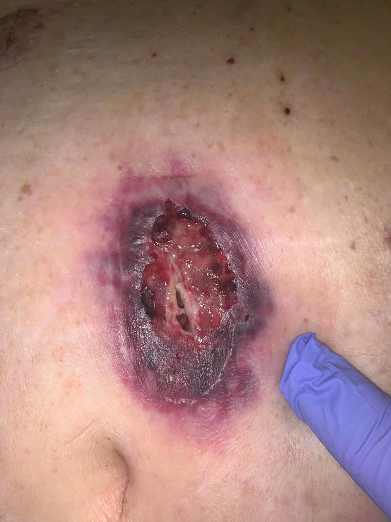
This clinical photograph displays a large, ulcerative skin lesion on the abdomen of a 66-year-old patient, characteristic of pyoderma gangrenosum (PG) manifesting at a post-operative port site. The central ulceration shows a deep, beefy red base with areas of necrotic tissue, yellow slough, and visible undermining or tunneling. The most distinctive feature is the well-demarcated, gunmetal grey to violaceous, undermined border that is surrounded by a halo of erythematous, inflamed skin. The lesion is located near the umbilicus, with a faint horizontal surgical scar visible superiorly. This image serves as a classic educational example of pathergy, where a skin condition is triggered or worsened by surgical trauma. The clinical context involves a patient with underlying inflammatory arthritis and myelodysplastic syndrome, highlighting the systemic associations often found with PG. The visual presentation is critical for distinguishing PG from infectious processes, emphasizing the characteristic 'violaceous border' and 'undermining' required for clinical diagnosis and subsequent management with immunosuppressive therapy rather than surgical debridement.
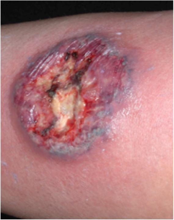
This clinical photograph displays a primary skin lesion characteristic of classic (ulcerative) pyoderma gangrenosum. The lesion presents as a deep, roughly circular ulcer with well-demarcated, irregular, and undermined borders. A defining diagnostic feature is the distinct violaceous (purple-hued) coloration of the peripheral margin. The central wound bed contains a mixture of yellowish-tan purulent material, areas of granulation tissue, and dark necrotic or hemorrhagic crusting. Immediately surrounding the violaceous border, there is a prominent erythematous inflammatory halo, indicating significant perilesional tissue infiltration and inflammation. This visual represents a neutrophilic dermatosis often associated with systemic conditions such as inflammatory bowel disease, rheumatoid arthritis, or hematologic malignancies. The image demonstrates key morphological signs used in dermatology for differentiating pyoderma gangrenosum from other chronic ulcers, such as venous stasis ulcers or cutaneous infections like leishmaniasis.
Differential Diagnosis of Sweet Syndrome
A. Clinical Differential Diagnosis
1. Pyoderma Gangrenosum (PG)
- Key overlap: Both are neutrophilic dermatoses, both show pathergy, both associated with IBD and hematologic malignancy
- Key difference: PG has a characteristic undermined, overhanging violaceous border with painful ulceration; Sweet lesions are non-ulcerated edematous plaques (though bullous Sweet can overlap)
- Bullous Sweet syndrome with leukemia closely mimics PG - the distinction can be very difficult clinically

2. Cellulitis / Erysipelas
- Key overlap: Red, warm, tender, spreading skin plaque with fever
- Key differences:
- Cellulitis is usually unilateral, lacks the "juicy" edematous plaques of Sweet
- Cultures positive in cellulitis; Sweet cultures are negative (sterile)
- Cellulitis does not respond dramatically to corticosteroids (which would worsen infection)
- Sweet often has multiple lesions at different sites; cellulitis is typically localized
3. Leukocytoclastic Vasculitis (LCV)
- Key overlap: Erythematous skin lesions with neutrophilic infiltrate on biopsy
- Key differences:
- LCV produces palpable purpura (non-blanching), not edematous plaques
- Histology: LCV shows fibrinoid necrosis of vessel walls + extravasated RBCs - absent in Sweet
- Focal leukocytoclasia can appear in Sweet biopsy but true vessel wall destruction is absent
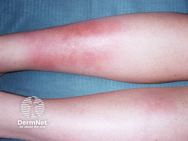
4. Erythema Nodosum (EN)
- Key overlap: Tender erythematous nodules with fever and elevated inflammatory markers
- Key differences:
- EN lesions are on the anterior shins (pretibial), deep, non-ulcerated
- Histology: EN is a septal panniculitis (subcutaneous) vs. Sweet is a dermal neutrophilic infiltrate
- EN lesions are never pseudovesicular or pustular
5. Behcet Disease
- Key overlap: Pathergy (koebnerization), oral lesions, fever, neutrophilic infiltrate
- Key difference: Behcet disease is associated with HLA-B51, recurrent oral and genital ulcers, uveitis, and thrombophlebitis - a broader systemic vasculitis picture
- Oral lesions in Sweet are aphthae-like but genital ulcers do not occur
6. Erythema Multiforme (EM)
- Key overlap: Acute-onset erythematous lesions, sometimes target-like morphology
- Key differences:
- EM has classic targetoid (iris) lesions with 3 zones (central dusky/blister, pale ring, red halo)
- EM lacks dense dermal neutrophilia on biopsy
- EM is typically triggered by HSV or Mycoplasma, not systemic malignancy
7. Leukemia Cutis / Chloroma
- Key overlap: Both occur in leukemia; histiocytoid Sweet can mimic leukemia cutis
- Key differences:
- Leukemia cutis infiltrate = malignant immature blast cells (myeloperoxidase+ immature myeloid cells)
- Sweet infiltrate = mature segmented neutrophils with karyorrhexis
- Immunohistochemistry (CD34, myeloperoxidase staining pattern) resolves the distinction
8. Neutrophilic Eccrine Hidradenitis (NEH)
- Key overlap: Erythematous plaques, fever, neutrophilic infiltrate; occurs in AML patients on chemotherapy
- Key difference: NEH histology shows neutrophils concentrated around eccrine glands, not diffusely throughout the dermis; papillary dermal edema is absent
9. Rheumatoid Neutrophilic Dermatitis
- Key overlap: Neutrophilic dermal infiltrate, plaques
- Key difference: History of rheumatoid arthritis with nodules; lesions are typically asymptomatic compared to the painful plaques of Sweet
10. Bowel Bypass Syndrome
- Occurs after jejunoileal bypass surgery for obesity
- Histologically identical to Sweet syndrome - can only be distinguished by clinical history
- Presents with fever, arthralgia, and neutrophilic skin lesions
11. Schnitzler Syndrome
- Key overlap: Urticarial plaques, fever, neutrophilic infiltrate, elevated inflammatory markers
- Key difference: Associated with monoclonal IgM (or IgG) gammopathy; urticarial morphology (non-tender wheals) rather than edematous plaques; biopsy shows neutrophilic urticarial pattern without papillary dermal edema
12. Erythema Elevatum Diutinum (EED)
- Key overlap: Neutrophilic dermal infiltrate
- Key difference: Lesions are asymptomatic, located on dorsal hands and elbows, evolve with dermal fibrosis and mucin; early lesions show leukocytoclastic vasculitis on biopsy
13. Halogenoderma (Iododerma / Bromoderma)
- Key overlap: Neutrophilic dermal infiltrate
- Key difference: History of iodide or bromide ingestion (or topical fluoride); biopsy shows neutrophilic infiltrate with pseudoepitheliomatous hyperplasia and intraepidermal abscesses + necrosis
14. Drug Eruptions
- Wide morphologic spectrum; temporal relationship to drug is the key clue
- Drug-induced Sweet itself is a subtype (requires all 5 criteria including resolution after withdrawal)
15. Other Conditions in the Clinical DD
| Condition | Key Distinguishing Feature |
|---|---|
| Bacterial sepsis | Culture-positive; skin findings differ |
| Lupus erythematosus | Photosensitive distribution, ANA+, interface dermatitis on biopsy |
| Dermatomyositis | Heliotrope rash, Gottron papules, proximal myopathy, elevated CK |
| Granuloma faciale | Asymptomatic facial lesions, grenz zone + eosinophils on biopsy |
| Lymphoma / metastatic tumor | Biopsy shows atypical lymphoid/carcinoma cells |
| Panniculitis | Subcutaneous nodules; septal or lobular changes on biopsy |
| Familial Mediterranean fever | Periodic fevers, autosomal recessive, MEFV gene mutation |
| Syphilis | Serology positive; varied morphology; spirochetes on biopsy |
| Systemic mycosis | Fungal cultures/stains positive |
| CANDLE syndrome | Chronic atypical neutrophilic dermatosis with lipodystrophy and elevated temperature; neonatal onset, autoinflammatory |
| Rosacea fulminans | Confined to face, no fever, no systemic neutrophilia |
| Acral erythema | Palmoplantar erythema; in chemotherapy patients |
B. Histologic Differential Diagnosis (Biopsy Mimics)
| Histologic Mimic | Distinguishing Feature on Biopsy |
|---|---|
| Leukemia cutis | Immature blast cells (vs. mature neutrophils); MPO staining pattern differs |
| Leukocytoclastic vasculitis | Fibrinoid necrosis of vessel walls, extravasated RBCs, neutrophils in vessel walls |
| Pyoderma gangrenosum | Ulcer histology; undermined border |
| Neutrophilic eccrine hidradenitis | Neutrophils around eccrine glands specifically |
| Neutrophilic urticarial dermatosis | Perivascular/interstitial infiltrate WITHOUT papillary dermal edema |
| Erythema elevatum diutinum | Older lesions: fibrosis + mucin; younger: LCV pattern |
| Granuloma faciale | Grenz zone, LCV features, numerous eosinophils |
| Bowel bypass syndrome | Histologically identical - purely a clinical distinction |
| Abscess / cellulitis | Culture-positive for infectious organism |
| Arthropod bite reaction | History of arthropod exposure; abundant eosinophils with neutrophils |
| Lobular neutrophilic panniculitis | Subcutaneous location: includes α-1-AT deficiency, infectious panniculitis, pancreatic panniculitis, subcutaneous Sweet |
| Halogenoderma | Pseudoepitheliomatous hyperplasia, intraepidermal abscesses, history of halide ingestion |
| Histiocytoid Sweet (its own sub-DD) | Must distinguish from leukemia cutis, interstitial granuloma annulare, interstitial granulomatous dermatitis with arthritis |
Summary: High-Yield Distinguishing Points
| Feature | Sweet Syndrome | Main Mimic |
|---|---|---|
| Dense mature neutrophils, papillary edema | ✅ | Absent in LCV, NEH |
| No vessel wall destruction | ✅ | LCV has fibrinoid necrosis |
| Sterile cultures | ✅ | Cellulitis/abscess culture-positive |
| Dramatic corticosteroid response | ✅ | Worsens infection |
| Pathergy | ✅ | Also in Behcet, PG |
| Undermined ulcer edge | ❌ | PG hallmark |
| Pretibial septal panniculitis | ❌ | EN hallmark |
| Monoclonal gammopathy | ❌ | Schnitzler hallmark |