
Lower jaw of th patient is outward. Tell me the prognosis of this patient
mandibular prognathism Class III malocclusion prognosis treatment
mandibular prognathism Class III malocclusion prognosis treatment outcome
mandibular prognathism Class III malocclusion jaw
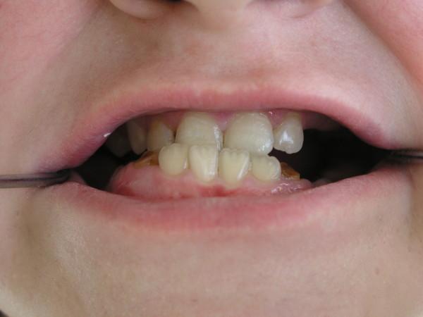
This intraoral clinical photograph demonstrates significant dental malocclusion and mandibular prognathism in a pediatric patient. The primary finding is a Class III malocclusion, where the lower jaw (mandible) and mandibular incisors are positioned anterior to the upper jaw (maxilla). The maxillary teeth exhibit moderate crowding, irregular alignment, and labial tilting. The lower teeth are noticeably protruded and angled forward. Additionally, there is evidence of gingival inflammation and potential dental caries, common oral manifestations in Hyper-IgE Syndrome (HIES). Two metal retractors are visible at the corners of the mouth to facilitate the clinical examination. The image serves as an educational example of craniofacial and dental abnormalities associated with primary immunodeficiency disorders.
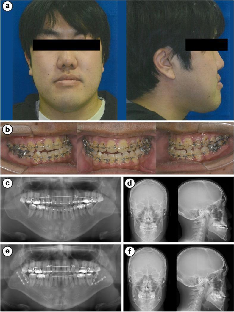
A multi-panel clinical resource documenting a case of mandibular prognathism and its surgical management. Panel (a) presents preoperative frontal and profile facial photographs showing a concave facial profile and prominent lower jaw. Panel (b) contains intraoral photographs displaying bilateral Angle Class III malocclusion, negative overjet (-8 mm), and fixed orthodontic appliances (braces). Panels (c) and (d) provide preoperative diagnostic imaging including a panoramic radiograph and lateral/frontal cephalometric radiograms, highlighting the skeletal discrepancy. Panels (e) and (f) show postoperative imaging one month after bilateral sagittal split ramus osteotomy (SSRO). The postoperative panoramic radiograph (e) reveals surgical fixation hardware (plates and screws) at the mandibular angles bilaterally, and the cephalometric radiogram (f) demonstrates the posterior repositioning of the mandible to achieve improved skeletal alignment. This series illustrates the comprehensive diagnostic and surgical sequence for treating severe jaw deformity and anterior crossbite.
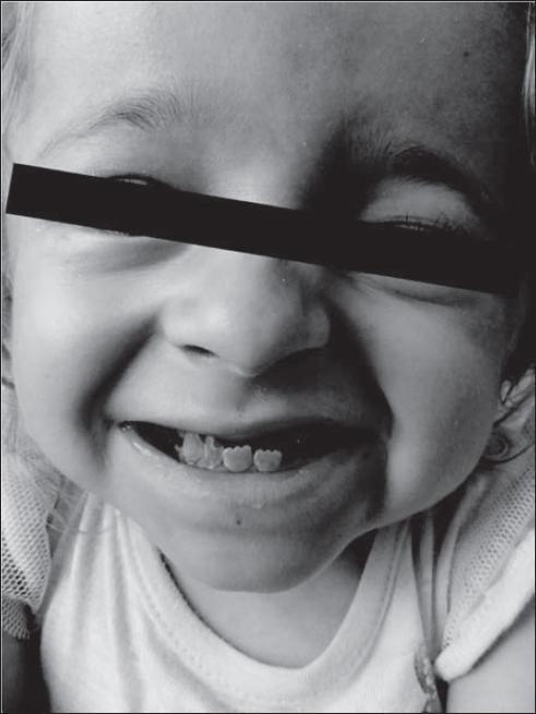
A monochrome clinical photograph of a 7-year-old child illustrating significant craniofacial and dental anomalies. The image demonstrates a prominent mandibular protrusion (prognathism) resulting in a Class III malocclusion. Key visible dental features include an anterior crossbite and an 'undershot' jaw where the mandibular incisors are positioned anterior to the maxillary teeth. The teeth themselves show structural abnormalities consistent with dentin dysplasia, characterized by irregular morphology, rounded or worn incisal edges, and atypical spacing. The facial skeleton displays structural disharmony, which is accentuated by the child's smile. This visual serves as a clinical example of the secondary skeletal and dental manifestations of dentin dysplasia and its impact on pediatric occlusion and maxillofacial development.
Mandibular Prognathism (Class III Malocclusion) - Prognosis
First: Rule Out a Systemic Cause
| Cause | Mechanism | Additional clues |
|---|---|---|
| Acromegaly (GH-secreting pituitary adenoma) | Excess GH/IGF-1 after epiphyseal closure causes jaw enlargement and protrusion, broadening of the lower face, enlarged hands/feet, soft tissue overgrowth | Sausage-like fingers, coarse facial features, hypertension, diabetes, gonadal dysfunction |
| Gigantism | Same cause, but GH excess occurs before epiphyseal closure | Disproportionately tall stature |
| Paget's disease | Bony enlargement of the jaw | Other sites of bony deformity |
| Primary skeletal/dental (most common) | Genetic, racial, or developmental mandibular overgrowth or maxillary deficiency | Isolated finding, family history |
From Robbins, Cotran & Kumar Pathologic Basis of Disease: "Enlargement of the jaw results in its protrusion (prognathism) and broadening of the lower face... changes may develop slowly over decades before being recognized." If acromegaly is the cause, achieving control of excess GH leads to gradual regression of tissue overgrowth and metabolic improvement.
Prognosis for Primary (Skeletal/Dental) Mandibular Prognathism
By Patient Age
| Age Group | Prognosis | Recommended Management |
|---|---|---|
| 5-10 years | Good to excellent with early intervention | Rapid maxillary expansion (RME) + facemask protraction; chin cap for pure mandibular excess |
| 10-12 years | Good with bone-anchored maxillary protraction | Bone anchor protraction, mini-plates + facemask |
| Adolescent (post-puberty) | Moderate - camouflage possible for mild cases | Orthodontic camouflage treatment |
| Adult | Skeletal growth complete; surgical correction is required for significant deformity | Orthognathic surgery (BSSO) + orthodontics |
By Subtype
- Maxillary deficiency (retrognathic maxilla) - Better prognosis with non-surgical and early orthodontic treatment
- Mandibular prognathism (excess mandibular growth) - Poorer prognosis with non-surgical management; 1.5x higher risk of treatment failure compared to maxillary deficiency subtypes; surgical correction typically required for definitive results
- Combination type (both) - intermediate prognosis
Key Prognostic Factors
- Severity of skeletal discrepancy - mild cases can be managed orthodontically; severe cases require surgery
- Patient age - early treatment in children (5-12 years) yields best skeletal outcomes; adults need orthognathic surgery
- Mandibular plane angle - high mandibular plane angle carries worse prognosis for chin cap/non-surgical approaches
- Surgical vs. non-surgical route - non-surgical treatment of mandibular prognathic subtypes has significantly higher relapse/failure rates
- Underlying cause - if acromegaly-driven, the jaw prognathism may improve with somatostatin analog therapy or surgical removal of the pituitary adenoma
Treatment Outcomes (What to Expect)
- Surgical correction (bilateral sagittal split ramus osteotomy, BSSO): Excellent functional and aesthetic outcomes. Resolves difficulty chewing, TMJ pain, speech issues (lisp). Requires pre- and post-surgical orthodontics for 12-24 months.
- Non-surgical/orthodontic camouflage: Adequate for mild cases; high relapse risk in moderate-severe mandibular prognathism. The underlying skeletal discrepancy is masked, not corrected.
- Early interceptive treatment in children: Facemask therapy has demonstrated mid-term effectiveness per recent systematic review (PMID 34305011), but long-term stability requires monitoring through adolescence.
Summary
| Scenario | Overall Prognosis |
|---|---|
| Child (5-10 yrs), maxillary deficiency | Good - facemask + expansion |
| Child (5-10 yrs), mandibular excess | Moderate - chin cap; may need surgery at maturity |
| Adult, mild | Fair - orthodontic camouflage |
| Adult, moderate-severe | Good with surgery - orthognathic surgery + orthodontics gives excellent results |
| Due to acromegaly | Depends on GH control - treat the pituitary tumor first |
Color Atlas of Human Anatomy Vol. 2: "Such anomalies [prognathism] can interfere with swallowing, nasal breathing, and speech" - highlighting why treatment is functional, not just cosmetic.