Structures of angle of eye
Reading File
Finding Sources
Finding Sources
Searching Images
angle of eye iridocorneal angle anatomy structures
Finding Sources
Finding Sources
Reading File
Reading File
Finding Sources
Loading Image
Loading Image
Finding Sources
Loading Image
Finding Sources
Reading File
Reading File
Reading File
Loading Image
Excellent — I now have comprehensive textbook content and images. Here is a complete, well-organized answer.
Structures of the Angle of the Eye (Iridocorneal Angle)
The angle of the anterior chamber (iridocorneal angle) is the recess formed at the junction of the peripheral cornea and the root of the iris. It is the principal site of aqueous humor drainage and is examined clinically by gonioscopy.
Anatomical Diagram & Gonioscopic Appearance
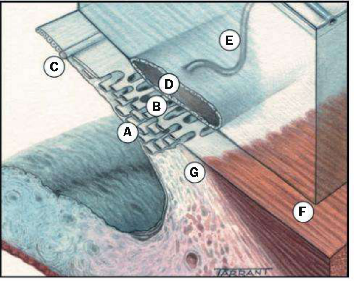
Fig. 11.2 — Anatomy of outflow channels. (A) Uveal meshwork; (B) Corneoscleral meshwork; (C) Schwalbe line; (D) Schlemm canal; (E) Collector channels; (F) Longitudinal ciliary muscle; (G) Scleral spur — Kanski's Clinical Ophthalmology, 10e
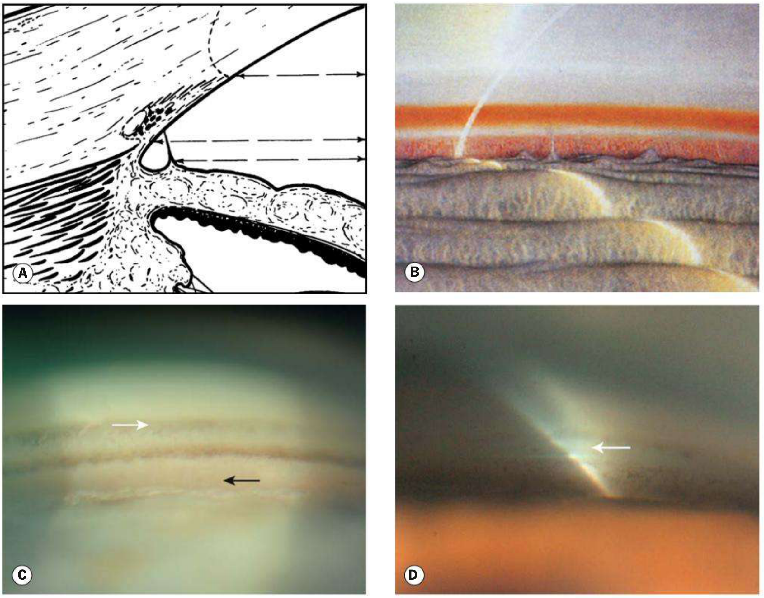
Fig. 1.52 — Normal angle structures. (A) Schematic, (B) corneal wedge, (C) goniophotograph showing Schwalbe line (white arrow), pigmented meshwork, scleral spur, and ciliary body (black arrow), (D) corneal wedge locating Schwalbe line — Kanski's Clinical Ophthalmology, 10e
Structures — Anterior to Posterior (as seen on gonioscopy)
1. Schwalbe Line
- The most anterior visible structure.
- Anatomically marks the peripheral termination of Descemet membrane and the anterior limit of the trabeculum.
- Appears whitish to variably pigmented; may be barely discernible in the young.
- Located using the corneal wedge: a narrow slit beam creates two linear corneal reflections that meet at the apex = Schwalbe line.
- Pigment deposits on/anterior to it = Sampaolesi line (seen in pseudoexfoliation, pigment dispersion).
2. Trabecular Meshwork (Trabeculum)
Extends from Schwalbe line to scleral spur; average width ~600 µm. Provides ~90% of aqueous outflow resistance. Has three layers (from inner to outer):
| Layer | Location | Spaces | Resistance |
|---|---|---|---|
| Uveal meshwork | Innermost; cord-like strands from iris/ciliary body stroma | Large intertrabecular spaces | Least |
| Corneoscleral meshwork | Middle; thickest part; layered connective tissue | Smaller spaces | Moderate |
| Juxtacanalicular (cribriform) meshwork | Outermost; links to Schlemm canal endothelium | Narrow intercellular spaces in dense extracellular matrix | Greatest (major site of normal outflow resistance) |
Gonioscopically:
- Anterior (non-functional) part — adjacent to Schwalbe line; whitish, ground-glass appearance
- Posterior (pigmented/functional) part — adjacent to scleral spur; greyish-blue translucent; pigmentation increases with age, most marked inferiorly
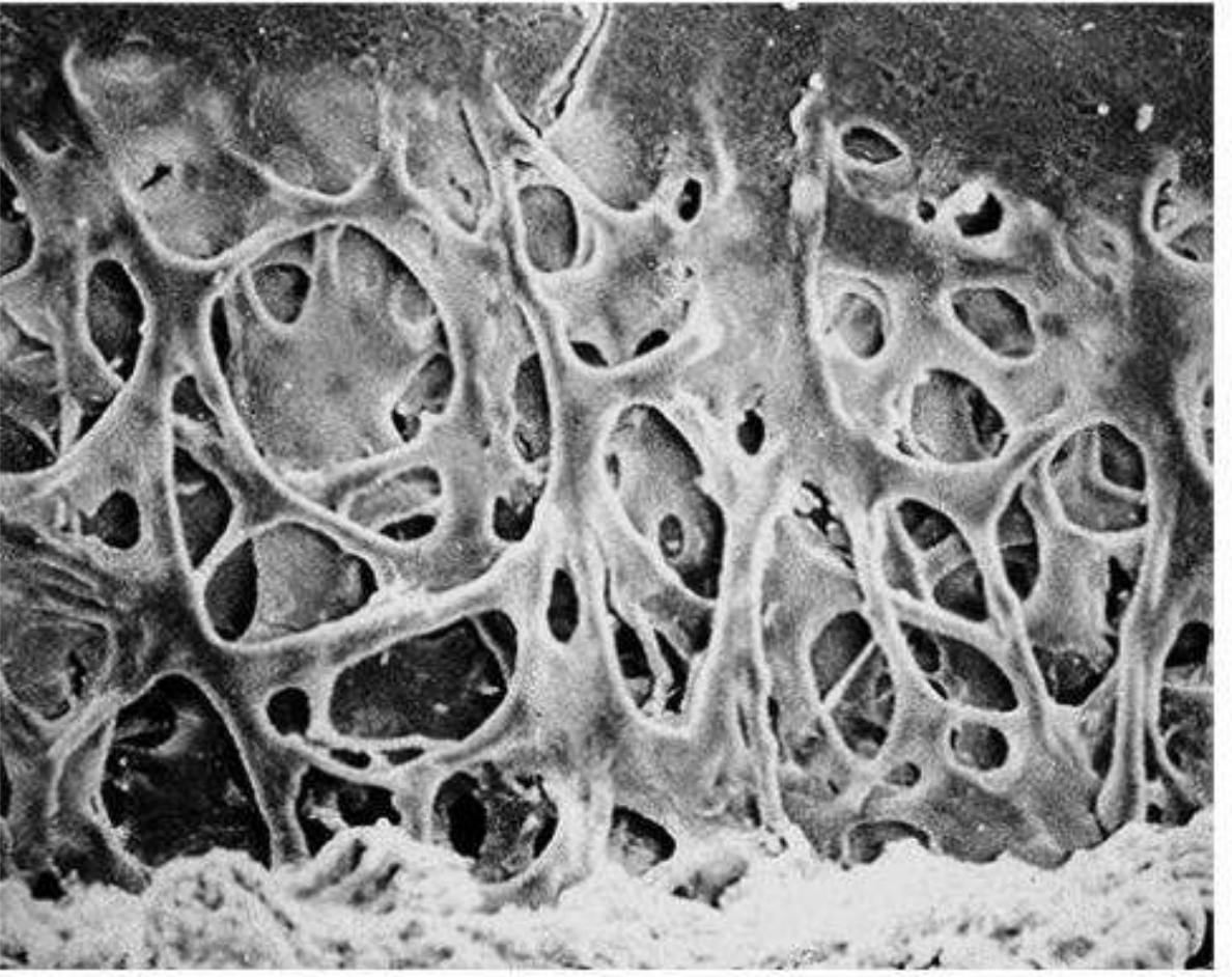
Fig. 11.1 — Scanning electron micrograph of the trabecular meshwork showing sieve-like interconnected spaces — Kanski's Clinical Ophthalmology, 10e
3. Schlemm Canal
- A circumferential channel within the perilimbal sclera, lying just external to the juxtacanalicular meshwork.
- Inner wall: irregular spindle-shaped endothelial cells with giant vacuoles (transcellular pores) — conveys aqueous into the canal.
- Outer wall: smooth flat cells; contains openings of collector channels connecting to episcleral veins.
- Septa commonly divide the lumen into 2–4 channels.
- Gonioscopically: a slightly darker line deep to the posterior trabeculum; blood can sometimes be seen within it (low IOP or raised episcleral venous pressure).
4. Scleral Spur
- The most anterior projection of the sclera.
- Site of attachment of the longitudinal muscle of the ciliary body.
- Gonioscopically: a narrow whitish band immediately posterior to the trabeculum (yellows with age).
- Key surgical landmark separating canal-based (trabecular) from suprachoroidal (ciliary body) surgery.
5. Ciliary Body Band
- Stands just posterior to the scleral spur.
- Appears pink, dull brown, or slate grey.
- Width depends on iris insertion position — narrower in hypermetropic eyes, wider in myopic eyes.
6. Angle Recess
- The posterior dipping of the iris as it inserts into the ciliary body.
- May not be visible in eyes with physiological anterior iris insertion.
7. Iris Processes (Pectinate Ligaments)
- Small, tenuous extensions of the anterior iris surface inserting at the scleral spur level.
- Present in ~one-third of normal eyes; most prominent in childhood and in brown eyes.
- Must be distinguished from peripheral anterior synechiae (PAS), which are more substantial and extend more anteriorly.
8. Blood Vessels
- Normal: radial vessels at the base of the angle recess.
- Abnormal: any vessel crossing the scleral spur onto the trabecular meshwork; randomly directed neovascular vessels (rubeosis).
Aqueous Outflow Routes
| Route | Contribution | Pathway |
|---|---|---|
| Trabecular (conventional) | ~90% | Trabeculum → Schlemm canal → Collector channels → Episcleral veins |
| Uveoscleral (unconventional) | ~10% | Ciliary body face → Suprachoroidal space → Choroidal/scleral venous drainage |
| Iris | Minor | Across iris stroma |
The trabecular route is pressure-sensitive (bulk flow increases with rising IOP); the uveoscleral route is largely pressure-independent.
Spaeth Classification of Iris Insertion
Assessed by gonioscopy; describes how anteriorly the iris root inserts:
| Grade | Description |
|---|---|
| A | Anterior to Schwalbe line |
| B | Between Schwalbe line and scleral spur |
| C | At scleral spur (common in Black/Asian populations) |
| D | Deep — ciliary body visible (common in White populations) |
| E | Extremely deep — >1 mm of ciliary body visible |
Iris curvature (bowing anteriorly, plateau, flat, concave) and posterior trabecular meshwork pigmentation (0–4+) are also graded.
Sources: Kanski's Clinical Ophthalmology: A Systematic Approach, 10e; The Wills Eye Manual, 8e
This is a shared conversation. Sign in to Orris to start your own chat.