आइए काम करूँ, थोड़ा डिटेल में समझाओ हिस्टोलॉजी ऑफ द फंडस ऑफ स्टमक थोड़ा बेसिक टू डिप लेवल समझाओ
Reading File
Finding Sources
Finding Sources
Reading File
Reading File
Reading File
Reading File
Reading File
Reading File
Loading Image
Loading Image
Reading File
Loading Image
Reading File
Loading Image
Reading File
Loading Image
अब सब जानकारी इकट्ठी है। एक comprehensive, detailed answer तैयार करते हैं।
Histology of the Fundus of Stomach - Basic to Deep Level
1. OVERVIEW - STOMACH KI REGIONS
Stomach को 4 major regions में divide किया गया है:
- Cardia - esophagus के पास, mostly mucous glands
- Fundus - ऊपरी dome-shaped हिस्सा
- Body (Corpus) - सबसे बड़ा हिस्सा
- Pylorus/Antrum - duodenum के पास
Fundus + Body = Oxyntic Gland Area - यह stomach का 75-80% हिस्सा है। इसी region की histology exams में सबसे ज्यादा पूछी जाती है।
2. STOMACH WALL की 4 LAYERS (Basic Structure)
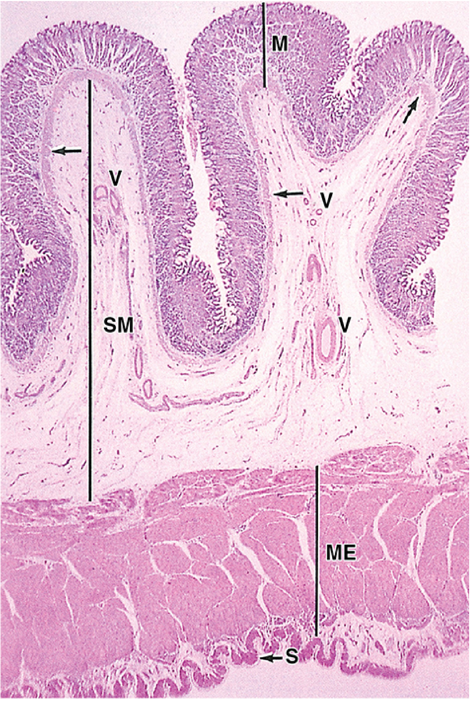
ऊपर की image में clearly दिखता है: Mucosa (M), Submucosa (SM), Muscularis Externa (ME), और Serosa (S) - बीच में रुगे (Rugae) भी दिख रही हैं।
Layer 1: MUCOSA (सबसे important)
इसे 3 sub-layers में divide करते हैं:
- Epithelium - Simple columnar
- Lamina Propria - loose connective tissue, gastric glands से भरी हुई
- Muscularis Mucosae - thin smooth muscle layer
Layer 2: SUBMUCOSA
- Loose connective tissue
- Large blood vessels (V) और lymphatics
- Meissner's (Submucosal) nerve plexus
- Fundus में कोई special glands नहीं होती (duodenum में Brunner's glands होती हैं)
Layer 3: MUSCULARIS EXTERNA (Muscularis)
Stomach में यह 3-layered होती है (बाकी GI tract में 2 layers होती हैं):
- Outer - Longitudinal
- Middle - Circular (सबसे thick, pyloric sphincter बनाती है)
- Inner - Oblique (only stomach में, churning में help)
- Auerbach's (Myenteric) nerve plexus इन layers के बीच होता है
Layer 4: SEROSA
- Thin layer of mesothelium
- Visceral peritoneum से covered
3. GASTRIC PITS (Foveolae) - MUCOSA का GATEWAY
जब खाली stomach होता है, तो mucosa + submucosa मिलकर longitudinal folds बनाते हैं जिन्हें Rugae कहते हैं। जब stomach भरता है तो ये flatten हो जाते हैं।
Mucosal surface पर:
- Millions of gastric pits होते हैं (invaginations)
- हर gastric pit में 4-5 gastric glands open होती हैं
- Pits की lining = Surface Mucous Cells
Fundus में pit depth = mucosa की depth का approximately 1/4 हिस्सा
(Antrum में pits ज्यादा deep होते हैं - mucosa का 1/2 से ज्यादा)
4. GASTRIC GLANDS (Oxyntic/Gastric Glands proper) - CORE TOPIC
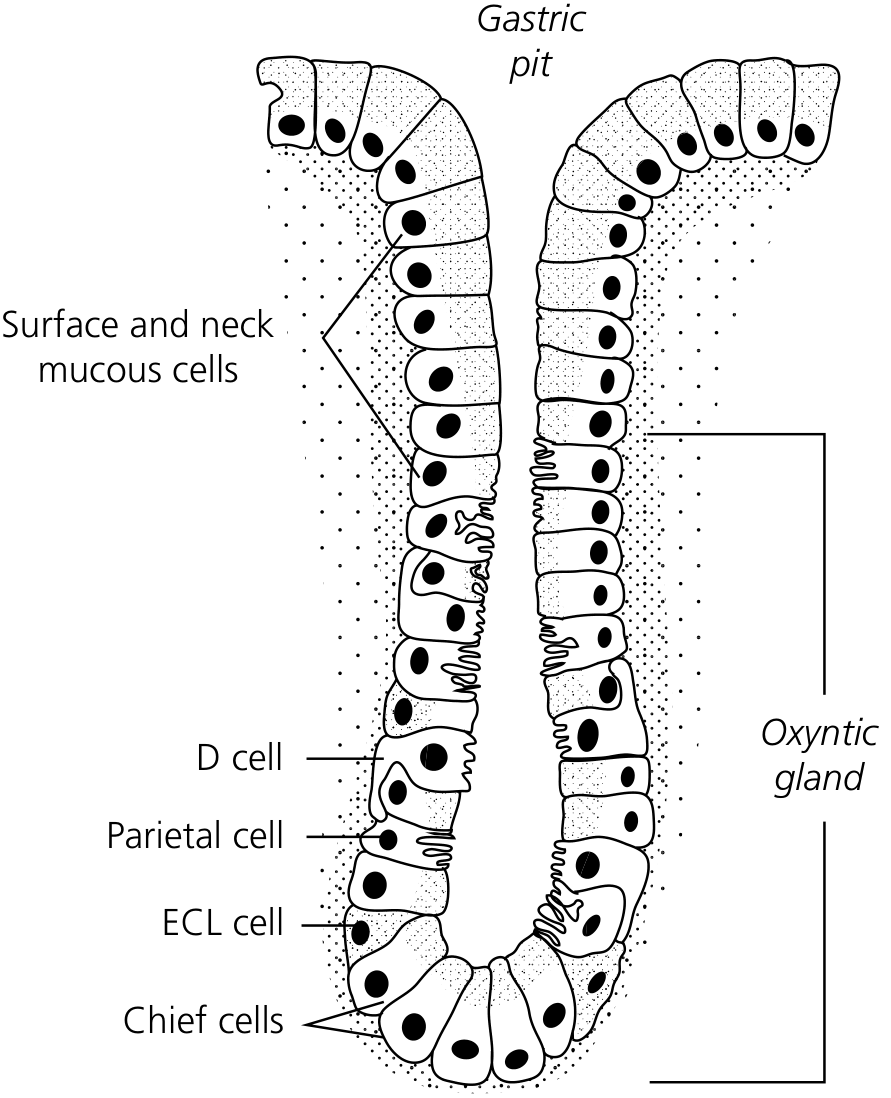
ऊपर के diagram में देखो: Gastric Pit ऊपर है, नीचे Oxyntic Gland है जिसमें D cell, Parietal cell, ECL cell, और Chief cells हैं।
Fundus की gastric glands branched tubular glands होती हैं। इन्हें 3 regions में divide करते हैं:
| Region | Location | Major Cells |
|---|---|---|
| Isthmus/Neck | Pit के नीचे | Stem cells, Mucous neck cells, कुछ Parietal cells |
| Body (Middle) | Middle portion | Parietal cells dominant |
| Base (Fundus of gland) | Muscularis mucosae के पास | Chief cells dominant |
5. CELL TYPES - DETAILED (MOST IMPORTANT)
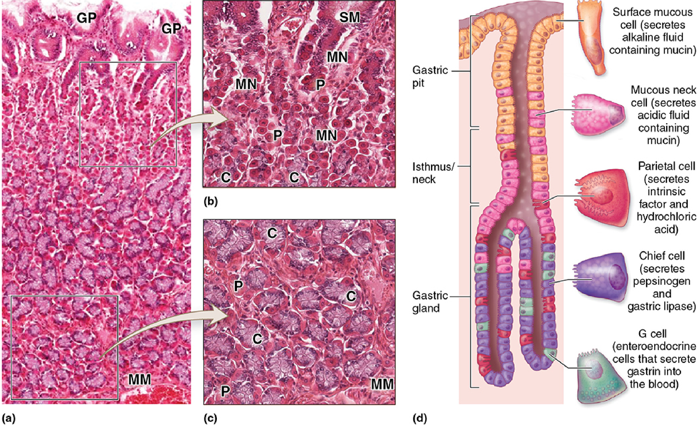
A. SURFACE MUCOUS CELLS (Foveolar cells)
- Location: Stomach surface + gastric pit lining
- Shape: Tall columnar cells
- Nucleus: Basal में, flattened
- Cytoplasm: Apical में pale, mucin granules से भरा
- Secretion: Thick, viscous, alkaline mucus + HCO₃⁻ ions
- Function: Mucosal protection - acid और mechanical abrasion से बचाती है
- Turnover: बहुत तेज़ - 4-7 days में replace होती हैं
B. MUCOUS NECK CELLS
- Location: Gland का neck/isthmus region (gastric pit और gland के junction पर)
- Shape: Surface mucous cells से छोटी, often distorted (neighboring cells के pressure से)
- Nucleus: Round, basal
- Cytoplasm: Apical में secretory granules
- Key difference from Surface mucous cells: इनका mucus कम alkaline होता है
- Important: Stem cells भी इसी region में होते हैं (pluripotent) जो सभी cell types को replace करते हैं
C. PARIETAL CELLS (Oxyntic Cells) ⭐ MOST IMPORTANT
- Location: Neck region में ज़्यादा, body में भी scattered
- Shape: Large, rounded/pyramidal cells - अक्सर tubule से बाहर bulge करती हैं
- Nucleus: Central, round (कभी-कभी 2 nuclei)
- Cytoplasm: Intensely eosinophilic (bright pink) - H&E में सबसे आसानी से identify होती हैं
- Eosinophilia का कारण: Abundant mitochondria (HCl production के लिए energy)
- Ultrastructure (EM पर):
- Numerous mitochondria
- Intracellular canaliculi (cell के अंदर channels)
- Resting state: Tubular vesicles apically
- Active state: Vesicles membrane में fuse होकर large canaliculi बनाते हैं + microvilli बढ़ते हैं
- Products:
- HCl (hydrochloric acid)
- Intrinsic Factor (Vitamin B12 absorption के लिए)
HCl Synthesis - Step by Step
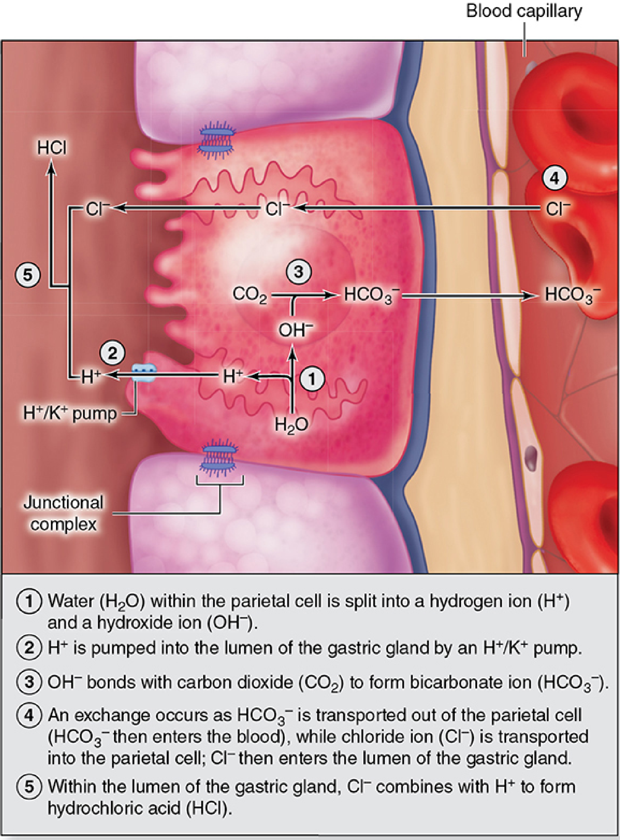
- H₂O → H⁺ + OH⁻ (cell के अंदर)
- H⁺ को H⁺/K⁺-ATPase pump द्वारा canaliculus में pump किया जाता है (K⁺ बाहर आता है)
- OH⁻ + CO₂ → HCO₃⁻ (carbonic anhydrase enzyme द्वारा)
- HCO₃⁻ को basal side से blood में भेजा जाता है, बदले में Cl⁻ अंदर आता है (antiport)
- Cl⁻ canaliculus में जाता है, वहाँ H⁺ से मिलकर HCl बनाता है
"Alkaline tide": खाने के बाद parietal cells इतना HCO₃⁻ blood में release करती हैं कि blood temporarily alkaline हो जाता है।
Control of Parietal Cell Secretion:
- Vagus (ACh) - stimulates
- Histamine (from ECL cells) - stimulates (H₂ receptor)
- Gastrin (from G cells) - stimulates
- Somatostatin (from D cells) - inhibits
Clinical: PPIs (Omeprazole) H⁺/K⁺-ATPase को block करते हैं। H₂ blockers (Ranitidine) histamine receptor block करते हैं।
D. CHIEF CELLS (Zymogenic/Peptic Cells) ⭐
- Location: Gland के lower/basal portion में predominant (clusters में)
- Shape: Columnar cells
- Nucleus: Basal में, round
- Cytoplasm: Basophilic (purple/blue) - H&E में lower gland में blue दिखती हैं
- Basophilia का कारण: Abundant RER (protein synthesis)
- Apical में zymogen granules (routine H&E में poorly stain होते हैं)
- Products:
- Pepsinogen → active pepsin (pH 1.8-3.5 पर activate, endoproteinase)
- Gastric lipase (lipid digestion)
- Protein-secreting cell की सभी characteristics: abundant RER, Golgi, apical granules
E. ENTEROENDOCRINE CELLS (DNES cells) ⭐
- Location: Gland के basal region में scattered
- Shape: Small cells, rarely visible on routine H&E
- Type: Mostly "Closed type" (lumen से contact नहीं, basally secrete करती हैं)
- Visualization: Chromium/Silver salts से - इसलिए "Enterochromaffin (EC) cells" या "Argentaffin cells" भी कहते हैं। आजकल immunohistochemistry use होती है।
- APUD cells = Amine Precursor Uptake and Decarboxylation
Fundus में important enteroendocrine cells:
| Cell | Hormone | Function |
|---|---|---|
| ECL cells (Enterochromaffin-like) | Histamine | Parietal cells को stimulate करती हैं HCl के लिए |
| D cells | Somatostatin | Local inhibition - अन्य DNES cells को inhibit |
| EC cells | Serotonin + Substance P | Gut motility बढ़ाती हैं |
(G cells = mainly pylorus में होती हैं, Gastrin secrete करती हैं)
6. STEM CELLS - Renewal का Source
- Location: Isthmus/Neck region (gastric pit और gland के junction)
- Type: Pluripotent stem cells
- Function: Asymmetric division करती हैं:
- ऊपर migrate → Surface mucous cells + pit lining cells (turnover: 4-7 days)
- नीचे migrate → Parietal, Chief, Enteroendocrine cells (turnover: बहुत slow)
7. LAMINA PROPRIA
- Loose connective tissue जो glands को surround करती है
- Scattered smooth muscle fibers
- Lymphocytes, plasma cells, macrophages, mast cells
- Fenestrated capillaries, lymphatics
8. MUSCULARIS MUCOSAE
- Thin smooth muscle layer जो mucosa को submucosa से separate करती है
- Fundus में gastric glands इसी तक पहुँचती हैं (full thickness of lamina propria)
9. COMPARISON TABLE - Fundus vs Antrum vs Cardia
| Feature | Fundus/Body | Antrum/Pylorus | Cardia |
|---|---|---|---|
| Gland type | Oxyntic/Gastric glands proper | Pyloric glands | Cardiac glands |
| Pit depth | Shallow (1/4 mucosa) | Deep (1/2+ mucosa) | Shallow |
| Parietal cells | +++ (abundant) | Absent | Absent |
| Chief cells | +++ | Absent (humans) | Absent |
| G cells | Absent/rare | +++ | Absent |
| Main secretion | HCl + Pepsinogen | Gastrin + Mucus | Mucus |
10. CLINICAL CORRELATES
- Pernicious Anemia: Autoimmune destruction of parietal cells → Intrinsic factor की कमी → B12 absorption नहीं → Megaloblastic anemia
- Gastric Ulcer: Fundus/body में mucosal barrier fail होने से (H. pylori, NSAIDs, excess HCl)
- Carcinoid Tumor (ECL cell tumor): ECL cells का neoplasia - hypergastrinemia में (Zollinger-Ellison syndrome)
- Gastric Atrophy: Parietal और chief cells का loss → Achlorhydria
Sources:
- Junqueira's Basic Histology - Text and Atlas, 17e (Ch. 15)
- Yamada's Textbook of Gastroenterology, 7e (Ch. 17)
- Mulholland and Greenfield's Surgery, 7e
This is a shared conversation. Sign in to Orris to start your own chat.