
What is nasopharyn
The nasopharynx is the uppermost part of the pharynx (throat), located behind the nasal cavity and above the soft palate. It connects the nasal passages to the oropharynx, serves as an air passageway, and houses the pharyngeal tonsil (adenoid). The Eustachian tubes open into it on either side, linking the middle ear to the throat.
Searching sources...
Reading File
Searching the Web
nasopharynx anatomy function
Finding Sources
Reading File
Reading File
Loading Image
The Nasopharynx
The nasopharynx is the uppermost division of the pharynx. Here is a full breakdown:
Location and Boundaries
- Anteriorly: opens into the nasal cavities via the posterior nasal apertures (choanae)
- Inferiorly: continuous with the oropharynx at the pharyngeal isthmus (marked by the palatopharyngeal sphincter fold)
- Superiorly/posteriorly: the roof is formed by the body of the sphenoid bone and the basal part of the occipital bone
- Level: extends from the skull base down to the upper border of the soft palate
Unlike the oro- and laryngopharynx, the nasopharynx is an air-only passage - it carries no food or drink.
Key Structural Features
| Structure | Description |
|---|---|
| Choanae | Posterior openings of the nasal cavities into the nasopharynx |
| Pharyngeal tonsil (Adenoids) | Lymphoid tissue on the roof/posterior wall; can enlarge in children to obstruct breathing |
| Pharyngotympanic (Eustachian) tube openings | Located on each lateral wall, connecting the nasopharynx to the middle ear |
| Torus tubarius | Cartilaginous ridge posterior to each Eustachian tube opening |
| Fossa of Rosenmüller (pharyngeal recess) | A recess posterior to the torus tubarius; clinically important as the most common site of nasopharyngeal carcinoma |
| Salpingopharyngeal fold | Mucosal fold descending from the torus tubarius |
| Sinus of Morgagni | A gap in the superior pharyngeal constrictor at the skull base where the Eustachian tube enters |
Functions
- Air conduction: Part of the upper airway, channeling air from the nose toward the larynx and trachea
- Pressure equalization: Muscles around the Eustachian tube openings (tensor and levator veli palatini) regulate middle ear pressure
- Immune surveillance: The pharyngeal tonsil (adenoid) filters pathogens entering via the nose
- Nasopharynx closure during swallowing: Elevation of the soft palate + contraction of the palatopharyngeal sphincter seals the nasopharynx off from the oropharynx, preventing food from entering the nasal cavity
Anatomy Diagram
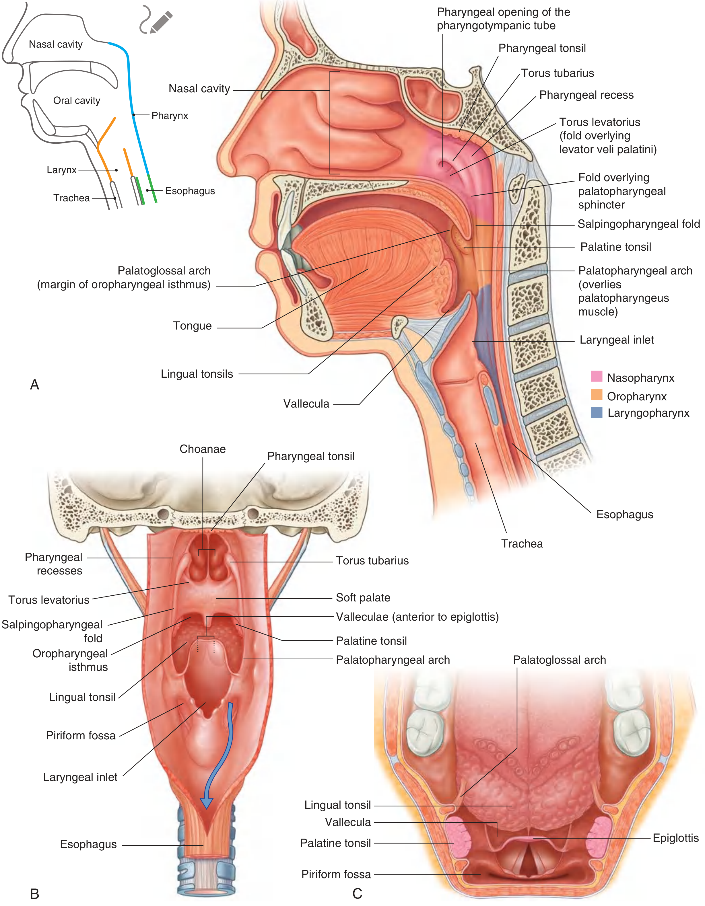
Fig. 8.200 - Mucosal features of the pharynx. The nasopharynx is shown in pink (A, lateral view; B, posterior view; C, superior view). - Gray's Anatomy for Students, p. 1186
Clinical Relevance
- Adenoid hypertrophy: Enlarged adenoids in children can obstruct the nasopharynx, causing mouth breathing, snoring, and middle ear effusion (glue ear)
- Nasopharyngeal carcinoma (NPC): Most commonly arises in the fossa of Rosenmüller; strongly associated with Epstein-Barr virus (EBV) and is more prevalent in Southeast Asian populations
- Otitis media: Nasopharyngeal masses or inflammation can block the Eustachian tube, leading to middle ear problems; all patients with unilateral otitis media should have their nasopharynx examined
- Nasopharyngeal angiofibroma: A benign but locally aggressive vascular tumor occurring in adolescent males, typically at the posterior choana
- Tornwaldt cyst: A benign midline cyst in the nasopharynx arising from a persistent embryological notochord remnant
Sources: Gray's Anatomy for Students, p. 1186-1187; Cummings Otolaryngology, p. 85; StatPearls - Nasopharynx Anatomy
This is a shared conversation. Sign in to Orris to start your own chat.