
Readh thi pdf and explain, Defination, criteria, indication, types of phakic iol, power calculation, advantage dis advantage, complications of phakis iol idividual complication and their management, make a long answr question for exam writing
python3 << 'EOF' import pdfplumber with pdfplumber.open("/tmp/workspace/attachments/36d95aac-b66c-4382-8cf5-5dd6d6d05aa5/Phakic IOL.pdf") as pdf: print(f"Total pages: {len(pdf.pages)}") for i, page in enumerate(pdf.pages): text = page.extract_text() print(f"\n===== PAGE {i+1} =====") if text: print(text) else: print("[No text extracted]") EOF
python3 << 'EOF' import base64 from pypdf import PdfReader, PdfWriter import io reader = PdfReader("/tmp/workspace/attachments/36d95aac-b66c-4382-8cf5-5dd6d6d05aa5/Phakic IOL.pdf") print(f"Pages: {len(reader.pages)}") # Try to extract images from first page page = reader.pages[0] if '/Resources' in page: resources = page['/Resources'] print(f"Resources keys: {list(resources.keys())}") if '/XObject' in resources: xobject = resources['/XObject'] print(f"XObject keys: {list(xobject.keys())}") EOF
python3 << 'EOF' import os from pypdf import PdfReader import base64 reader = PdfReader("/tmp/workspace/attachments/36d95aac-b66c-4382-8cf5-5dd6d6d05aa5/Phakic IOL.pdf") os.makedirs("/tmp/workspace/phakic_pages", exist_ok=True) for i, page in enumerate(reader.pages): resources = page['/Resources'] xobject = resources['/XObject'] img_obj = xobject['/Im0'] img_obj = img_obj.get_object() # Get raw image data data = img_obj._data # Save as file # Check filter filters = img_obj.get('/Filter', None) print(f"Page {i+1}: Filter={filters}, Width={img_obj.get('/Width')}, Height={img_obj.get('/Height')}, ColorSpace={img_obj.get('/ColorSpace')}") with open(f"/tmp/workspace/phakic_pages/page_{i+1:02d}.raw", "wb") as f: f.write(data) print("Done extracting raw image data") EOF
python3 << 'EOF' import os from pypdf import PdfReader reader = PdfReader("/tmp/workspace/attachments/36d95aac-b66c-4382-8cf5-5dd6d6d05aa5/Phakic IOL.pdf") os.makedirs("/tmp/workspace/phakic_pages", exist_ok=True) for i, page in enumerate(reader.pages): resources = page['/Resources'] xobject = resources['/XObject'] img_obj = xobject['/Im0'] img_obj = img_obj.get_object() data = img_obj._data with open(f"/tmp/workspace/phakic_pages/page_{i+1:02d}.jpg", "wb") as f: f.write(data) print("Done saving JPEGs") import os files = os.listdir("/tmp/workspace/phakic_pages") print(files) EOF
python3 << 'EOF' from PIL import Image import os input_dir = "/tmp/workspace/phakic_pages" output_dir = "/tmp/workspace/phakic_small" os.makedirs(output_dir, exist_ok=True) for i in range(1, 13): fname = f"page_{i:02d}.jpg" img = Image.open(os.path.join(input_dir, fname)) # Resize to max width 1024 keeping aspect ratio w, h = img.size new_w = 1024 new_h = int(h * new_w / w) img_resized = img.resize((new_w, new_h), Image.LANCZOS) out_path = os.path.join(output_dir, fname) img_resized.save(out_path, "JPEG", quality=85) size = os.path.getsize(out_path) print(f"Page {i}: {w}x{h} -> {new_w}x{new_h}, {size} bytes") print("Done") EOF
PHAKIC INTRAOCULAR LENSES (Phakic IOLs)
Comprehensive Exam Answer
1. DEFINITION
- Aphakic = no natural lens present
- Pseudophakic = natural lens replaced by an IOL
- Phakic IOL = natural lens retained + additional lens implanted
2. CRITERIA / PATIENT SELECTION CRITERIA
Refractive Criteria:
- Myopia: -6 D to -20 D (or higher - up to -25 D)
- Hyperopia: +3 D to +10 D
- Astigmatism: up to 6 D (toric phakic IOLs)
Age Criteria:
- Minimum age: 21 years (refractive error must be stable)
- Maximum age: generally <45-50 years (before significant presbyopia and early cataract)
- Refraction must be stable for at least 1-2 years (change < 0.5 D per year)
Corneal Criteria:
- Inadequate corneal thickness for LASIK (< 480-500 µm residual stromal bed)
- No significant corneal pathology (keratoconus, dystrophies)
Anterior Chamber Criteria:
- Anterior chamber depth (ACD): minimum 2.8 mm (angle-supported) or 3.0 mm (iris-fixated) from endothelium
- Endothelial cell count (ECC): must be adequate - typically > 2000-2500 cells/mm²
- White-to-white (WTW) distance: 11.0-12.5 mm (for sizing)
Other Criteria:
- IOP normal (< 21 mmHg)
- No history of uveitis, glaucoma, or significant retinal pathology
- No pupil abnormalities
- Pupil diameter in scotopic conditions (if anterior chamber lens - to avoid glare/halos)
- No systemic contraindications (autoimmune, immunocompromise)
3. INDICATIONS
- High myopia: > -6 D where LASIK is contraindicated due to thin cornea or inadequate residual stromal bed
- High hyperopia: > +4 D
- High astigmatism: when corneal correction alone is inadequate
- Thin corneas (inadequate corneal thickness for ablative procedures)
- Dry eyes that preclude LASIK/surface ablation
- Large optical zones required (large pupils)
- Patient preference for reversible procedure
- Anisometropia correction when LASIK cannot achieve adequate correction
- Patients with occupations at risk for corneal trauma (military, contact sports) - reversible phakic IOL preferred
- Refractive error in the "LASIK gray zone" where outcome is uncertain
4. TYPES OF PHAKIC IOLs
A. ANTERIOR CHAMBER PHAKIC IOLs
i. Angle-Supported (Angle-Fixated) IOLs
- The haptics rest in the iridocorneal angle (trabecular meshwork)
- Examples: NuVita MA20 (Bausch & Lomb), ZB5M
- Design: Rigid PMMA or flexible (PMMA/acrylic/silicone)
- Sizing: Based on white-to-white diameter
- Problems: Endothelial cell loss (haptics touch angle), pupil ovalization, glaucoma, chronic inflammation
- Status: Largely abandoned due to unacceptable complication rates
ii. Iris-Fixated (Iris-Claw) IOLs
- The lens is clipped/enclavated onto the anterior iris stroma (mid-peripheral iris) using a claw mechanism
- Examples:
- Artisan IOL (Ophtec, Netherlands) - rigid PMMA lens
- Verisyse IOL (Abbott/AMO) - PMMA version of Artisan
- Artiflex - foldable version (silicone optic, PMMA haptics)
- Toric Artisan - for astigmatism correction
- Position: Between cornea and iris
- Key feature: Enclavation at 3 and 9 o'clock positions on iris
- Advantage: Does not touch trabecular meshwork; less risk of glaucoma
- Requires: Peripheral iridectomy (PI) to prevent pupillary block
B. POSTERIOR CHAMBER PHAKIC IOLs
Implantable Collamer Lens (ICL) / Implantable Contact Lens
- Implanted in the posterior chamber between the iris posteriorly and crystalline lens anteriorly (in the ciliary sulcus)
- Material: Collamer (a biocompatible hydrophilic collagen co-polymer with UV-blocking ability)
- Examples:
- ICL (STAAR Surgical, EVO ICL) - most widely used globally
- TICL - toric ICL for astigmatism correction
- EVO+ ICL - newer version with central port (KS-AquaPORT) - no peripheral iridectomy required
- PRL (Phakic Refractive Lens) - silicone posterior chamber lens (less common)
- Size: Available in 12.1, 12.6, 13.2, 13.7 mm sizes based on horizontal white-to-white and anterior chamber depth
- Range: -0.5 D to -20 D (myopic), +0.5 D to +10 D (hyperopic), with toric options
Summary Table of Types:
| Type | Location | Fixation | Example |
|---|---|---|---|
| Angle-supported AC | Anterior chamber | Iridocorneal angle | NuVita MA20 |
| Iris-claw | Anterior chamber | Iris stroma | Artisan / Verisyse |
| Posterior chamber | Posterior chamber | Ciliary sulcus | ICL (EVO ICL) |
5. POWER CALCULATION
Formula Used:
P (phakic IOL power) = [Spectacle Rx / (1 - d × Spectacle Rx)]
Van der Heijde Formula (for anterior chamber IOLs):
P = (n / (d - (n/P_total))) - (n / (d - 0))
Manufacturer-Specific Nomograms:
- ICL (STAAR): Uses proprietary online calculator incorporating:
- Manifest refraction (sphere and cylinder)
- ACD (anterior chamber depth)
- WTW (white-to-white = corneal diameter)
- Keratometry readings
- Vault prediction algorithms (EVO ICL uses myopic astigmatic formula)
Biometric Parameters Needed:
- Manifest refraction (sphere, cylinder, axis)
- Cycloplegic refraction
- Keratometry (K readings)
- Anterior chamber depth (ACD - from endothelium to anterior lens surface)
- Axial length
- White-to-white (WTW) corneal diameter
- Endothelial cell count (ECC)
- Sulcus-to-sulcus (STS) distance - measured by UBM or OCT
Sizing (for ICL):
- Undersized ICL: low vault, risk of cataract (lens touching natural lens)
- Oversized ICL: high vault, risk of pupillary block, angle closure, raised IOP
6. ADVANTAGES AND DISADVANTAGES
ADVANTAGES
| Advantage | Explanation |
|---|---|
| Reversible | Lens can be removed; unlike LASIK which is irreversible |
| No corneal tissue removed | Corneal biomechanics preserved |
| Wider range of correction | Corrects up to -20 D myopia; beyond LASIK range |
| Excellent optical quality | High contrast sensitivity, minimal aberrations |
| Rapid visual recovery | Vision improves within 24-48 hours |
| Stable long-term results | No regression (unlike LASIK) |
| UV protection | Collamer material blocks UV radiation |
| Night vision preserved | Larger optic zone than LASIK |
| Suitable for thin corneas | Does not require corneal stroma |
| Toric options available | Corrects astigmatism simultaneously |
DISADVANTAGES
| Disadvantage | Explanation |
|---|---|
| Intraocular surgery | Higher risk than surface procedure (infection, hemorrhage) |
| Endothelial cell loss | Particularly with anterior chamber IOLs |
| Cataract risk | ICL can induce anterior subcapsular cataract if vault too low |
| Glaucoma risk | Pupillary block, pigment dispersion |
| Halos/glare | Especially with large pupils and smaller optic zones |
| Cost | Expensive; not covered by insurance in most countries |
| Requires PI or iridotomy | (older ICL designs) |
| Sizing challenges | No perfect method; under/over-vault complications |
| No presbyopia correction | Accommodation not addressed |
| Long-term unknown | 20-30 year data limited |
| Cannot correct all astigmatism | Some residual astigmatism may remain |
7. COMPLICATIONS - INDIVIDUAL COMPLICATIONS AND THEIR MANAGEMENT
A. INTRAOPERATIVE COMPLICATIONS
1. Hyphema
- Cause: Trauma to iris vessels during manipulation, enclavation, or haptic placement
- Management:
- Small hyphemas: conservative (head elevation, cycloplegia, topical steroids)
- Large hyphemas: observation; surgical washout if IOP uncontrolled or clot persistent > 7 days
- Prevent: gentle handling of iris, avoid iris trauma
2. Pupillary Block (Acute)
- Cause: Aqueous trapped behind lens obstructing flow through pupil
- Prevention: Peripheral iridectomy (PI) - performed at surgery or pre-operatively with Nd:YAG laser
- Management: Emergent Nd:YAG peripheral iridotomy; if not possible, surgical PI
B. EARLY POSTOPERATIVE COMPLICATIONS
3. Elevated Intraocular Pressure (IOP)
- Causes:
- Retained viscoelastic in anterior chamber (most common - resolves within 24-48h)
- Pupillary block
- Pigment dispersion
- Steroid-induced
- Management:
- Temporary: oral/topical IOP-lowering agents (acetazolamide, timolol, brimonidine)
- Residual viscoelastic: resolves spontaneously; consider paracentesis if severe
- Pupillary block: Nd:YAG iridotomy
- Steroid response: switch to lower-potency steroid
4. Corneal Edema
- Cause: Endothelial cell trauma during surgery (particularly with rigid lenses)
- Management:
- Mild: topical hypertonic saline (NaCl 5%), lubricants
- Persistent/severe: evaluate for bullous keratopathy - may need DSAEK/DMEK
- Prevention: careful technique, use of OVDs, smaller incisions
5. Infection / Endophthalmitis
- Incidence: Rare (< 0.1%) but vision-threatening
- Organisms: Staphylococcus epidermidis, S. aureus, gram-negative
- Management:
- Intravitreal vancomycin (1 mg/0.1 mL) + ceftazidime (2.25 mg/0.1 mL)
- Vitreous tap/injection or pars plana vitrectomy if no improvement at 48h
- Systemic antibiotics if severe
- IOL removal may be necessary
C. LATE POSTOPERATIVE COMPLICATIONS
6. Cataract Formation (Anterior Subcapsular Cataract)
- Most common serious complication of posterior chamber phakic IOLs (ICL)
- Cause:
- Low vault (< 250 µm): ICL contacts the anterior lens capsule, disrupting metabolism
- Chronic subclinical contact
- Mechanical trauma during implantation
- Grading: Anterior subcapsular opacity to nuclear sclerosis
- Management:
- Prevention: adequate sizing, maintain vault 250-750 µm
- Mild lens opacity: observation
- Visually significant cataract: ICL removal + phacoemulsification + posterior chamber IOL
- ICL power can sometimes be incorporated into the replacement IOL calculation
7. Glaucoma
- Types:
- Pupillary block glaucoma: inadequate PI
- Pigment dispersion glaucoma: ICL rubbing on posterior iris surface releasing pigment
- Angle closure: oversized lens
- Steroid-induced glaucoma
- Mechanical angle crowding (angle-supported lenses)
- Incidence: More common with angle-supported lenses (up to 10-40%)
- Management:
- Medical: topical prostaglandins, beta blockers, CAIs
- Laser: Nd:YAG iridotomy for pupillary block; SLT for open angle
- Surgical: trabeculectomy, tube shunt if uncontrolled
- ICL removal: if vault is pathologically high or angle crowding
- Lens rotation (for oversized ICL)
8. Endothelial Cell Loss (ECL)
- Cause: Proximity of phakic IOL to corneal endothelium (especially iris-claw and angle-supported lenses)
- Normal physiological ECL: ~0.6%/year
- Phakic IOL ECL: 1-2%/year or more (anterior chamber lenses)
- Significance: Endothelial cell count < 500 cells/mm² leads to bullous keratopathy
- Management:
- Regular monitoring: specular microscopy every 6-12 months
- If ECC falling rapidly or < 1500 cells/mm²: explant the phakic IOL
- Established bullous keratopathy: penetrating keratoplasty (PK) or lamellar (DSAEK, DMEK)
- ICL has significantly less ECL than anterior chamber lenses
9. Pigment Dispersion Syndrome
- Mechanism: ICL haptics or optic rubs against posterior iris surface → iris pigment released → trabecular meshwork blockage → IOP rise
- Signs: Pigment on corneal endothelium (Krukenberg spindle), on anterior IOL surface, in angle
- Management:
- Miosis (pilocarpine) to reduce iris-ICL contact
- IOP-lowering medications
- ICL exchange for smaller size or different model
- Nd:YAG iridotomy for pupillary block component
10. Decentration / Rotation / Dislocation
- Causes: Incorrect sizing, trauma, inadequate sulcus support, poor surgical technique
- For Toric ICL: Rotation leads to residual astigmatism (every 1° off-axis = ~3.3% loss of cylindrical correction)
- Management:
- Decentration: surgical repositioning under topical anesthesia
- Rotation of toric ICL: reposition to correct axis
- Complete dislocation: surgical retrieval and replacement or removal
11. Halos and Glare (Dysphotopsia)
- Cause: Edge of phakic IOL within the pupillary zone in mesopic/scotopic conditions
- Risk factors: Large pupils, undersized optic diameter, high refractive error correction
- Management:
- Most cases improve with time (neural adaptation)
- Pupil-constricting drops (pilocarpine) at night - temporary
- IOL exchange for larger optic or repositioning
- Reassurance - majority resolve within 3-6 months
12. Incorrect Vault (ICL-specific)
- Low vault (< 250 µm):
- Risk of cataract, lens contact
- Management: Exchange for larger ICL size
- High vault (> 750-1000 µm):
- Risk of angle closure, elevated IOP, glaucoma, pupillary block
- Management: Exchange for smaller ICL size; Nd:YAG iridotomy if pupillary block
13. Uveitis / Chronic Inflammation
- Cause: Foreign body reaction, mechanical contact with uveal tissue, residual lens material
- Management:
- Topical corticosteroids (prednisolone acetate 1%)
- NSAIDs
- Cycloplegics (atropine 1%, homatropine)
- If persistent: IOL explantation
14. Pupil Ovalization
- More common with angle-supported lenses
- Cause: Haptics in the angle pulling the iris tissue, distorting the pupil
- Management:
- Observation if mild and asymptomatic
- IOL explantation if progressive or causing visual symptoms
- Largely eliminated with modern posterior chamber ICLs
15. Retinal Detachment (RD)
- Risk: Increased in high myopes (inherent risk of retinal pathology)
- Not directly caused by ICL but myopic eyes have thin retinas and lattice degeneration
- Management:
- Preoperative: treat lattice degeneration and holes with laser retinopexy
- Established RD: pneumatic retinopexy, scleral buckle, or pars plana vitrectomy (PPV)
8. LONG-FORM EXAM QUESTION AND MODEL ANSWER
EXAM QUESTION:
MODEL ANSWER:
(a) Definition and Rationale (5 marks)
- Estimated ablation depth ≈ 14 × 12 µm = ~168 µm
- Minimum flap thickness: 100-110 µm (microkeratome) or 90 µm (femtosecond)
- Corneal thickness: 490 µm
- Residual stromal bed (RSB) = 490 - 100 (flap) - 168 (ablation) = 222 µm
- This is below the safe minimum of 250-300 µm RSB, risking post-LASIK ectasia
- Preserves corneal integrity
- Reversible procedure
- Excellent predictability for high myopia
- No risk of regression
- Age 28 is ideal (stable refraction, no presbyopia, no early cataract)
(b) Classification of Phakic IOLs (8 marks)
- Haptics rest in the iridocorneal angle
- Examples: NuVita MA20, ZB5M (Bausch & Lomb)
- Largely abandoned - associated with progressive endothelial cell loss, pupil ovalization, and glaucoma
- Enclavated onto the anterior iris stroma at mid-periphery (3 and 9 o'clock)
- Examples: Artisan (PMMA), Artiflex (foldable), Toric Artisan
- Requires peripheral iridectomy
- Risk of endothelial cell loss (due to proximity to cornea)
- Placed in the posterior chamber, between iris and natural crystalline lens in the ciliary sulcus
- Material: Collamer (hydrophilic collagen co-polymer with UV blocker)
- Examples: STAAR ICL, EVO ICL (KS-AquaPORT - central port eliminates need for PI), Toric ICL (TICL)
- Gold standard phakic IOL
- ACD of 3.2 mm is adequate (minimum requirement 3.0 mm)
- ECC of 2800 is excellent (>2000 cells/mm²)
- ICL has the lowest endothelial cell loss rate (~0.5-1.5%/year vs. 2-3% for AC lenses)
- No peripheral iridectomy required with EVO ICL
- Highest long-term safety profile
- Corrects up to -20 D (this patient needs -14 D)
- Toric option available (check for astigmatism)
- Reversible if complications arise
(c) Power Calculation (7 marks)
- Manifest refraction (sphere, cylinder, axis)
- Cycloplegic refraction
- Keratometry (K1, K2) - to determine corneal power
- Axial length
- Anterior chamber depth (ACD) from endothelium - 3.2 mm (given)
- White-to-white (WTW) diameter - 12.0 mm (given) - for ICL sizing
- Sulcus-to-sulcus (STS) distance - by UBM or anterior OCT - most accurate for ICL sizing
- Endothelial cell count - 2800 cells/mm² (given)
- Pupil diameter (photopic and scotopic)
P (IOL) = Pspectacle / (1 - d × Pspectacle)
P = -14.0 / (1 - 0.012 × (-14.0)) P = -14.0 / (1 + 0.168) P = -14.0 / 1.168 P ≈ -11.99 D ≈ -12.0 D
- Spherical equivalent refraction
- Cylinder and axis
- ACD
- WTW
- Keratometry
- Desired postoperative refraction (usually plano or slight myopia if bilateral)
- WTW + 0.5 to 1.0 mm = approximately 12.5-13.0 mm → select 13.2 mm ICL
- Vault target: 250-750 µm (monitored postoperatively by OCT)
(d) Complications and Management (20 marks)
- Management: Head elevation, cycloplegia (atropine 1%), topical steroids; surgical washout if clot is persistent or IOP rises
- Prevention: Use of adequate ophthalmic viscosurgical device (OVD); foldable IOL through a smaller incision; careful insertion technique
- Causes: Retained OVD, pupillary block, steroid response
- Management: IOP-lowering drops (brimonidine, timolol, oral acetazolamide); Nd:YAG iridotomy for pupillary block; switch steroids if steroid responder; resolves within 48h if OVD-related
- Due to surgical trauma to endothelium
- Management: Hypertonic NaCl 5% drops/ointment; lubricants; monitor ECC; if persistent - decompensated - DSAEK/DMEK
- Mechanism: Aqueous accumulates behind IOL, pushes iris forward, blocks trabecular outflow
- Management: Urgent Nd:YAG peripheral iridotomy; surgical PI if Nd:YAG fails
- Rare but sight-threatening (< 0.1%)
- Organisms: Staphylococcus epidermidis, S. aureus
- Management: Urgent vitreous tap + intravitreal antibiotics (vancomycin + ceftazidime); PPV if no improvement; remove IOL if focus of infection
- Grade 0: No opacity (vault > 250 µm) - ideal
- Grade 1-2: Anterior subcapsular punctate opacities (vault 100-250 µm) - monitor
- Grade 3-4: Progressive opacification (vault < 100 µm) - visually significant
- Management:
- Prevention: correct sizing; target vault 250-750 µm
- Mild: observation, ICL exchange to larger size to increase vault
- Visually significant cataract: ICL removal + phacoemulsification + PC IOL implantation
- Types: Pupillary block, pigment dispersion, angle closure (oversized ICL), steroid-induced
- Management:
- Pupillary block: Nd:YAG PI (prophylactic in older ICL; not needed in EVO ICL with KS-AquaPORT)
- Pigment dispersion: pilocarpine 1%, IOP-lowering drops; ICL exchange if persistent
- Angle closure (oversized ICL): exchange for smaller ICL
- Medically uncontrolled: trabeculectomy, tube shunt
- Steroid-induced: substitute fluorometholone or prednisolone with tapering
- Rate: ~0.5-1.5%/year (ICL); up to 2-3%/year (iris-claw); up to 5%/year (angle-supported)
- Monitoring: Specular microscopy every 6-12 months
- Management: If rapid decline or ECC < 1500-1800 cells/mm², explant the phakic IOL to prevent corneal decompensation. Established bullous keratopathy: corneal transplant (DSAEK/DMEK preferred over PK)
- ICL haptics contact posterior iris pigment epithelium → pigment granules released → Krukenberg spindle, elevated IOP
- Management: Pilocarpine (reduces iris-ICL contact); IOP-lowering agents; ICL exchange for smaller/different size; Nd:YAG iridotomy
- Decentration: visual symptoms (halos, decentered reflex); surgical repositioning
- Toric ICL rotation: each degree of off-axis rotation causes ~3.3% reduction in cylindrical correction; if > 30° rotation or visually significant: surgical reposition to correct axis
- Particularly in dim light when pupil dilates beyond the optic edge
- Management: Neural adaptation (most improve in 3-6 months); pilocarpine at night (temporary); IOL exchange for larger optic diameter if persistent and disabling
- Low vault (< 250 µm): exchange for larger ICL
- High vault (> 750-1000 µm): exchange for smaller ICL; Nd:YAG PI for pupillary block component; monitor IOP
- Chronic low-grade inflammation due to mechanical irritation
- Management: Topical prednisolone 1%; NSAIDs; cycloplegics; if persistent: explant IOL
- Historically common with angle-supported lenses (now obsolete)
- Management: Observation if mild; IOL removal if progressive
- High myopes are at increased risk of lattice degeneration, retinal holes, and detachment (independent of ICL)
- Preoperative fundus examination; laser prophylaxis of lattice/holes
- Established RD: pneumatic retinopexy, scleral buckle, or PPV based on extent and location
SUMMARY TABLE OF COMPLICATIONS AND MANAGEMENT:
| Complication | Timing | Management |
|---|---|---|
| Hyphema | Intraoperative | Head elevation, steroids; surgical washout if severe |
| Corneal edema | Early | Hypertonic NaCl; DSAEK if decompensation |
| Elevated IOP | Early | IOP-lowering drops; Nd:YAG PI if pupillary block |
| Endophthalmitis | Early | Intravitreal antibiotics ± PPV |
| Cataract | Late | ICL exchange (early) or phaco + PC IOL (if opaque) |
| Glaucoma | Late | Medical/laser/surgical; ICL exchange |
| Endothelial cell loss | Late (progressive) | Regular specular microscopy; explant if declining rapidly |
| Pigment dispersion | Late | Pilocarpine; IOP drops; ICL exchange |
| Decentration/rotation | Late | Surgical repositioning |
| Halos/glare | Late | Reassurance; adapt; exchange if disabling |
| Low vault | Late | Exchange for larger ICL |
| High vault | Late | Exchange for smaller ICL |
KEY POINTS TO REMEMBER FOR EXAMS:
- Phakic IOL = IOL + natural lens retained (phakos = lens in Greek)
- ICL (EVO) is the gold standard - posterior chamber, sulcus-placed, collamer material
- Minimum ACD for ICL = 3.0 mm; for iris-claw = 2.8 mm
- ECC must be > 2000 cells/mm² before surgery
- Target vault = 250-750 µm (too low = cataract; too high = glaucoma/AC angle closure)
- Most common serious late complication = anterior subcapsular cataract (low vault)
- Angle-supported lenses are largely abandoned due to unacceptable ECL and glaucoma rates
- EVO ICL has a central port (KS-AquaPORT) - eliminates need for peripheral iridectomy
- Power conversion: spectacle power is always less than IOL power required at the ocular plane (vertex effect)
- Contraindications: ACD < 3.0 mm, ECC < 2000, glaucoma, uveitis, unstable refraction, age < 21
Explain Osteoodento keratoprostesis for example in detail
osteo-odonto-keratoprosthesis OOKP tooth cornea prosthesis
keratoprosthesis corneal blindness artificial cornea surgery
osteo-odonto-keratoprosthesis OOKP procedure steps indications complications review
https://eyewiki.org/Modified_Osteo-Odonto-Keratoprosthesis
osteo-odonto-keratoprosthesis OOKP outcomes complications
https://eyewiki.org/Modified_Osteo-Odonto-Keratoprosthesis
OSTEO-ODONTO-KERATOPROSTHESIS (OOKP)
"Tooth-in-Eye" Surgery - Comprehensive Explanation
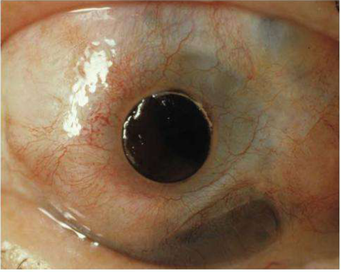
1. DEFINITION AND CONCEPT
- The entire diseased cornea and conjunctival surface is replaced by oral mucosa (which tolerates dryness and survives without a lacrimal film)
- A tooth-bone lamina acts as a natural, biocompatible scaffold
- A PMMA optical cylinder drilled through the lamina provides the refracting window for vision
2. HISTORICAL BACKGROUND
| Year | Milestone |
|---|---|
| 1963 | Prof. Benedetto Strampelli (Rome, Italy) - pioneer, first described and performed OOKP |
| 1970s | Falcinelli (Italy) - modified technique (MOOKP - Modified OOKP), improved outcomes |
| 2001-2002 | Rome-Vienna Protocol - international standardization, became the gold standard |
| 2004 | First OOKP performed in UK (Christopher Liu, Sussex Eye Hospital) |
| 2022 | 30-year retrospective data published: 94% anatomical survival, 81% maintained vision >20/200 at 10 years |
3. RATIONALE - WHY USE A TOOTH?
- Autologous tissue - no immune rejection
- Bone + periodontal ligament - provides structural rigidity AND a vascularized, living scaffold
- Highly biocompatible - integrates with surrounding ocular and mucosal tissues
- Long-term stability - bone remodels and survives decades in the cheek pouch
- Resists infection - living tissue with blood supply, unlike synthetic haptics which get infected
- PDL cells prevent resorption and promote integration
- It is non-keratinized (like conjunctiva)
- It produces mucus and is designed to survive without a continuous liquid tear film
- It is a wet epithelium - ideal for the ocular surface
- It is easily harvested with minimal donor site morbidity
4. INDICATIONS
- All other surgical rehabilitation has failed or is deemed futile
- Conventional penetrating keratoplasty (corneal graft) is contraindicated or has failed repeatedly
Specific Conditions:
- Stevens-Johnson Syndrome (SJS) / Toxic Epidermal Necrolysis (TEN) - most common indication
- Ocular Cicatricial Pemphigoid (OCP)
- Graft-versus-Host Disease (GvHD) after bone marrow transplant
- Lyell syndrome
- Severe alkali burns (lime, sodium hydroxide) with total limbal stem cell deficiency
- Acid burns
- Radiation burns
- Trachoma (Chlamydia trachomatis - worldwide leading infectious cause of corneal blindness)
- Multiple failed corneal grafts following HSV, bacterial, or fungal keratitis
- Aniridia with severe keratopathy (congenital)
- Multiple (>3) failed penetrating keratoplasties
- Severe dry eye with keratinization of corneal surface
Vision Criteria:
- Visual acuity of counting fingers (CF) or worse in the better eye
- Proven intact optic nerve and retinal function (light projection and perception in all four quadrants; ERG and VEP if needed)
- The patient must have potential for functional vision if the optical axis is cleared
5. CONTRAINDICATIONS
Absolute Contraindications:
- Active, uncontrolled inflammation (disease must be in the inactive phase)
- No light perception bilaterally (no retinal/optic nerve function - no point in surgery)
- Glaucoma with severe optic neuropathy (advanced field loss)
- Retinal detachment (untreated)
- Edentulous patient with no suitable donor tooth (no tooth = no surgery in original technique)
- Uncontrolled systemic disease precluding multi-stage major surgery
- Very young children (technically and ethically complex)
Relative Contraindications:
- Active systemic autoimmune disease (flare) - defer until controlled
- Severe psychiatric illness / inability to comply with follow-up
- Systemic corticosteroid dependence causing immune suppression
- Very advanced glaucoma with some residual field
- Uncontrolled diabetes
6. PREOPERATIVE ASSESSMENT (Multidisciplinary)
A. Ophthalmological Assessment:
- Visual acuity and light projection in all quadrants
- B-scan ultrasonography - assess posterior segment (vitreous, retina, optic nerve)
- Electroretinogram (ERG) - retinal function
- Visual Evoked Potentials (VEP) - optic nerve and cortical function
- IOP measurement (Schiotz or pneumotonometer - no corneal contact possible)
- Ultrasound biomicroscopy (UBM) - anterior segment anatomy, presence of angle
- Assess conjunctival and eyelid status
B. Dental Examination:
- Identify the best available tooth (canine or premolar preferred - see section on tooth selection)
- OPG (Orthopantomogram) - panoramic X-ray of jaw to assess tooth root length and bone quality
- Root length must be sufficient to fashion a lamina of adequate size
- No endodontic treatment ideally (root canal teeth are weaker)
- Edentulous patients: consider allograft (donor) tooth as alternative (higher resorption risk)
C. Pre-anesthetic Evaluation:
- Two-stage major general anesthesia required
- Cardiac, respiratory, hepatic, renal workup
D. Psychological Evaluation:
- Patient must understand the complexity, multistage nature, long-term commitment
- High patient motivation is listed as a key factor by Kanski's for surgical success
- Counselling about realistic expectations (functional, not always perfect vision)
7. THE OOKP PROSTHESIS - ANATOMY OF THE DEVICE
         [PMMA OPTICAL CYLINDER]
                  |
         [TOOTH ROOT LAMINA]  ← drilled through
                  |
    [ALVEOLAR BONE surrounding root]
Tooth Selection:
- Canine (eyetooth) - preferred: longest root, most alveolar bone
- Premolar - acceptable alternative
- Lower teeth preferred over upper (more bone)
- Root length: ideally > 14 mm
- Root width: > 5 mm to allow drilling
- Single-rooted tooth preferred (simpler lamina preparation)
PMMA Optical Cylinder:
- Made of polymethyl methacrylate (PMMA)
- Refractive index ~1.49
- Diameter: 3-4 mm (optic zone)
- Power: +10 D to +50 D (usually ~+20 to +30 D, compensating for removal of natural cornea/lens)
- Can be customized for each patient's target refraction
- The patient typically wears spectacles or contact lenses postoperatively for fine-tuning
8. SURGICAL TECHNIQUE - STEP BY STEP
STAGE 1A: Preparation of the Eye
- Conjunctival and corneal surface stripping: All keratinized epithelium, pannus, scar tissue, and diseased conjunctiva are surgically removed from the entire ocular surface including the inner eyelid surfaces (tarsal conjunctiva)
- Lysis of symblephara: Adhesions between eyelid and globe are released
- Keratectomy: The scarred, vascularized cornea is superficially excised (not penetrating at this stage)
- Buccal mucosal graft harvest: Full-thickness oral mucosa is harvested from the inner cheek. The graft must be large enough to cover the entire ocular surface including the fornices and tarsal surfaces
- Mucosal transplantation: The buccal mucosal graft is sutured over the entire newly prepared ocular surface, creating a wet, vascularized epithelial cover that will nourish the eventual lamina
INTERMEDIATE STAGE (2-4 months after Stage 1A): Preparation of the Osteo-Odonto-Alveolar Lamina (OOAL)
- Tooth extraction: The selected canine or premolar is extracted with its surrounding alveolar bone (a segment of the jaw), keeping the periodontal ligament intact
- Lamina preparation (by oral surgeon/dentist):
- The tooth-bone block is shaped into a rectangular lamina approximately 18 × 12 × 4 mm
- Excess bone is trimmed, preserving the tooth root centrally
- A cylindrical hole is drilled perpendicular through the lamina, centered on the tooth root
- The PMMA optical cylinder of appropriate power is fitted and cemented into this hole using dental adhesive or PMMA cement
- The final structure looks like a rectangular bone plate with a tooth root in the middle and a transparent plastic cylinder protruding from it
- Sub-periosteal cheek implantation: The tooth-bone-cylinder lamina (OOAL) is placed under the mucosal lining of the lower cheek, between the oral mucosa and the masseter muscle
- Over 2-4 months: The lamina becomes vascularized and integrated within the cheek pouch. Blood vessels from surrounding tissue grow into the periodontal ligament. This vascularization is CRITICAL - it ensures the lamina will survive when later transferred to the eye.
STAGE 2: Implantation onto the Eye (4 months after Stage 1A)
- Cheek re-opening: The cheek is re-opened and the tooth-bone-cylinder complex is retrieved with its surrounding vascularized tissue
- Ocular surface preparation: The buccal mucosa graft on the eye (placed in Stage 1A) is carefully incised. A central circular window is cut through the mucosa and underlying corneal tissue at the visual axis
- Penetrating keratectomy / anterior segment surgery:
- The central cornea is trephined (cut through full thickness) at the visual axis
- The iris is surgically excised (iridectomy) to prevent future pupil block
- The lens is removed (lensectomy) - a vitrectomy may also be performed if the vitreous is abnormal
- The pupil area is cleared
- OOAL placement and suturing: The vascularized tooth-bone lamina is placed over the anterior surface of the eye, with the optical cylinder positioned exactly at the visual axis (centered over the opening made in the cornea). The lamina is sutured to the sclera with multiple sutures
- Mucosal flap closure: The surrounding buccal mucosa (from the Stage 1A graft) is brought over the edges of the lamina and sutured, covering and sealing the entire construct. The mucosa provides ongoing vascular support to the lamina
- The optical cylinder now protrudes through the central mucosa, providing the patient's new "cornea"
9. POSTOPERATIVE CARE AND FOLLOW-UP
- Antiseptic mouthwash (Chlorhexidine 0.2%) - cheek care
- Systemic broad-spectrum antibiotics (amoxicillin-clavulanate or ciprofloxacin) after each stage
- Topical antibiotics to the eye
- Systemic corticosteroids (prednisolone) - reduce inflammation
- Acetazolamide (oral) - IOP reduction (glaucoma prevention is priority)
- Scleral shield - protective eye cover
- Strict bed rest (supine) after Stage 2 until intraocular air bubble absorbs
- Daily hygiene: irrigate optical cylinder with balanced salt solution (BSS) using a syringe - keeps it clear and prevents mucus buildup
- Regular hygiene of oral mucosa overlying the lamina
Long-term Follow-up Schedule:
- First 6 months: monthly examinations
- Thereafter: 6-monthly (lifelong)
- Each visit: assess IOP (digital tonometry or pneumotonometer), visual acuity, mucosal health, lamina position, optical cylinder clarity
10. OUTCOMES
| Parameter | Result |
|---|---|
| Anatomic success (KPro retention) at last follow-up | 88-94% |
| Visual acuity ≥ 20/400 | 78% of all patients |
| Any visual improvement after MOOKP | 91.2% |
| 30-year anatomic survival (Strampelli technique, 2022 data) | 94% |
| VA > 20/200 at 10 years | 81% |
| Best outcomes | Bullous keratopathy, post-infectious scarring |
| Worst outcomes | Chemical burns, dry eye disease, SJS |
11. COMPLICATIONS
A. INTRAOPERATIVE COMPLICATIONS
- Adjacent root exposure during lamina preparation
- Oral mucosa flap perforation (mucosal tear - repair with additional mucosal graft)
- Mouth numbness and paresthesias (inferior alveolar nerve damage during extraction)
- Mouth tightness and trismus
- Submucosal scar band formation
- Infection at the cheek grafting site
- Mucosal graft defect, overgrowth, or necrosis/melting (inadequate vascularization)
- Vitreous hemorrhage (during penetrating keratectomy or lensectomy)
- Retinal detachment (from vitreous traction)
- Expulsive hemorrhage (rare, catastrophic)
- Incorrect optical cylinder positioning (off-center - requires intraoperative adjustment)
B. POSTOPERATIVE COMPLICATIONS
1. Glaucoma (most common serious complication)
- Incidence: up to 40% (Kanski's), the single most dreaded late complication
- Mechanism: The iris is excised and pupil is abolished, but the absence of normal aqueous outflow dynamics, chronic inflammation, and angle abnormalities predispose to raised IOP. The optical cylinder also prevents standard IOP monitoring
- Challenge: IOP cannot be measured by standard applanation (no cornea). Digital palpation and non-contact tonometers over the sclera are used
- Management:
- Oral acetazolamide (first-line prophylaxis and treatment)
- Topical timolol, dorzolamide, brimonidine
- If medically uncontrolled: drainage tube implant (Ahmed or Baerveldt valve) - most reliable surgical option
- Trabeculectomy is less effective due to scarred conjunctiva
- Cyclodiode laser (diode laser to ciliary body) - reduces aqueous production
- Prevention: acetazolamide at Stage 2, ongoing monitoring
2. Retroprosthetic Membrane (RPM)
- Incidence: ~25% (Kanski's)
- Mechanism: Fibrous proliferative tissue forms behind the optical cylinder, occluding the visual axis. Similar to posterior capsule opacification after cataract surgery but harder to treat
- Symptoms: Gradual visual deterioration (painless)
- Management:
- Nd:YAG laser membranectomy - first-line (but the cylinder may scatter the beam; modified technique needed)
- If thick/vascular: surgical membranectomy via pars plana approach
- Prevention: avoid residual vitreous (perform thorough vitrectomy at Stage 2)
3. Laminar Resorption / Bone Resorption
- Mechanism: The tooth-bone lamina undergoes osteoclastic resorption over years. This is more common in:
- Allografts (donor tooth - higher resorption rate than autograft)
- Young patients with active bone remodeling
- Chronic inflammation
- Consequences: The lamina thins and can no longer support the cylinder. The cylinder may tilt, dislocate, or extrude
- Management:
- Grade 1-2: Bone morphogenetic protein (BMP) + bone grafting to restore lamina volume
- Advanced: Explant and redo OOKP with new tooth/bone, or switch to another keratoprosthesis
- A 2025 BJO study (50-year data) showed lamina resorption as the leading late failure cause
4. Optical Cylinder Problems
- Tilting (~2.5% in long-term data): cylinder becomes displaced from the visual axis due to laminar asymmetry or resorption
- Management: surgical repositioning
- Instability (~1.4%): cylinder loosens in the lamina
- Management: re-cementation; exchange cylinder; or lamina revision
- Expulsion (~1.4%): complete loss of the cylinder from the lamina (emergency)
- Management: emergency surgical replacement; antibiotic prophylaxis
5. Mucosal Complications
- Mucous membrane overgrowth: buccal mucosa overgrows the optical cylinder, obscuring vision
- Management: surgical trimming of excess mucosa
- Mucosal trophic alteration / necrosis: inadequate blood supply to the mucosal graft
- Management: regrafting with fresh buccal mucosa (or alternative - vaginal mucosa in women, nasal mucosa)
- Cyst formation under the mucosa
- Dryness and crusting if mucosa atrophies over time - daily BSS irrigation essential
6. Endophthalmitis
- Incidence: ~7-8% (long-term data)
- Life-threatening to the eye; can spread to give meningitis/panophthalmitis
- Organisms: Staphylococcus, gram-negatives; anaerobes from oral flora (Streptococcus, Bacteroides)
- The oral mucosa and tooth create a direct communication risk from mouth flora to the eye
- Management:
- Intravitreal antibiotics (vancomycin + ceftazidime)
- Systemic IV antibiotics (vancomycin + meropenem for severe cases)
- Pars plana vitrectomy (PPV)
- Cylinder removal if the cylinder itself is a nidus
- Prevention: chlorhexidine mouthwash daily; strict prosthesis hygiene; dental prophylaxis
7. Retinal Detachment
- Incidence: ~5% in long-term data
- Predisposed by: underlying high myopia (many OOKP patients are highly myopic), surgical manipulation during Stage 2 lensectomy/vitrectomy
- Management:
- PPV + gas/silicone oil tamponade
- Scleral buckle (if accessible)
- Challenge: limited visualization through the PMMA cylinder
8. Vitreous Hemorrhage
- ~4.3% in long-term data
- Occurs from retinal neovascularization, surgical trauma, or retinal break
- Management: observation (usually clears), PPV if persistent
9. Choroidal Detachment
- ~1.4%
- Usually resolves with systemic steroids and cycloplegics
10. Prosthesis Extrusion / Total Loss
- Complete anatomical failure with expulsion of the entire OOAL
- ~0.7% in large series
- Requires full revision surgery or alternative KPro
12. COMPARISON: OOKP vs. OTHER KERATOPROSTHESES
| Feature | OOKP/MOOKP | Boston KPro Type 1 | Boston KPro Type 2 |
|---|---|---|---|
| Material | Biological (tooth/bone) + PMMA | PMMA | PMMA |
| Ocular surface | Buccal mucosa | Native conjunctiva needed | Requires mucosal graft |
| Best indication | SJS, chemical burns, trachoma | Failed grafts, dry eye | Severe OCP, SJS |
| Immune rejection | None (autologous) | Possible (allograft carrier) | Possible |
| Long-term retention | ~88-94% | ~85-90% | ~60-70% |
| Complexity | Very high (multistage) | Moderate (single stage) | High |
| Glaucoma risk | Up to 40% | 20-40% | High |
13. KEY POINTS / EXAM SUMMARY
- OOKP = "tooth in eye" surgery - autologous tooth root + alveolar bone + PMMA optical cylinder, covered by buccal mucosa
- Pioneer: Strampelli (1963, Italy); Modified by Falcinelli; standardized by Rome-Vienna Protocol (2001-2002)
- Last resort - for bilateral end-stage ocular surface disease with CF or worse vision
- Best indications: Stevens-Johnson Syndrome, ocular cicatricial pemphigoid, chemical burns, trachoma, multiple failed grafts
- Key prerequisite: intact retina and optic nerve function (ERG + VEP must be performed)
- Two-stage surgery with an intermediate cheek implantation phase (total process: ~4-6 months)
- Tooth of choice: canine or premolar (longest root, most alveolar bone)
- Intermediate stage = lamina incubated in cheek pouch for vascularization (2-4 months)
- Most common serious complication: Glaucoma (up to 40%) - managed with acetazolamide, drainage tubes
- Second most common complication: Retroprosthetic membrane (~25%) - Nd:YAG laser membranectomy
- Long-term outcomes: 78-91% visual improvement; 88-94% anatomic retention
- Edentulous patients: can use donor tooth (allograft) but higher resorption risk
- Daily hygiene: BSS irrigation of the cylinder, chlorhexidine mouthwash - lifelong
- High patient motivation is considered essential for success (Kanski's)
- Oral flora (anaerobes) create unique endophthalmitis risk not seen with other KPros