Diverticulitis

Reading File
Finding Sources
Searching PubMed

"diverticulitis" AND "management"

Searching Images

diverticulitis CT scan colon diverticula inflammation

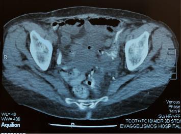
Axial contrast-enhanced computed tomography (CT) scan of the pelvis demonstrating findings of chronic diverticulitis. The image shows significant circumferential wall thickening of the sigmoid colon, located in the lower-left quadrant of the pelvic cavity. Multiple small, air-filled outpouchings (diverticula) are visible protruding from the colonic wall. Distinct pericolic fat stranding is observed, characterized by increased attenuation and a hazy, streaky appearance of the surrounding mesenteric fat, indicating active or chronic inflammation. The anatomical region is defined by the visualization of the iliac bones and pelvic musculature. These radiological features are classic markers of diverticular disease and are used to assess the severity of inflammatory changes and potential complications such as fistulization or abscess formation in a clinical setting.

Axial contrast-enhanced computed tomography (CT) scan of the pelvis demonstrating findings of chronic diverticulitis. The image shows significant circumferential wall thickening of the sigmoid colon, located in the lower-left quadrant of the pelvic cavity. Multiple small, air-filled outpouchings (diverticula) are visible protruding from the colonic wall. Distinct pericolic fat stranding is observed, characterized by increased attenuation and a hazy, streaky appearance of the surrounding mesenteric fat, indicating active or chronic inflammation. The anatomical region is defined by the visualization of the iliac bones and pelvic musculature. These radiological features are classic markers of diverticular disease and are used to assess the severity of inflammatory changes and potential complications such as fistulization or abscess formation in a clinical setting.

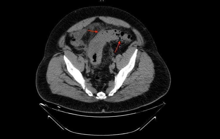
A diagnostic imaging study featuring a transverse section of a non-contrast Computed Tomography (CT) scan of the abdomen and pelvis. The image demonstrates a localized inflammatory process in the left lower quadrant, primarily affecting the descending/sigmoid colon. Key findings include significant circumferential colonic wall thickening and the presence of several small, air-filled outpouchings characteristic of colonic diverticula. Red arrows highlight prominent pericolic fat stranding, identified by increased attenuation and a hazy, reticular appearance in the surrounding mesenteric adipose tissue, which contrasts with the darker, homogenous appearance of healthy fat elsewhere in the scan. These radiological features are classic indicators of acute uncomplicated diverticulitis. No gross evidence of abscess formation or free intraperitoneal air is visible in this specific slice. The scan is an essential educational resource for identifying the hallmarks of acute colonic inflammation and understanding the radiographic diagnostic criteria for diverticular disease.

A diagnostic imaging study featuring a transverse section of a non-contrast Computed Tomography (CT) scan of the abdomen and pelvis. The image demonstrates a localized inflammatory process in the left lower quadrant, primarily affecting the descending/sigmoid colon. Key findings include significant circumferential colonic wall thickening and the presence of several small, air-filled outpouchings characteristic of colonic diverticula. Red arrows highlight prominent pericolic fat stranding, identified by increased attenuation and a hazy, reticular appearance in the surrounding mesenteric adipose tissue, which contrasts with the darker, homogenous appearance of healthy fat elsewhere in the scan. These radiological features are classic indicators of acute uncomplicated diverticulitis. No gross evidence of abscess formation or free intraperitoneal air is visible in this specific slice. The scan is an essential educational resource for identifying the hallmarks of acute colonic inflammation and understanding the radiographic diagnostic criteria for diverticular disease.

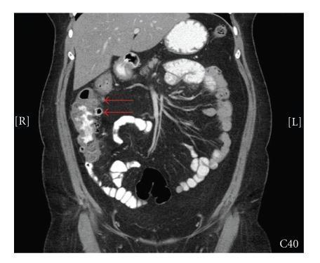
This coronal contrast-enhanced computed tomography (CT) scan of the abdomen and pelvis illustrates findings pathognomonic for right-sided colonic diverticulitis. The primary focus is on the ascending colon, which displays significant circumferential wall thickening and multiple small, air-filled outpouchings representing diverticula. Distinctive periluminal features include prominent 'fat stranding'—visualized as increased attenuation and hazy, streaky densities in the surrounding pericolic fat—indicating acute inflammatory changes. Red arrows specifically highlight the affected segment and associated diverticular structures. The image also depicts the normal anatomy of the transverse and descending colon, small bowel loops with visible valvulae conniventes, and the liver and spleen in the upper abdomen. This diagnostic image is intended for medical education regarding gastrointestinal emergencies and the radiological differentiation of right-sided abdominal pain, emphasizing the key signs of diverticular inflammation in a non-traditional anatomical location.

This coronal contrast-enhanced computed tomography (CT) scan of the abdomen and pelvis illustrates findings pathognomonic for right-sided colonic diverticulitis. The primary focus is on the ascending colon, which displays significant circumferential wall thickening and multiple small, air-filled outpouchings representing diverticula. Distinctive periluminal features include prominent 'fat stranding'—visualized as increased attenuation and hazy, streaky densities in the surrounding pericolic fat—indicating acute inflammatory changes. Red arrows specifically highlight the affected segment and associated diverticular structures. The image also depicts the normal anatomy of the transverse and descending colon, small bowel loops with visible valvulae conniventes, and the liver and spleen in the upper abdomen. This diagnostic image is intended for medical education regarding gastrointestinal emergencies and the radiological differentiation of right-sided abdominal pain, emphasizing the key signs of diverticular inflammation in a non-traditional anatomical location.

Reading File
Reading File
Reading File
Reading File
Reading File
Reading File
Reading File
I now have rich content from multiple authoritative sources. Let me compile the full answer.

Diverticulitis

Definition

Diverticulitis is inflammation and/or infection of a diverticulum - a small outpouching of the colonic wall. It is classified as uncomplicated (localized inflammation, with or without a small confined abscess) or complicated (abscess, fistula, obstruction, or free perforation). Uncomplicated disease accounts for approximately 80% of first attacks.
  • Sleisenger and Fordtran's Gastrointestinal and Liver Disease

Epidemiology

  • Diverticulitis was historically thought to affect 10-25% of patients with diverticulosis, but more recent population-based data suggest only ~4% risk over an 11-year period.
  • Incidence of hospitalization plateaued around 2008 in the US; the proportion requiring surgery declined from 25% to 15% between 2000-2010.
  • In Western countries, diverticula - and therefore diverticulitis - predominantly affect the sigmoid colon. In Asian populations, right-sided colonic involvement is far more common.

Pathophysiology

The mechanism resembles appendicitis:
  1. A fecolith obstructs the neck of a diverticulum
  2. The fecolith abrades the mucosa, causing low-grade inflammation
  3. Bacterial overgrowth, localized ischemia, and impaired mucosal defenses develop
  4. Bacteria breach the mucosa - the process extends transmurally, leading to microperforation or frank perforation
  5. The extent of perforation determines clinical severity:
    • Microperforation - contained by pericolic fat - small pericolic abscess
    • Larger perforation - large inflammatory mass, fibrosis, fistula to adjacent organs
    • Free perforation - bacterial or fecal peritonitis (4 per 100,000/year - uncommon)
Recent evidence also implicates CMV reactivation in local inflammatory activity (found in >2/3 of affected bowel segments).
  • Sleisenger and Fordtran's GI and Liver Disease

Hinchey Classification of Diverticular Perforation

StageDefinition
IConfined pericolic abscess
IIDistant abscess (retroperitoneal or pelvic)
IIIGeneralized peritonitis from rupture of a pericolic/pelvic abscess (no communication with colonic lumen)
IVFecal peritonitis from free perforation (communicating with colonic lumen)

Clinical Features

Symptoms:
  • Left lower quadrant (LLQ) pain - persistent, often >24 hours - most characteristic
  • Fever (low-grade in uncomplicated; high fever/sepsis in complicated disease)
  • Change in bowel habits (diarrhea or constipation)
  • Anorexia, nausea, vomiting
  • Dysuria/urinary frequency ("sympathetic cystitis" from adjacent inflamed sigmoid colon)
  • Feculent vaginal discharge (colovaginal fistula)
  • BRBPR is NOT a feature of acute diverticulitis
Examination:
  • LLQ tenderness, with guarding and rebound if complicated
  • Palpable cylindrical tender mass possible
  • Depressed bowel sounds (or increased with obstruction)
  • Rectal tenderness or palpable mass (low-lying pelvic abscess)
Special populations: Elderly and immunocompromised patients present more subtly but have higher rates of perforation and mortality.

Differential Diagnosis

ConditionKey distinguishing features
Acute appendicitisRLQ pain; most common misdiagnosis for right-sided diverticulitis
Inflammatory bowel diseaseYounger age; chronic/recurrent course; bloody diarrhea
Colorectal cancerWeight loss; progressive obstruction; mass on imaging
Ischemic colitisSudden pain; bloody stool; vascular risk factors
Pelvic inflammatory disease / ovarian pathologyFemale patients; pelvic exam findings; no diverticula on imaging
Ureteral stonesColicky pain; hematuria
Clinical diagnosis alone is unreliable - misdiagnosis rates of 34-68% have been reported even with "classic" presentations.

Diagnosis

Imaging

CT scan (with IV contrast, ideally with oral/rectal contrast too) is the standard of care:
  • Sensitivity 98%, specificity 99%
  • Findings: colonic wall thickening, pericolic fat stranding, localized perforation (micro-perforation), abscess, free air/fluid
  • Also the modality of choice for identifying complications and guiding percutaneous abscess drainage
Barium enemas, contrast enemas, and plain radiography are no longer recommended for acute evaluation.

Labs

  • CBC: leukocytosis is common but not mandatory - up to 46% have a normal WBC with no left shift
  • Urinalysis: if colovesical fistula suspected
  • Clinical diagnosis alone may suffice in patients with prior confirmed diverticulitis presenting with identical symptoms
Here are CT imaging examples of diverticulitis:
Axial CT showing chronic sigmoid diverticulitis with wall thickening, diverticula, and pericolic fat stranding
Axial contrast-enhanced CT: sigmoid colon wall thickening, multiple air-filled diverticula, and pericolic fat stranding - classic findings of diverticulitis.
Non-contrast CT of acute uncomplicated diverticulitis - left lower quadrant colonic wall thickening with fat stranding
Non-contrast CT showing acute uncomplicated diverticulitis: circumferential colonic wall thickening and prominent pericolic fat stranding (red arrows), no abscess or free air.

Management

Uncomplicated Diverticulitis

Outpatient (mild, no comorbidities, able to tolerate oral intake):
  • Liquid diet advancing as tolerated
  • Antibiotics: now selective, not routine - The AVOD and DIABOLO randomized trials (>1,100 patients) showed antibiotics do not accelerate recovery or prevent complications/recurrence in uncomplicated disease. The AGA (2015) recommends selective antibiotic use. If used, coverage for gram-negative aerobes AND anaerobes is required.
  • Common oral regimens: ciprofloxacin + metronidazole, or amoxicillin-clavulanate
Inpatient (elderly, immunocompromised, multiple comorbidities, unable to tolerate oral intake):
  • IV antibiotics + bowel rest
  • Improvement expected within 2-4 days; diet advanced once improving
  • Discharge with 7-10 day oral antibiotic course (if antibiotics deemed necessary)

Complicated Diverticulitis

ComplicationManagement
Abscess (Hinchey I-II)IV antibiotics; CT-guided percutaneous drainage for abscesses >3-4 cm
Fistula (colovesical, colovaginal)Elective surgical resection
ObstructionConservative initially; surgery if unresolved
Free perforation (Hinchey III-IV)Emergency surgery

Surgical Management

  • Only 15-25% of hospitalized patients require surgery.
  • Indications for elective surgery now evaluated on a case-by-case basis (not automatically after 2 episodes as was previously recommended). The updated ASCRS (2014) and AGA guidelines moved away from the "2-episode rule."
  • A 2025 network meta-analysis (EAES/ESCP) examined surgical options for complicated diverticulitis (PMID: 39733170).
  • Standard approach: sigmoid colectomy (2-stage: Hartmann's procedure with later stoma reversal - but reversal only occurs ~50% of the time, even less in elderly patients).
  • There is growing discussion about primary anastomosis vs. colostomy at the time of emergency surgery.

Prevention / Lifestyle

  • High-fiber diet reduces abdominal symptoms in diverticulosis
  • Physical activity is associated with reduced risk of acute diverticulitis
  • Previously-advised food restrictions (nuts, seeds, popcorn) have been discredited - no evidence supports them

Recurrence

  • Risk of recurrent symptoms after a first attack: 15-30%
  • Half of second attacks occur within 1 year of the first

Recent Evidence (2024-2026)

A 2025 systematic review (PMID 41160390) reviewed current evidence on prevention of onset and recurrence of acute diverticulitis. A 2025 network meta-analysis (PMID 39733170) analyzed surgical approaches to complicated diverticulitis from the EAES/ESCP. A 2025 meta-analysis (PMID 41261215) specifically addressed Hinchey III perforated diverticulitis management. These do not contradict the textbook framework above but continue to refine surgical technique selection and antibiotic stewardship guidelines.

Sources: Sleisenger and Fordtran's Gastrointestinal and Liver Disease; Rosen's Emergency Medicine; Bailey and Love's Short Practice of Surgery, 28th ed.; Grainger & Allison's Diagnostic Radiology

Bitte für Kenntnisse Prüfung Vorbereitung mit Fragen auch

Hier ist eine vollständige Prüfungsvorbereitung zum Thema Divertikulitis - kompakte Zusammenfassung + Übungsfragen mit Antworten.

📚 Divertikulitis - Prüfungsvorbereitung


🔷 KOMPAKTZUSAMMENFASSUNG

Definition

  • Divertikulose = Vorhandensein von Divertikeln (Ausstülpungen der Darmwand)
  • Divertikulitis = Entzündung/Infektion eines Divertikels
  • Unkompliziert: lokale Entzündung ± kleiner Abszess (~80% der Fälle)
  • Kompliziert: Abszess, Fistel, Obstruktion, freie Perforation

Lokalisation

PopulationHäufigste Lokalisation
WestlichSigma (linkes Kolon)
AsiatischRechtes Kolon

Pathophysiologie (Kurzformel)

Koprolithobstruktion → Mukosaläsion → Bakterienüberwuchs → Ischämie → transmurale Entzündung → Perforation

Hinchey-Klassifikation

StadiumBefund
IPerikolischer Abszess (lokalisiert)
IIFerner Abszess (retroperitoneal/pelvin)
IIIGeneralisierte Peritonitis (rupturierter Abszess, kein Kontakt mit Darmlumen)
IVKotperitonitis (freie Perforation, Kontakt mit Darmlumen)

Klinik - MERKSATZ: "Links - Fieber - Stuhlveränderung"

  • Schmerz: linker Unterbauch (LUB), persistierend >24h
  • Fieber (subfebrile bis hohes Fieber je nach Schweregrad)
  • Stuhlveränderungen: Diarrhö oder Obstipation
  • Dysurie/Pollakisurie: "sympathische Zystitis"
  • KEIN hellrotes Blut per rectum (BRBPR) bei akuter Divertikulitis!
  • Ältere/Immunsupprimierte: subtile Klinik, aber höhere Perforationsrate

Diagnostik

MethodeBedeutung
CT Abdomen (mit KM)Goldstandard - Sensitivität 98%, Spezifität 99%
Labor (CRP, Leukozyten)Leukozyten können in 46% normal sein!
UrinanalyseBei Verdacht auf kolovesikale Fistel
KolonoskopieNicht akut! Nach Abklingen (altersgerechtes Screening)
BariumeinlaufNicht mehr empfohlen
CT-Befunde: Wandverdickung, perikolisches Fettgewebe-Stranding, Abszess, freie Luft/Flüssigkeit

Therapie - Übersicht

Unkompliziert → ambulant möglich
  ├─ Flüssigkost → Kostaufbau
  ├─ Antibiotika: SELEKTIV (nicht routinemäßig!)
  │   └─ Wenn nötig: Gramneg. Aerobier + Anaerobier abdecken
  │       z.B. Ciprofloxacin + Metronidazol
  └─ Kein Nutzen von Bettruhe / NPO allein

Kompliziert → stationär + chirurgische Konsultation
  ├─ Abszess: IV Antibiotika ± CT-gesteuerte Drainage (>3-4 cm)
  ├─ Fistel: elektive Resektion
  ├─ Obstruktion: konservativ → OP wenn nötig
  └─ Perforation Hinchey III/IV: Notfall-OP

Chirurgische Therapie

  • Nur 15-25% der hospitalisierten Patienten benötigen OP
  • Hartmann-Operation: Resektion + Kolostoma (Standard bei Notfall-OP)
  • Stoma-Rückverlagerung: nur in ~50% der Fälle möglich
  • Elektive OP: Nicht mehr automatisch nach 2 Episoden - individuelle Entscheidung (ASCRS 2014, AGA)
  • Antibiotika verhüten keine Komplikationen/Rezidive (AVOD-Studie, DIABOLO-Studie)

Differenzialdiagnosen - MERKLISTE

  1. Appendizitis (v.a. bei rechtsseitiger Divertikulitis)
  2. CED (Morbus Crohn, Colitis ulcerosa)
  3. Kolorektales Karzinom
  4. Ischämische Kolitis
  5. Adnexitis / Ovarialzyste / Extrauteringravidität
  6. Ureterstein

✅ ÜBUNGSFRAGEN


Frage 1 - Multiple Choice

Welches Symptom spricht GEGEN eine akute Divertikulitis?
A) Linker Unterbauchschmerz B) Subfebrile Temperaturen C) Hellrotes Blut per rectum (BRBPR) D) Dysurie E) Übelkeit und Erbrechen
💡 Antwort anzeigen
Antwort: C
BRBPR ist kein Merkmal der akuten Divertikulitis. Es spricht eher für eine divertikuläre Blutung, Kolitis, oder Karzinom. Die Divertikulitis ist eine entzündliche, keine blutende Erkrankung.

Frage 2 - Multiple Choice

Ein 65-jähriger Patient hat Fieber 38,5°C, LUB-Schmerz seit 2 Tagen, Leukozyten 14.000/µl. Das CT zeigt Wandverdickung des Sigmas, perikolisches Fat-Stranding, aber keinen Abszess und keine freie Luft. Was ist die beste initiale Therapie?
A) Sofortige Operation B) Stationäre Aufnahme mit IV-Antibiotika und Nahrungskarenz C) Ambulante Therapie mit oraler Flüssigkost, selektiver Antibiose D) Koloskopie zum Ausschluss eines Karzinoms E) Bariumeinlauf zur Bestätigung
💡 Antwort anzeigen
Antwort: C
Unkomplizierte Divertikulitis bei einem Patienten ohne schwere Komorbiditäten und mit Fähigkeit zur oralen Aufnahme: ambulante Therapie möglich. Antibiotika selektiv (nicht routinemäßig). Koloskopie ist akut kontraindiziert. Bariumeinlauf nicht mehr empfohlen. Sofortige OP nur bei Hinchey III/IV.

Frage 3 - Multiple Choice

Welches Hinchey-Stadium beschreibt eine Kotperitonitis durch freie Perforation eines Divertikels (mit direktem Kontakt zum Darmlumen)?
A) Stadium I B) Stadium II C) Stadium III D) Stadium IV E) Stadium V
💡 Antwort anzeigen
Antwort: D - Stadium IV
  • Stadium I = perikolischer Abszess
  • Stadium II = ferner (pelviner/retroperitonealer) Abszess
  • Stadium III = generalisierte Peritonitis durch rupturierten Abszess (kein Lumenkontakt)
  • Stadium IV = Kotperitonitis, freie Perforation mit Lumenkontakt → Notfall-OP!

Frage 4 - Wahr/Falsch

Beurteilen Sie folgende Aussagen als WAHR oder FALSCH:
AussageW/F
A) Leukozytose ist immer bei Divertikulitis vorhanden?
B) CT mit KM ist der Goldstandard (Sens. 98%, Spez. 99%)?
C) Antibiotika verhindern nachweislich Komplikationen bei unkomplizierter Divertikulitis?
D) Die Hartmann-OP beinhaltet Resektion + Kolostoma?
E) Bei Asiaten sind Divertikel häufiger im rechten Kolon?
💡 Antwort anzeigen
AussageAntwortBegründung
AFALSCHIn 46% der Fälle normales Blutbild ohne Linksverschiebung
BWAHRCT mit IV±orales/rektales KM: Goldstandard
CFALSCHAVOD- und DIABOLO-Studien: Antibiotika beschleunigen Heilung nicht und verhindern keine Komplikationen/Rezidive
DWAHRHartmann = Resektion + endständiges Kolostoma; Rückverlagerung nur in ~50%
EWAHRIm Gegensatz zur westlichen Population (Sigma-dominant)

Frage 5 - Kurzantwort

Nennen Sie 4 Komplikationen der Divertikulitis (= "Komplizierte Divertikulitis").
💡 Antwort anzeigen
  1. Abszess (perikolisch oder distant)
  2. Fistel (kolovesikal, kolovaginal, koloenterisch)
  3. Obstruktion (entzündliche Stenose)
  4. Freie Perforation mit Peritonitis

Frage 6 - Klinisches Szenario

Eine 72-jährige Frau mit bekannter Divertikulose kommt mit LUB-Schmerz, Fieber 39°C, Pneumaturie und Fäkalurie beim Wasserlassen. Was ist die wahrscheinlichste Komplikation und wie wird sie behandelt?
💡 Antwort anzeigen
Komplikation: Kolovesikale Fistel
  • Verbindung zwischen entzündetem Sigma und Harnblase
  • Klinik: Pneumaturie (Luft im Urin), Fäkalurie, rezidivierende Harnwegsinfekte
  • Diagnose: CT Abdomen/Becken
  • Therapie: Elektive chirurgische Resektion (nach Abklingen der akuten Entzündung)

Frage 7 - Multiple Choice

Welche Aussage zur Chirurgie bei unkomplizierter Divertikulitis ist aktuell KORREKT?
A) Elektive OP ist nach jeder Episode indiziert B) Elektive OP ist standardmäßig nach der 2. Episode empfohlen (ASCRS 2000) C) Die Entscheidung zur elektiven OP erfolgt individuell, nicht nach fixer Episodenzahl (ASCRS 2014) D) Alle Patienten mit Divertikulitis benötigen eine OP E) Eine Notfall-OP ist bei unkomplizierter Divertikulitis immer erforderlich
💡 Antwort anzeigen
Antwort: C
Die alten ASCRS-Leitlinien (2000) empfahlen OP nach 2 Episoden. Die aktuellen Leitlinien (ASCRS 2014, AGA) empfehlen eine individuelle Entscheidung basierend auf Patientenfaktoren - nicht mehr die starre "2-Episoden-Regel".

Frage 8 - Kurzantwort

Welche Differenzialdiagnose ist die häufigste Fehldiagnose bei rechtsseitiger Divertikulitis?
💡 Antwort anzeigen
Appendizitis
In einer Studie aus Hongkong wurden 34 von 35 Patienten mit rechtsseitiger Divertikulitis initial als Appendizitis eingestuft. Das CT ist entscheidend zur Differenzierung.

🧠 LERNKARTEN - Schnellwiederholung

FrageAntwort
Goldstandard Diagnostik?CT Abdomen mit KM
Hinchey IV?Kotperitonitis, Notfall-OP
BRBPR bei Divertikulitis?NEIN - spricht dagegen
Antibiotika bei unkompliziert?Selektiv, nicht routinemäßig (AGA 2015)
Häufigste Lokalisation (westlich)?Sigma
Häufigste Fehldiagnose (rechtsseitig)?Appendizitis
Rezidivrate nach 1. Episode?15-30%
Stoma-Rückverlagerung nach Hartmann?Nur ~50%
CT-Spezifität?99%
Kolonoskopie akut?Kontraindiziert - erst nach Abklingen

Viel Erfolg bei der Prüfung! 🎓
This is a shared conversation. Sign in to Orris to start your own chat.