
Conventional culture technique
conventional bacterial culture technique agar plate streaking
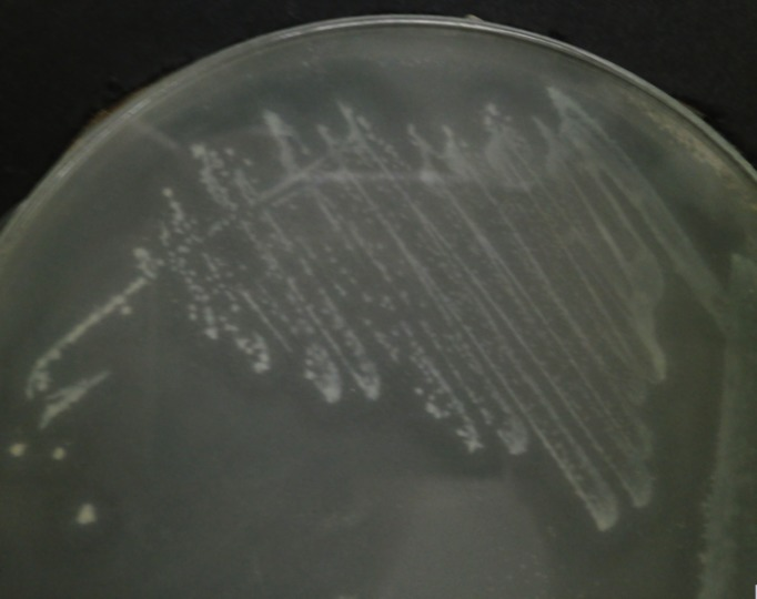
**Imaging Modality:** Laboratory photograph of a microbiological culture on an agar plate. **Anatomical/Biological Entity:** Bacterial culture of *Cedecea neteri* (strain SSMD04). **Observed Pathology/Phenotype:** The image displays bacterial colonies streaked onto a specialized agar medium supplemented with 0.5% corn oil. The primary diagnostic feature is the presence of distinct "halo" zones (zones of clearance or precipitation) surrounding the bacterial growth. **Characteristic Visual Features:** * **Colony Morphology:** Small, circular, opaque white to light-gray colonies distributed along streak lines. * **Enzymatic Activity:** Clear translucency or a discernible halo effect in the agar adjacent to the colonies, indicative of extracellular lipase activity and lipid hydrolysis. * **Growth Pattern:** Standard quadrant streaking technique used to isolate individual colonies. **Diagnostic Significance:** This biochemical assay demonstrates the metabolic capability of *C. neteri* to utilize lipids as a carbon source. The visible halo serves as a positive indicator for lipase production, a key phenotypic trait used in microbial classification and the study of bacterial virulence or industrial potential.
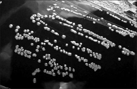
**Modality:** Microbiology culture photograph (grayscale). **Specimen:** *Corynebacterium pseudotuberculosis* (formerly *Corynebacterium ovis*) isolated on blood agar medium. **Morphological Features:** The image displays a streak plate demonstrating isolated bacterial colonies. The colonies are characterized by a distinct circular morphology with an entire margin. They exhibit a convex elevation and appear opaque with a grayish-white coloration against the dark background of the agar. The surface texture of the colonies appears slightly dry or waxy, which is characteristic of this genus. **Distribution:** Streaking pattern shows confluent growth in the primary sectors transitioning to well-defined, discrete individual colonies in the peripheral sectors, allowing for the observation of specific colony macro-morphology. **Diagnostic Context:** These visual features are consistent with the growth characteristics of *Corynebacterium* species, typically found in veterinary clinical microbiology samples. Key differentiating features include the specific convex profile and circular uniformity of the colonies on enriched media.
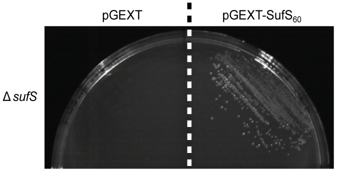
**Imaging Modality:** Laboratory photograph of a microbiological culture on an agar plate. **Specimen and Context:** Comparative growth assay of *Escherichia coli* ΔsufS (cysteine desulfurase null) mutants under iron-starvation conditions induced by the chelator 2,2′-dipyridyl. **Observed Findings:** The image displays a split Petri dish divided by a vertical dashed line, illustrating a functional complementation (rescue) assay. - **Left Sector (pGEXT):** The control group transformed with an empty vector shows a complete absence of bacterial growth, indicating that the endogenous *sufS* deficiency is lethal under iron-restricted conditions. - **Right Sector (pGEXT-SufS60):** The experimental group transformed with a plasmid expressing *Plasmodium falciparum* SufS displays robust bacterial colony formation and streaking patterns. **Key Diagnostic Features:** Clear phenotypic rescue visible through the restoration of colony-forming units (CFUs) in the presence of the *P. falciparum* gene. This differential growth pattern serves as visual evidence of the heterologous gene's ability to fulfill the essential enzymatic role of cysteine desulfurase in the bacterial host.
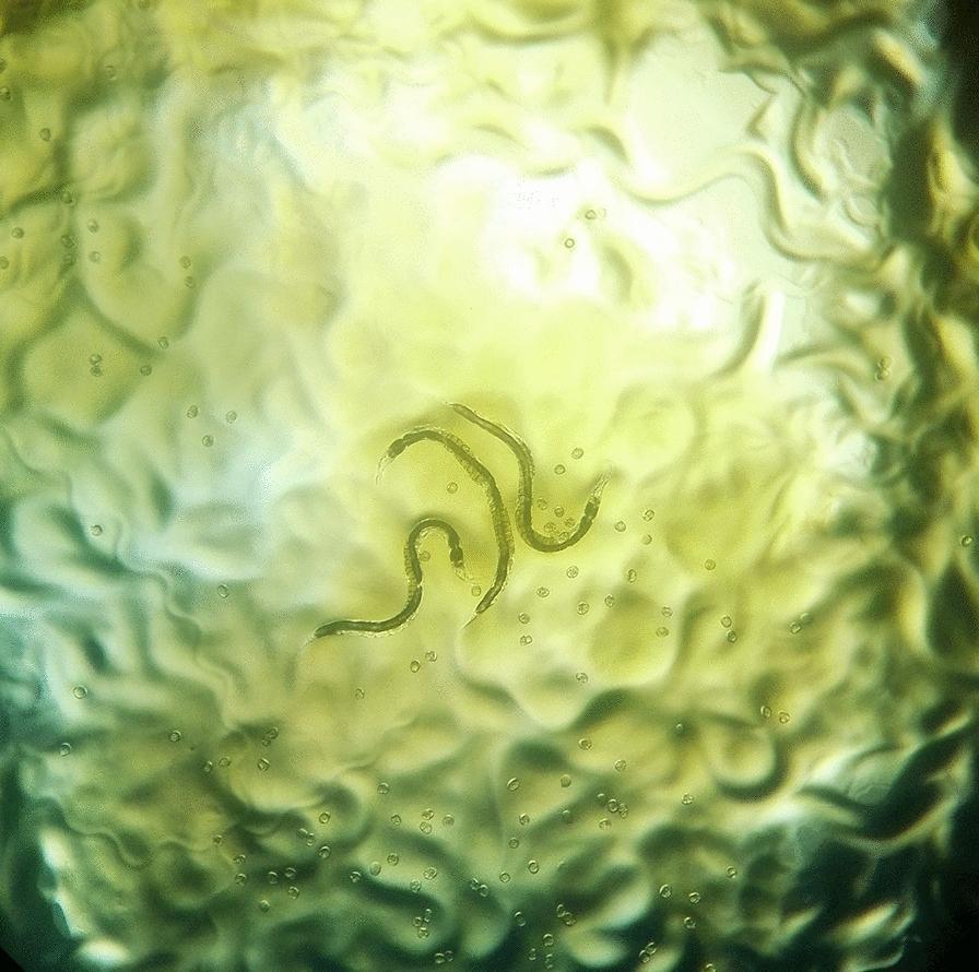
This diagnostic image shows a macroscopic view of an agar plate culture used for the detection of Strongyloides stercoralis, a human pathogenic nematode. The culture medium has a translucent, yellowish-green appearance with a highly irregular, wrinkled surface texture. These surface furrows and grooves represent characteristic 'larval tracks,' which are formed as the motile larvae migrate across the agar surface, often carrying bacteria along their path that then grow into visible trails. Centrally located are two slender, elongated adult worms exhibiting a curved morphology. Scattered throughout the medium are numerous small, circular, translucent structures consistent with parasite ova. This agar plate culture technique is a highly sensitive diagnostic method for identifying strongyloidiasis in clinical stool samples, as the visualization of these tracks and parasite life stages (larvae, adults, and eggs) provides definitive evidence of infection.
culture media types blood agar MacConkey nutrient broth microbiology
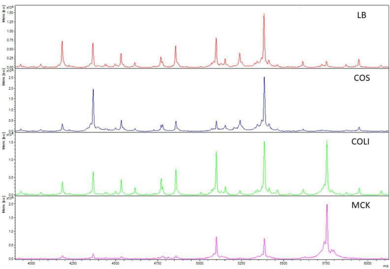
A multi-panel comparison chart of Matrix-Assisted Laser Desorption/Ionization Time-of-Flight (MALDI-TOF) mass spectra used in clinical microbiology. The image displays the spectral profiles of an Escherichia coli (E. coli) isolate grown on four different solid agar media: Luria Broth (LB, red), Columbia agar with sheep blood (COS, blue), Coliformen agar (COLI, green), and MacConkey agar (MCK, pink). The x-axis represents the mass-to-charge ratio (m/z) in the 4000–6000 range, while the y-axis indicates relative intensity. The LB spectrum shows the highest peak density and resolution, serving as the reference for reproducibility. In contrast, the COS, COLI, and MCK spectra demonstrate variations in peak presence and signal intensity, illustrating how different growth media influence the proteomic fingerprint of bacterial isolates. This diagnostic imaging comparison highlights the importance of standardized culture protocols for accurate microbial characterization and subspecies-level identification beyond simple species detection.
**Modality:** Microbiology culture photograph (grayscale). **Specimen:** *Corynebacterium pseudotuberculosis* (formerly *Corynebacterium ovis*) isolated on blood agar medium. **Morphological Features:** The image displays a streak plate demonstrating isolated bacterial colonies. The colonies are characterized by a distinct circular morphology with an entire margin. They exhibit a convex elevation and appear opaque with a grayish-white coloration against the dark background of the agar. The surface texture of the colonies appears slightly dry or waxy, which is characteristic of this genus. **Distribution:** Streaking pattern shows confluent growth in the primary sectors transitioning to well-defined, discrete individual colonies in the peripheral sectors, allowing for the observation of specific colony macro-morphology. **Diagnostic Context:** These visual features are consistent with the growth characteristics of *Corynebacterium* species, typically found in veterinary clinical microbiology samples. Key differentiating features include the specific convex profile and circular uniformity of the colonies on enriched media.

This clinical diagram illustrates a systematic inoculation pattern used in dental microbiology studies. The visual depicts a top-down view of a circular agar culture medium within a Petri dish. A continuous, serpentine, or meandering path is shown spread across the entire surface of the medium, representing the application of dental impression tray adhesive using a cap-attached brush. The path is characterized by a uniform thickness and a textured, coarse stroke appearance, consistent with a brush-applied liquid sample. This illustration demonstrates the standardized method for inoculating culture media to test for cross-contamination of oral microorganisms (bacteria and fungi) between patients via reusable adhesive application brushes. The clinical relevance focuses on infection control and hygiene protocols within prosthetic dentistry and dental laboratory procedures.
Conventional Culture Technique
1. Specimen Collection and Transport
- Collect from the site of infection before antibiotic therapy begins.
- Use sterile technique to avoid contaminating organisms.
- Transport promptly in appropriate containers (sterile swabs, transport media such as Amies or Stuart's medium).
- Volume matters - larger volumes increase sensitivity (e.g., 10 mL per blood culture bottle in adults).
2. Types of Culture Media
A. General-Purpose (Non-selective) Media
| Medium | Purpose |
|---|---|
| Nutrient broth (e.g., soybean-casein digest / tryptic soy broth) | Blood culture and sterile-site specimens - no selective agents, maximizes recovery of fastidious organisms |
| Thioglycolate broth | Anaerobes and facultative bacteria; sodium thioglycolate reduces oxygen tension |
| Blood agar (5% sheep blood + nutrient agar base) | Most bacteria; shows hemolysis patterns (see below) |
| Chocolate agar | Fastidious organisms (e.g., Haemophilus influenzae, Neisseria); red cells lysed at ~80°C releasing hemin and NAD |
B. Selective Media
| Medium | Selective Agents | Target Organisms |
|---|---|---|
| MacConkey agar | Bile salts + crystal violet (inhibit Gram-positives) | Gram-negative rods; lactose fermenters appear red/pink |
| Martin-Lewis / Thayer-Martin | Vancomycin, colistin, trimethoprim, anisomycin | Pathogenic Neisseria spp. |
| Hektoen enteric agar | Bile salts, thiosulfate, citrate | Salmonella and Shigella from stool |
| Anaerobic blood agar | Aminoglycosides + reducing agents (L-cysteine) | Strict anaerobes |
3. Inoculation Technique
Streak Plate Method (for solid media)
- Primary streak: Inoculate one quadrant of the plate.
- Secondary streak: Flame loop, rotate plate 90°, and streak through the primary zone into fresh agar.
- Tertiary and quaternary streaks: Repeat, diluting the inoculum progressively.
- Result: Isolated colonies in the final quadrant, each derived from a single organism (colony-forming unit, CFU).
Broth Inoculation
- Used for specimens from normally sterile sites (blood, CSF, pleural fluid).
- Broth provides maximum sensitivity - even 1-10 organisms per mL can be detected.
- Selective/indicator agents are omitted to avoid inhibiting fastidious pathogens.
4. Incubation Conditions
| Parameter | Standard | Special Conditions |
|---|---|---|
| Temperature | 35-37°C for most pathogens | 30°C for M. marinum, M. haemophilum; 42°C for Campylobacter |
| Atmosphere | Aerobic (ambient air) | CO₂ (5-10%) for fastidious organisms; anaerobic for strict anaerobes |
| Duration | 18-24 hours for most bacteria | 6 weeks for mycobacteria on solid media; up to 5 days for blood cultures |
| Humidity | Moist environment | Prevents desiccation of agar plates |
5. Detection and Reading of Growth
Colony Morphology Assessment
- Size: Pinpoint (<1 mm), small (1-2 mm), large (>2 mm)
- Shape: Circular, irregular, filamentous
- Elevation: Flat, raised, convex, umbonate
- Surface: Smooth (glistening), rough, dry, mucoid
- Color: Pigmentation (e.g., Pseudomonas aeruginosa - blue-green; Staphylococcus aureus - golden)
- Opacity: Transparent, translucent, opaque
- Edge (margin): Entire, undulate, lobate, filamentous
- Hemolysis on blood agar:
- α-hemolysis: Incomplete lysis - green hazy zone (e.g., viridans streptococci, S. pneumoniae)
- β-hemolysis: Complete clear zone (e.g., S. pyogenes, S. agalactiae)
- γ (non-hemolytic): No lysis (e.g., Enterococcus)
Broth Cultures
- Check daily for turbidity (= growth).
- Subculture positive bottles onto solid media for colony isolation.
6. Identification of Isolates
- Gram stain - morphology and staining reaction (Gram-positive vs. negative; coccus, rod, spiral)
- Biochemical tests - catalase, oxidase, coagulase, urease, indole, citrate, etc.
- Automated systems - e.g., VITEK 2, Phoenix (miniaturized biochemical panels)
- MALDI-TOF MS - now the gold standard for rapid identification from pure colonies; identifies proteins by mass spectrometry within minutes
- Molecular methods (16S rRNA sequencing) for unusual or fastidious organisms
7. Antibiotic Susceptibility Testing (AST)
- Disk diffusion (Kirby-Bauer): Antibiotic-impregnated disks placed on inoculated Mueller-Hinton agar; zone of inhibition measured.
- Broth microdilution / E-test: Determines the minimum inhibitory concentration (MIC).
- Results guide therapy as Susceptible (S), Intermediate (I), or Resistant (R).
8. Conventional Culture vs. Modern Methods
| Feature | Conventional Culture | Modern Alternatives |
|---|---|---|
| Sensitivity | High (if broth used) | PCR/NAA - higher for some pathogens |
| Speed | 18 hours to 6 weeks | MALDI-TOF: minutes; PCR: 1-4 hours |
| AST possible | Yes | Usually not from molecular tests |
| Live organism | Yes (for epidemiology, storage) | No |
| Cost | Low | Higher |
| Detects novel organisms | Limited | Metagenomics (mNGS) |
9. Special Conventional Culture Scenarios
Blood Culture
- Inoculate aerobic and anaerobic bottles (typically 8-10 mL each per set).
- Draw 2-3 sets from different sites to distinguish true bacteremia from contamination.
- Continuously monitored automated systems (e.g., BACTEC, BacT/ALERT) detect CO₂ production.
Mycobacterial Culture (Henry's Clinical Diagnosis, 22nd Ed)
- Inoculate both solid and broth media for optimal recovery.
- Solid: Löwenstein-Jensen (egg-based) or Middlebrook 7H10/7H11 (agar-based)
- Broth: MGIT 960, BACTEC 9000 (automated liquid systems - faster detection)
- Incubate at 37°C in 5-10% CO₂ for up to 6 weeks.
- Second set at 30°C if cutaneous infection (M. marinum, M. haemophilum) is suspected.
Fungal Culture
- General medium: Sabouraud dextrose agar (pH 5.5, designed for dermatophytes) or Emmons modification (pH 6.8, more widely used).
- Selective medium: Sabouraud + cycloheximide + antibacterial agent (inhibits bacteria and saprobic fungi).
- Yeast differentiation: CHROMagar Candida - C. albicans (green), C. krusei (pink), C. tropicalis (metallic blue).
- Sherris & Ryan's Medical Microbiology, 8th Edition - Appendix 4-1 and 4-2
- Henry's Clinical Diagnosis and Management by Laboratory Methods, 22nd Ed - Culture Methods (mycobacteria, fungi)
- Jawetz, Melnick & Adelberg's Medical Microbiology, 28th Ed