

17935638999665887659834967252575.jpg
Red palm reason
Reading File
analyse_media
Finding Sources
Searching PubMed
"palmar erythema"
The photo shows patchy, fairly well-defined pink-red discoloration over the heel of the palm near the wrist (the hypothenar area). It has a few faint darker purple dots, and the rest of the palm looks normal. I can't diagnose from one photo, but this pattern has a short list of likely causes.
Likely causes
Harmless or local causes (the most common)
- Physiologic palmar erythema. Warm hands, exercise, heat, or a hot shower can all cause it. It is also common in pregnancy.
- Pressure, friction, or irritation. Leaning on the heel of the hand, using tools, a gym, a keyboard or mouse, or a wrist brace can do this.
- Contact dermatitis or allergy. Soaps, sanitizers, latex, plants, and cleaning products are typical triggers. This is more likely if the area itches, flakes, or has tiny blisters.
- Insect bite or other local reaction. The small darker dots could fit this.
Systemic causes (palmar erythema proper)
Andrews' says erythema palmare "is usually most marked on the hypothenar areas and is associated with an elevated level of circulating estrogen. Cirrhosis, hepatic metastases, and pregnancy are common causes" - Andrews' Diseases of the Skin, Erythema Palmare. Your photo shows this same hypothenar distribution. Other recognised associations are:
- Chronic liver disease or cirrhosis. Palmar erythema appears in the standard lists of signs of chronic liver disease, usually alongside spider naevi, jaundice, and easy bruising (Bailey and Love; Sleisenger and Fordtran).
- Hyperthyroidism (Fitzpatrick's Dermatology).
- Pregnancy and estrogen excess.
- Rheumatoid arthritis, other autoimmune disease, and polycythaemia.
- Medications. Examples are amiodarone and some other drugs.
- Hereditary palmar erythema (Lane disease), which is rare.
Other causes to keep in mind
- Vasculitis or small embolic lesions. Tender, purple, or non-blanching spots on the palms and fingers can be a sign of infective endocarditis (Janeway lesions) or small-vessel vasculitis. The faint dark dots in your photo are not enough to say this, but see below for when it matters.
- Palmar fasciitis or polyarthritis, complex regional pain syndrome, cellulitis, and hand-foot syndrome (from some chemotherapy drugs).
- Erythromelalgia, which causes burning redness with heat.
Questions that narrow it down
- Is it one hand or both? Both hands, symmetric, points to systemic causes. One hand points to a local cause.
- How long has it been there, and is it new or long-standing?
- Does it blanch when you press on it? Dots that stay put when pressed are more worrying.
- Is it itchy, painful, warm, or swollen?
- Do you drink alcohol heavily, have liver disease or hepatitis risk, or take new medications?
- Are you pregnant, or do you have thyroid symptoms (palpitations, weight loss, heat intolerance)?
- Do you have fever, joint pain, or spots on the fingertips or nails?
When to see a doctor soon
- Both palms are red and it lasts more than a couple of weeks.
- You also have yellow skin or eyes, easy bruising, abdominal swelling, or spider-like vessels on your chest.
- The area is hot, swollen, spreading, or painful, or you have a fever. This could be infection.
- You have tender or non-blanching purple spots, especially with fever or a new heart murmur. Get urgent assessment.
A doctor would usually check liver function, thyroid function, and a blood count first, and add other tests depending on your symptoms.
If you tell me the answers to the questions above, I can narrow this down further.
A quick PubMed check of recent meta-analyses and systematic reviews found nothing that changes the textbook picture of palmar erythema.
Both hands, many years, not itchy, warm, swollen, fatty liver, heat intolerance due to hot flashes, no fever, no pain, spotted with small more red spots and lighter spots. When hot, same on fingertips and another side of palm
Finding Sources
Searching PubMed
"palmar erythema" AND "fatty liver"
Your history makes the most likely explanation a long-standing, benign form of palmar erythema. Hot flashes and fatty liver are both plausible contributors. I can't confirm that from here, and a few simple tests would settle it.
Why this pattern fits
1. A mottled look that gets worse with heat points to vasomotor change.
Yamada's Textbook of Gastroenterology says the red of true liver-type palmar erythema is "fixed rather than transient," without a mottled look. A mottled look is more typical of physiologic hyperemia, which is blood-flow change in the skin. Your red and pale patches that spread to the fingertips and the sides of the palm when you are hot fit that. They also fit the vascular instability of menopausal hot flashes.
2. Fatty liver can cause palmar erythema, but it usually does so only when the disease is advanced.
Yamada's says that in NAFLD, palmar erythema and spider angiomas tend to appear with advanced fibrosis or cirrhosis. Goldman-Cecil lists palmar erythema among the possible findings in fatty liver disease. Simple steatosis is benign and usually causes no signs. Palmar erythema from the liver is thought to come from altered estrogen metabolism and vasodilation.
3. Other possible contributors
- Menopause or hormonal change. Estrogen shifts affect hand blood flow.
- Thyroid. Heat intolerance can also come from an overactive thyroid, which is a recognised cause of palmar erythema. Hot flashes and hyperthyroidism overlap, so this is worth ruling out with a blood test.
- Rheumatoid arthritis and other autoimmune disease. This is less likely without joint pain.
- Medications and familial palmar erythema. Long-standing, symmetric redness with no other problem is sometimes just a family trait.
What would separate benign from liver-related
| Points to a benign cause | Points to significant liver disease |
|---|---|
| Normal liver tests and platelets | Raised ALT/AST, low platelets, low albumin, or high bilirubin |
| No spider veins on the chest, neck, or face | Spider angiomas |
| Normal FIB-4 score or elastography (FibroScan) | High FIB-4 or stiff liver on elastography |
| Stable for years, no jaundice or abdominal swelling | Yellow eyes, easy bruising, swollen belly, or enlarged breasts in men |
What I would suggest
- See your doctor and ask for the following:
- Liver panel and a full blood count.
- A FIB-4 score, which is calculated from age, AST, ALT, and platelets. If it is intermediate or high, ask for a FibroScan.
- TSH, to exclude thyroid disease.
- Glucose or HbA1c and a lipid profile, because fatty liver often travels with metabolic syndrome.
- Consider checking inflammatory markers or rheumatoid factor if you have any joint stiffness.
- Look for spider veins on your upper chest, neck, and face, and check your eyes for yellowing.
- Treat the fatty liver itself. Weight loss of 7 to 10% when overweight, a Mediterranean-style diet, regular exercise, little or no alcohol, and good control of diabetes and cholesterol all help. If hot flashes are bothering you, discuss options with your doctor.
When to seek care sooner
- Yellow skin or eyes, a swelling belly, black or bloody stools, or confusion.
- The redness becomes painful, one-sided, swollen, or blistered.
- Fever, joint pain, or purple non-blanching spots on the fingertips.
The PubMed search I ran (palmar erythema with fatty liver) returned only older reviews and case reports, with no recent large studies or guidelines that change this approach. I didn't find a good source for how often hot flashes alone cause this pattern, so that part is a reasoned inference rather than an established fact.
Do you know your recent liver test results or FIB-4? If you share them, I can help you interpret them.
Normal thyroid, blood tests, Normal cholesterol with statins, no spider veins. Normal inflammation factors. BMI 29
Those results make a serious cause much less likely. The most likely explanation is now benign, long-standing palmar erythema driven by blood-vessel reactivity (vasomotor change).
What your results rule out
- Thyroid disease. Normal thyroid tests exclude hyperthyroidism as the cause of the heat intolerance and the red palms.
- Active autoimmune or inflammatory disease. Normal inflammation markers make rheumatoid arthritis and vasculitis unlikely.
- Advanced liver disease. No spider veins, normal blood tests, and no symptoms make cirrhosis unlikely. In fatty liver, palmar erythema and spider angiomas generally show up with advanced fibrosis or cirrhosis (Yamada's Textbook of Gastroenterology).
What remains most likely
- Physiologic or idiopathic palmar erythema. It is bilateral, long-standing, painless, and not itchy. It is mottled, with red and pale patches that spread to the fingertips and the sides of the palm when you are hot. Yamada's notes that a mottled look suggests physiologic hyperemia rather than the fixed red of liver disease.
- Menopausal vasomotor instability. Hot flashes involve the same kind of blood-vessel reactivity, and hormonal change also affects the circulation of the hands. This is an inference. I found no study showing that menopause alone causes this pattern.
- Mild fatty liver as a minor contributor. This is possible but not proven. The "normal blood tests" would need to include liver enzymes and platelets to support it.
- Familial tendency. Ask whether your mother, sisters, or children have red palms.
Remaining checks
- Confirm which tests were normal. A normal liver panel (ALT, AST, bilirubin, albumin) and platelet count matter most. With those, you can calculate a FIB-4 score to estimate liver fibrosis. A BMI of 29 plus fatty liver is a reason to do this once, even without symptoms. Elastography (FibroScan) is the next step if FIB-4 is not low.
- Statins. These can raise liver enzymes slightly, but they are generally safe in fatty liver. Statins are not a typical cause of palmar erythema.
- Hot-flash treatment. If the flushing bothers you, this is the area to treat. Options include menopausal hormone therapy (where suitable), SSRIs/SNRIs, or fezolinetant. Hormone therapy can change flushing and palm color, so review it with your clinician.
Management and outlook
- No treatment is needed for the redness itself. It does not harm the hands.
- Lose around 5 to 10% of body weight, keep alcohol low, and exercise regularly. This protects the liver, and some people find flushing improves too.
- Avoid hot water and heavy alcohol, which worsen the redness and flushing.
- Have an annual liver check, or follow your doctor's advice.
Worth seeing a doctor for
- Yellow eyes or skin, a swelling belly, easy bruising, or new spider veins.
- Red spots that don't fade when pressed, are painful, or are accompanied by fever.
- Burning pain with heat, which would suggest erythromelalgia.
I don't think you need further urgent investigation. If you can share the actual liver enzyme and platelet values, I can calculate your FIB-4 for you.
Pilonidal cyst treatment
Finding Sources
Searching PubMed
"pilonidal sinus"[MeSH Terms]
Searching the Web
pilonidal disease clinical practice guideline treatment 2026
Reading File
Reading File
Reading File
Treatment depends on the stage of the disease: no symptoms, an acute abscess, or chronic or recurrent sinus disease. Pilonidal disease is a foreign-body reaction to hair driven into the skin of the natal cleft (the crease between the buttocks). It usually affects young adults and is more common in men (Sabiston; Sleisenger and Fordtran).
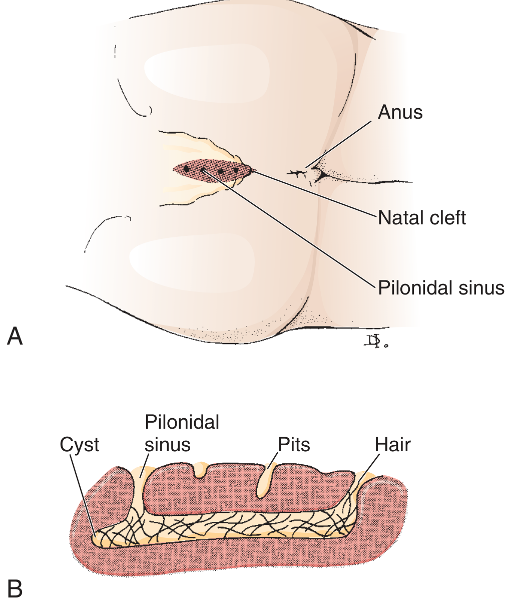
1. No symptoms (pits only)
- Leave it alone. Sleisenger notes that pilonidal disease can present "as an asymptomatic physical finding", and surgery is not needed.
- Prevention applies here too (see below).
2. Acute abscess (painful, swollen, tender lump)
- Incision and drainage is the standard treatment (Sabiston).
- Make the incision off the midline, over the most fluctuant part. This helps the wound heal.
- Curette the cavity to remove hair and dead tissue.
- Pack or leave the wound open, with regular dressing changes.
- Antibiotics are not routine after good drainage. They are considered for cellulitis, diabetes, immune suppression, or systemic illness. Cultures typically grow mixed fecal flora, mostly anaerobes, plus Staphylococcus aureus.
- Many abscesses recur after drainage alone, so definitive treatment is often needed later.
3. Chronic or recurrent sinus
Less invasive options are usually tried first. Sleisenger says the results of less extensive surgery are as good as more extensive surgery.
| Approach | Notes |
|---|---|
| Pit excision with lateral drainage (Bascom I, or the Gips procedure) | Excise the midline pits and clear the tracts through small incisions. Minimally invasive with quick recovery. |
| Unroofing or lay-open of tracts | The wound heals from the base. Healing takes about 40 to 50 days and needs frequent dressing changes. |
| Phenol injection | Recurrence about 20%, per Sleisenger. |
| Endoscopic pilonidal sinus treatment (EPSiT) and laser ablation | Newer minimally invasive options. A 2025 systematic review of laser ablation for recurrent disease and a review of endoscopic treatment versus excision are available. Long-term evidence is still limited. |
| Wide excision, left open | Wide excision with the wound left open has a low recurrence rate but a long healing time. |
| Wide excision with flap closure (Limberg, Karydakis, cleft lift, V-Y, Z-plasty) | For complex or recurrent disease. Flaps heal faster but have more wound complications, with no clear long-term benefit over simpler surgery (Sleisenger). |
Principles from the textbooks:
- Avoid midline closure. Off-midline wounds heal better and recur less (Sabiston; Sleisenger).
- Wounds healing by secondary intention have a lower recurrence rate than primary closure (Sleisenger).
- Disease severity tends to fade after age 30, which argues against radical surgery except in the most complicated cases (Sleisenger).
4. Hair control and prevention
Hair removal reduces recurrence (Sabiston). That means shaving, trimming, waxing, or laser hair removal of the area, kept up for months to years. Also:
- Keep the cleft clean and dry.
- Avoid prolonged sitting, and lose weight if overweight.
- Treat any new tender spot early.
5. Aftercare
- Dressing changes, keeping the wound clean, and regular hair removal around it.
- Complications include delayed healing and recurrence, which Sleisenger calls the two most troubling outcomes.
Recent evidence
A 2026 systematic review of pediatric pilonidal surgery exists, and the ASCRS guideline summarizes recommendations. I didn't find anything from the last few years that overturns the textbook approach above.
When to seek care urgently
- Rapidly spreading redness, fever, or severe pain.
- Diabetes or immune suppression with any signs of infection.
- Repeated drainage or a wound that won't heal.
- A long-standing sinus (over 10 years) with a non-healing ulcer, which rarely can signal malignancy and should be biopsied.
If you have this problem now, tell me whether it is a first episode, an abscess, or a recurrent draining sinus. I can then be more specific.
This is a shared conversation. Sign in to Orris to start your own chat.