
General physical examination
clubbing of fingers physical examination sign
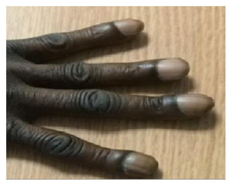
This clinical photograph displays the hand and fingers of a patient with severe digital clubbing, a clinical sign often associated with chronic hypoxia or certain systemic diseases. The image demonstrates characteristic 'drumstick' appearances of the distal phalanges, characterized by bulbous swelling and enlargement of the fingertips. The nails exhibit increased convexity in both the longitudinal and transverse planes, along with a significant increase in the Lovibond angle (the angle between the nail plate and the proximal nail fold). The skin overlying the interphalangeal joints shows marked thickening and deep, horizontal wrinkling, consistent with pachydermoperiostosis (primary hypertrophic osteoarthropathy). These visual findings are diagnostic markers for advanced clubbing (Grade 4). The content is relevant for medical education focusing on physical examination signs, rheumatology, and pulmonology.
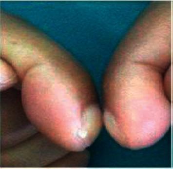
This clinical photograph displays a close-up view of two fingers positioned dorsal surface to dorsal surface to assess for digital clubbing. The image demonstrates a classic presentation of clubbing, characterized by the bulbous, drumstick-like enlargement of the distal phalanges. There is a visible increase in the longitudinal and transverse curvature of the nails, along with the softening of the nail bed. Crucially, the image illustrates a positive Schamroth's sign, identified by the complete absence of the normal diamond-shaped window (Schamroth's window) between the opposed nail beds. This physical finding is a hallmark of hypertrophic osteoarthropathy (HOA) and can be associated with chronic underlying conditions such as pulmonary malignancy, cystic fibrosis, or cyanotic heart disease. The photograph serves as an educational reference for medical trainees to recognize morphological fingertip changes and the clinical examination technique for thoracic and systemic disease markers.
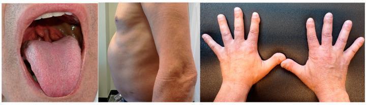
This composite of three clinical photographs displays hallmark physical signs associated with Tangier disease (ABCA1 deficiency). The left panel illustrates the oropharynx, showing characteristic hyperplastic, orange-colored tonsils, a pathognomonic sign resulting from the accumulation of cholesteryl esters in lymphoid tissue. The middle panel presents a lateral view of the patient's torso, demonstrating a significantly distended abdomen indicative of massive hepatosplenomegaly. The right panel displays the dorsal surface of both hands, revealing digital clubbing (hippocratic fingers), characterized by the loss of the Lovibond angle and increased curvature of the nail bed. Collectively, these images illustrate the systemic clinical manifestations of a severe lipid metabolism disorder, highlighting key diagnostic landmarks in the physical examination including reticuloendothelial organomegaly and characteristic mucosal pigmentary changes.
pitting pedal edema clinical examination
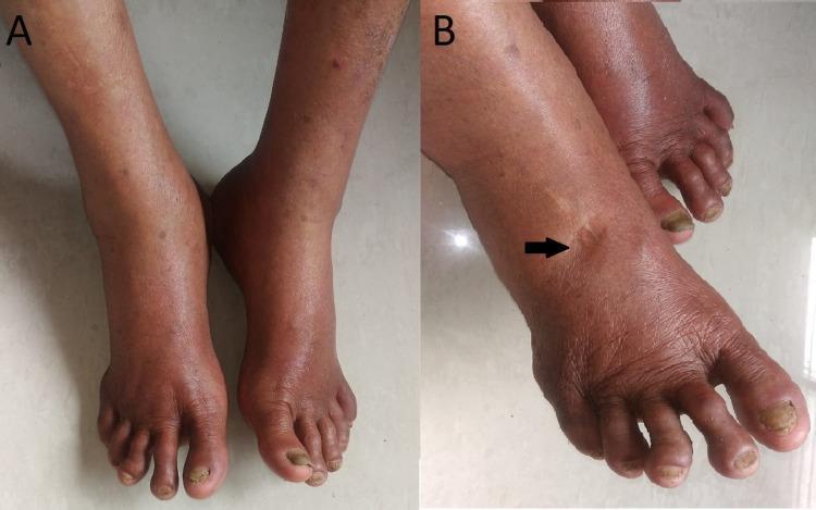
A two-panel clinical photograph illustrating bilateral lower extremity edema. Image A shows a superior view of both feet and ankles, demonstrating significant, symmetrical swelling (pedal edema) that obscures normal bony landmarks. The skin appears taut with subtle reddish-brown hyperpigmentation and scattered macules. Image B provides a lateral-oblique view of the dorsal foot where a black arrow points to a distinct, localized indentation, confirming the 'pitting' phenomenon. The skin in this region is stretched and shiny. Secondary findings include thickening and yellowish discoloration of the toenails, characteristic of onychomycosis. This visual documentation is used in medical education to teach the clinical physical examination of fluid retention and peripheral edema associated with systemic or inflammatory conditions such as post-viral sequelae (e.g., Chikungunya).
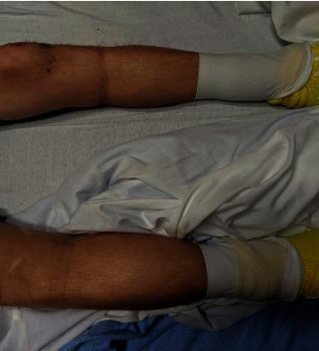
Clinical photograph of a patient's lower extremities in a supine position, demonstrating significant bilateral pedal edema. The swelling is symmetrical, extending from the feet proximally to the level of the knees. The skin over the shins and calves appears taut, shiny, and distended, characteristic of high-grade pitting edema (clinically noted as 4+). Both feet are covered with white medical compression wraps or dressings, with yellow non-slip hospital socks visible. This image serves as a clinical example of generalized peripheral edema, which in this educational context is associated with insulin-induced edema. Key landmarks include the visible distension of the pretibial area and the ankle region, contrasting with the proximal thigh area above the knee where the swelling appears to terminate. This visual is relevant for training in physical examination, fluid volume assessment, and recognizing secondary side effects of endocrine therapies.
General Physical Examination (GPE)
1. General Appearance / General Survey
- Level of consciousness and mental status - alert, drowsy, confused, mood and affect
- Build and nutrition - well-built/emaciated/obese, signs of wasting or cachexia (may suggest chronic heart failure, malignancy, or another systemic disorder)
- Posture, demeanor, and body position - e.g., a patient avoiding certain positions to reduce chest pain (as in acute pericarditis), or sitting upright to relieve dyspnea
- Facial expression and apparent distress - in pain, resting quietly, dyspneic, diaphoretic
- Decubitus/posture clues - a barrel-chest deformity, tachypnea, and pursed-lip breathing can point to a pulmonary cause of dyspnea
- Gait and mobility
- Obvious syndromic features - e.g., trisomy 21, Marfan syndrome, or other genetic syndromes are often recognizable on general inspection (Harrison's Principles of Internal Medicine 22E, p. 1903)
2. Anthropometry
- Height and weight, with calculation of BMI and body surface area
- Waist circumference and waist-to-hip ratio - used to predict long-term cardiovascular risk (Harrison's Principles of Internal Medicine 22E, p. 1903)
3. Vital Signs
- Temperature
- Pulse - rate, rhythm, volume, character, condition of vessel wall, symmetry (compare both radial/femoral pulses)
- Respiratory rate, depth, and pattern
- Blood pressure (both arms if indicated)
- Oxygen saturation (SpO2), where available
4. Skin and Mucous Membrane Signs
- Pallor - conjunctival, palmar, or generalized pallor suggesting anemia
- Icterus (jaundice) - best seen in the sclera; may be an early sign of advanced right heart failure with congestive hepatomegaly, hemolysis, or hepatobiliary disease
- Cyanosis
- Central cyanosis - from right-to-left cardiac or pulmonary shunting, causing deoxygenated blood to reach the systemic circulation
- Peripheral cyanosis/acrocyanosis - from reduced extremity blood flow (severe heart failure, shock, peripheral vascular disease); can worsen with beta-blocker use
- Differential cyanosis - isolated to the lower extremities in large PDA with reversed shunt (Harrison's Principles of Internal Medicine 22E, p. 1904)
- Clubbing - bulbous "drumstick" enlargement of the distal phalanges with increased nail curvature and loss of the Lovibond angle/Schamroth's window; associated with chronic hypoxia, cyanotic heart disease, pulmonary malignancy, cystic fibrosis, and other chronic suppurative or hepatic conditions
- Lymphadenopathy - palpation of cervical, axillary, inguinal, and other accessible nodal groups for size, tenderness, consistency, and mobility
- Edema - pitting or non-pitting, its distribution (pedal, sacral, generalized/anasarca) and grade
- Other skin findings: telangiectasias, xanthomas (tendon sheath, palmar crease, eruptive), bronze skin discoloration (hemochromatosis), petechiae/ecchymoses, hydration status (skin turgor, mucous membrane moisture)
5. Head and Neck Screening
- Dentition and oral hygiene (a source of infection and index of general health)
- High-arched palate, bifid uvula - connective tissue disease clues
- Eyes - arcus senilis, fundoscopic exam (important in hypertension, diabetes, suspected endocarditis), scleral color
- Neck - lymph nodes, thyroid, jugular venous pressure, tracheal position (Harrison's Principles of Internal Medicine 22E, p. 1904)
6. Chest and Abdomen (screening level)
- Surgical scars, chest wall deformities (pectus carinatum/excavatum, barrel chest), respiratory pattern and use of accessory muscles
- Abdominal contour, visible masses, organomegaly, tenderness on a screening palpation
Typical Sequence Used at the Bedside
- Introduce yourself, obtain consent, ensure privacy and adequate exposure/lighting
- General survey (consciousness, build, nutrition, posture, distress)
- Vital signs
- Pallor, icterus, cyanosis, clubbing, lymphadenopathy, edema (often remembered by trainees as "PICCLE")
- Head-to-toe screening as above
- Proceed to system-specific examination