
Brief one about karyotyping with examples
Reading File
Now let me search the library for karyotyping content.
Finding Sources
Finding Sources
Good results. Let me read the most relevant sections - Thompson & Thompson Genetics (best genetics textbook) and Henry's Clinical Pathology for the procedural detail.
Reading File
Reading File
Now let me get the clinical examples (chromosomal disorders) and the karyotype image.
Reading File
Loading Image
Finding Sources
I now have enough to write a solid, well-sourced brief on karyotyping. Let me also fetch the karyogram image for visual illustration.
Loading Image
Here is a concise but complete overview of karyotyping, drawn from authoritative sources.
Karyotyping
Definition
A karyotype is the organized visual display of all chromosomes from a single cell, arranged in homologous pairs from largest to smallest, with sex chromosomes placed last. The word also refers to the chromosomal constitution of an individual (e.g., "a normal female karyotype") and, as a verb, to the process of preparing that display.
- Thompson & Thompson Genetics and Genomics in Medicine, 9th ed.
The Chromosome Spread (G-Banding)
Chromosomes are best visualized at metaphase or prometaphase of mitosis, when they are maximally condensed. Cells are most commonly obtained from a peripheral blood lymphocyte culture, then treated with colchicine to arrest division at metaphase.
The standard staining method is Giemsa banding (G-banding), which produces a characteristic alternating light-and-dark band pattern along each chromosome. This pattern reflects underlying DNA features (GC vs. AT content, repetitive elements). Up to 1,000 bands can be resolved across all chromosomes; each band contains 50 or more genes.
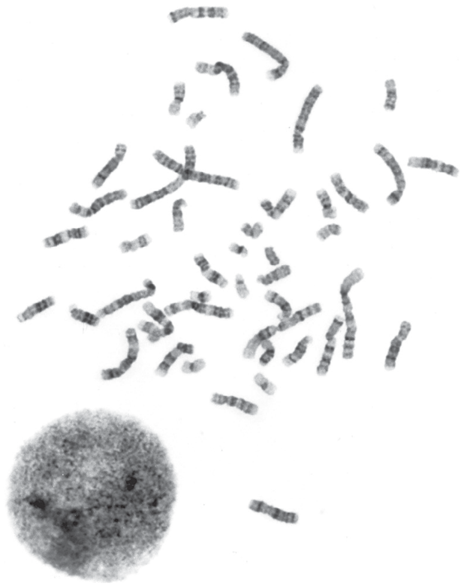
G-banded chromosome spread from a human lymphocyte culture. (Thompson & Thompson Genetics, 9th ed.)
Chromosome Identification
Each chromosome is identified by three features:
- Overall size - chromosomes are ranked 1 (largest) to 22 plus X and Y
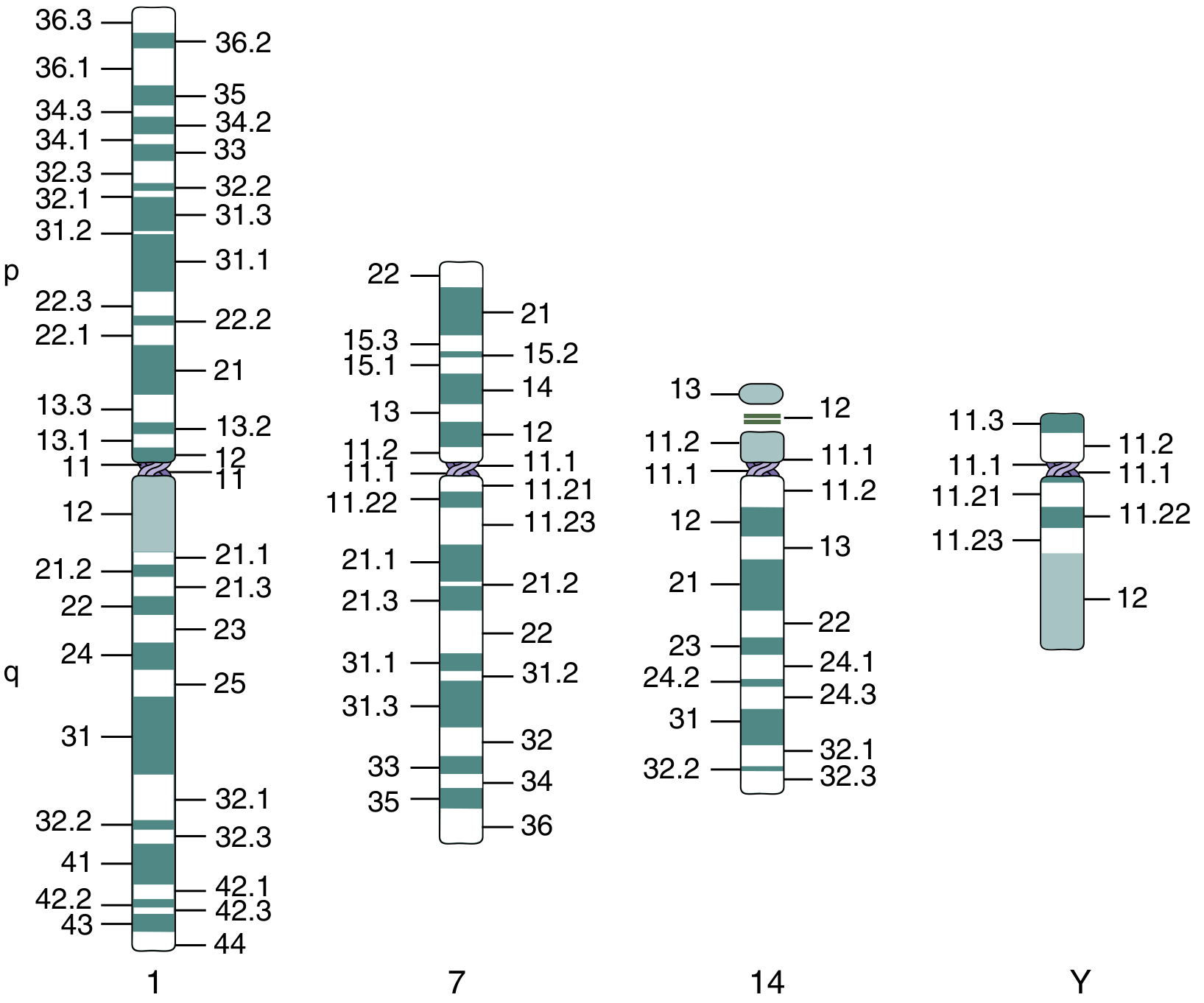
- Centromere position - divides each chromosome into a short arm (p, for petit) and a long arm (q)
- Banding pattern - unique to each chromosome pair
The ideogram below shows example chromosomes (1, 7, 14, Y) with their labeled p and q arm band positions:
The Procedure - Step by Step
| Step | Detail |
|---|---|
| Cell source | Peripheral blood (most common), amniotic fluid, chorionic villi, bone marrow |
| Culture | Cells grown in vitro; mitosis stimulated with phytohemagglutinin (lymphocytes) |
| Arrest | Colchicine/colcemid arrests cells at metaphase |
| Harvest & staining | Hypotonic solution swells cells; cells fixed, spread on slides, then G-banded |
| Analysis | 15-20 cells examined; single-cell anomalies = artifacts; 3+ cells with same loss OR 2+ with same gain = true abnormality |
| Resolution | Conventional karyotype can detect deletions/duplications down to ~3 Mb |
| High resolution | Cells harvested at prometaphase (less condensed) for smaller abnormalities |
| Output | A karyogram is generated (chromosomes arranged in pairs) using computer-assisted imaging |
- Henry's Clinical Diagnosis and Management by Laboratory Methods
Karyotype Nomenclature (ISCN)
| Notation | Meaning |
|---|---|
46,XX | Normal female |
46,XY | Normal male |
47,XX,+21 | Female with trisomy 21 (Down syndrome) |
45,X | Monosomy X (Turner syndrome) |
47,XXY | Klinefelter syndrome |
46,XX,rob(14;21)(q10;q10),+21 | Down syndrome by Robertsonian translocation |
46,XY,del(5p) | Cri-du-chat syndrome (5p deletion) |
Common Clinical Examples
1. Down Syndrome (Trisomy 21) - 47,XX,+21 or 47,XY,+21
- Extra copy of chromosome 21 due to meiotic nondisjunction
- ~95% of cases: standard trisomy (47 chromosomes)
- ~4%: Robertsonian translocation (46 chromosomes but trisomic for 21q)
- ~2%: mosaicism (two cell populations - normal + trisomy 21)
- Risk increases with maternal age; meiotic error is maternal in ~90%
- Karyotyping is essential to confirm diagnosis AND determine recurrence risk (standard trisomy vs. translocation vs. mosaic have very different recurrence risks)
2. Turner Syndrome - 45,X
- Monosomy X; lack of contribution of a sex chromosome (usually paternal)
- ~50% are 45,X; ~25% have structural X abnormality; ~25% are mosaic (45,X/46,XX)
- Features: short stature, gonadal dysgenesis, lymphedema, coarctation of aorta
3. Klinefelter Syndrome - 47,XXY
- Extra X chromosome in a male; most common sex chromosome aneuploidy in males
- Features: tall stature, hypogonadism, infertility, gynecomastia
4. Edwards Syndrome (Trisomy 18) - 47,XX,+18 or 47,XY,+18
- Second most common autosomal trisomy compatible with live birth
- Features: severe intellectual disability, clenched fists, rocker-bottom feet, heart defects
- Most die within the first year
5. Patau Syndrome (Trisomy 13) - 47,XX,+13 or 47,XY,+13
- Third most common autosomal trisomy compatible with live birth
- Features: holoprosencephaly, cleft lip/palate, polydactyly, cardiac defects
6. Cri-du-Chat Syndrome - 46,XX,del(5p) or 46,XY,del(5p)
- Deletion of the short arm of chromosome 5
- Features: high-pitched cat-like cry in infancy, microcephaly, intellectual disability
Indications for Karyotyping
- Suspected chromosomal syndrome (dysmorphic features, intellectual disability)
- Prenatal diagnosis (advanced maternal age, abnormal screening, structural fetal anomalies)
- Recurrent pregnancy loss
- Primary amenorrhea or infertility
- Hematologic malignancies (e.g., Philadelphia chromosome
t(9;22)in CML) - Ambiguous genitalia / differences of sex development
Limitations
-
Cannot detect single-gene mutations (e.g., cystic fibrosis, sickle cell)
-
Minimum detectable deletion/duplication is ~3 Mb
-
For submicroscopic abnormalities, FISH, chromosomal microarray (CMA), or NGS are required
-
Thompson & Thompson Genetics and Genomics in Medicine, 9th ed.; Henry's Clinical Diagnosis, Laboratory Methods; Robbins Pathologic Basis of Disease
This is a shared conversation. Sign in to Orris to start your own chat.