Maternal sepsis and its management. Long question for ms obg exam with possible table viva questions s . New guidelines

Reading File
Finding Sources
Finding Sources
Searching PubMed

"maternal sepsis"[MeSH Terms] AND management

Searching the Web

maternal sepsis management guidelines 2024 2025 WHO ACOG FIGO

Reading File
Reading Web Page

https://publications.smfm.org/publications/507-society-for-m…

Reading File
Reading File
Finding Sources
Finding Sources
Reading File
Reading File
Searching Images

maternal sepsis management algorithm flowchart

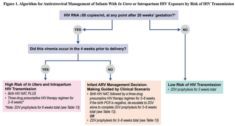
Summary : This flowchart presents an algorithm for antiretroviral management of infants with in utero or intrapartum HIV exposure, stratified by risk of HIV transmission based on maternal HIV RNA levels and timing of viremia.

flowchart:
# Nodes :
  • Start (rectangle): "HIV RNA ≥50 copies/mL at any point after 20 weeks’ gestation?"
  • Decision (diamond): "YES" / "NO" branches.
  • Decision (rectangle): "Did this viremia occur in the 4 weeks prior to delivery?"
  • Decision (diamond): "YES" / "NO" branches.
  • High Risk (rectangle, pink): "High Risk of In Utero and Intrapartum HIV Transmission"
    – "Birth HIV NAT, PLUS"
    – "Three-drug presumptive HIV therapy regimen for 2–6 weeks*"
    – "*Note: ZDV prophylaxis for 6 weeks total (see Table 13)"
  • Intermediate Risk (rectangle, peach): "Infant ARV Management Decision-Making Guided by Clinical Scenario"
    – "Birth HIV NAT, followed by a three-drug presumptive HIV therapy regimen for 2–6 weeks."
    – "If the birth PCR is negative, de-escalate to ZDV alone to complete ZDV prophylaxis for 2–6 weeks total (see Table 13)."
    – "OR"
    – "ZDV prophylaxis for 2–6 weeks total (see Table 13)"
  • Low Risk (rectangle, green): "Low Risk of HIV Transmission"
    – "ZDV prophylaxis for 2 weeks total"

# Connectors :
  • Arrow from Start node splits to YES and NO.
  • YES branch leads to "Did this viremia occur in the 4 weeks prior to delivery?" node.
  • NO branch leads directly to "Low Risk of HIV Transmission" node.
  • From "Did this viremia occur..." node, YES leads to "High Risk..." node, NO leads to "Infant ARV Management..." node.

# Layout :
  • Top-down hierarchical flow.
  • Initial decision splits into two main branches (YES/NO).
  • YES branch has a secondary decision, further splitting into two risk categories.
  • NO branch leads directly to low risk management.

# Analysis :
  • The algorithm stratifies infants into high, intermediate, or low risk for HIV transmission based on maternal HIV RNA levels and timing of viremia.
  • High risk infants receive more intensive ARV therapy (three-drug regimen and longer ZDV prophylaxis).
  • Intermediate risk infants have management tailored by clinical scenario, with possible de-escalation based on birth PCR results.
  • Low risk infants receive only short-course ZDV prophylaxis.
  • The flowchart ensures that infants at greatest risk receive the most aggressive prophylaxis, while minimizing unnecessary treatment for low-risk cases.

Summary : This flowchart presents an algorithm for antiretroviral management of infants with in utero or intrapartum HIV exposure, stratified by risk of HIV transmission based on maternal HIV RNA levels and timing of viremia. flowchart: # Nodes : • Start (rectangle): "HIV RNA ≥50 copies/mL at any point after 20 weeks’ gestation?" • Decision (diamond): "YES" / "NO" branches. • Decision (rectangle): "Did this viremia occur in the 4 weeks prior to delivery?" • Decision (diamond): "YES" / "NO" branches. • High Risk (rectangle, pink): "High Risk of In Utero and Intrapartum HIV Transmission" – "Birth HIV NAT, PLUS" – "Three-drug presumptive HIV therapy regimen for 2–6 weeks*" – "*Note: ZDV prophylaxis for 6 weeks total (see Table 13)" • Intermediate Risk (rectangle, peach): "Infant ARV Management Decision-Making Guided by Clinical Scenario" – "Birth HIV NAT, followed by a three-drug presumptive HIV therapy regimen for 2–6 weeks." – "If the birth PCR is negative, de-escalate to ZDV alone to complete ZDV prophylaxis for 2–6 weeks total (see Table 13)." – "OR" – "ZDV prophylaxis for 2–6 weeks total (see Table 13)" • Low Risk (rectangle, green): "Low Risk of HIV Transmission" – "ZDV prophylaxis for 2 weeks total" # Connectors : • Arrow from Start node splits to YES and NO. • YES branch leads to "Did this viremia occur in the 4 weeks prior to delivery?" node. • NO branch leads directly to "Low Risk of HIV Transmission" node. • From "Did this viremia occur..." node, YES leads to "High Risk..." node, NO leads to "Infant ARV Management..." node. # Layout : • Top-down hierarchical flow. • Initial decision splits into two main branches (YES/NO). • YES branch has a secondary decision, further splitting into two risk categories. • NO branch leads directly to low risk management. # Analysis : • The algorithm stratifies infants into high, intermediate, or low risk for HIV transmission based on maternal HIV RNA levels and timing of viremia. • High risk infants receive more intensive ARV therapy (three-drug regimen and longer ZDV prophylaxis). • Intermediate risk infants have management tailored by clinical scenario, with possible de-escalation based on birth PCR results. • Low risk infants receive only short-course ZDV prophylaxis. • The flowchart ensures that infants at greatest risk receive the most aggressive prophylaxis, while minimizing unnecessary treatment for low-risk cases.

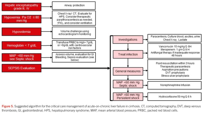
Summary : This figure presents a suggested algorithm for the critical care management of acute-on-chronic liver failure in cirrhosis, outlining stepwise assessment and interventions for various clinical scenarios including hepatic encephalopathy, hypoxemia, hypovolemia, anemia, and septic shock, with specific recommendations for investigations, infection treatment, and general supportive measures.

flowchart:
# Nodes :
  • Hepatic encephalopathy grade III, IV (rectangle, red)
  • Hypoxemia Pa O2 ≤ 80 mm Hg (rectangle, red)
  • Hypovolemia (rectangle, red)
  • Hemoglobin < 7 g/dL (rectangle, red)
  • MAP < 60 mm Hg see Septic shock (rectangle, red)
  • SEPSIS Evaluation (rectangle, red)
  • Airway protection (rectangle, white)
  • Chest X-ray/CT. Evaluate for HPS, Consider therapeutic para/thoracentesis as needed, ↑FiO2, and consider ventilation (rectangle, white)
  • Volume challenge using echocardiogram monitoring (rectangle, white)
  • Transfuse PRBC to Hgb > 7g/dL or > 9g/dL with cardiovascular risk factors (rectangle, white)
  • Assess volume, evaluation for GI Bleeding, Sepsis evaluation (rectangle, white)
  • Investigations (rectangle, pink)
  • Paracentesis, Culture blood, ascites, urine, Chest X-ray, Lactate (rectangle, white)
  • Treat infection (rectangle, pink)
  • Vancomycin 15 mg/kg Q 6H, Meropenem 1 gm Q 8H, Antifungal therapy if inadequate response 48 hours (rectangle, white)
  • General measures (rectangle, pink)
  • Fluid resuscitation within 3 hours, Therapeutic paracentesis, Aspiration precautions, DVT prophylaxis, Stress ulcer prophylaxis (rectangle, white)
  • MAP < 60 mm Hg Septic shock (rectangle, pink)
  • Norepinephrine Infusion (rectangle, white)
  • MAP < 60 mm Hg Persistent shock (rectangle, pink)
  • Hydrocortisone 50 mg Q 6 h (rectangle, white)

# Connectors :
  • Each red rectangle (clinical scenario) connects rightward to a specific white rectangle (intervention or assessment).
  • SEPSIS Evaluation leads to a vertical sequence of pink rectangles: Investigations → Treat infection → General measures → MAP < 60 mm Hg Septic shock → MAP < 60 mm Hg Persistent shock.
  • Each pink rectangle is followed by a white rectangle detailing specific actions.
  • The flow is primarily left-to-right for initial assessment, then top-to-bottom for sepsis management.

# Layout :
  • The diagram is organized in three main vertical columns:
    – Left: Red rectangles for initial clinical findings.
    – Middle: White rectangles for immediate interventions.
    – Right: Pink rectangles for sepsis evaluation and management, with white rectangles for detailed steps.
  • The sepsis management column is a vertical sequence, with each step leading to the next.

# Analysis :
  • The algorithm provides a structured, stepwise approach for managing acute-on-chronic liver failure in cirrhosis, prioritizing airway protection, oxygenation, volume status, anemia correction, and sepsis evaluation.
  • Sepsis management is detailed, with specific recommendations for investigations, antimicrobial therapy, supportive measures, and escalation to vasopressors and steroids for persistent shock.
  • The flowchart emphasizes early identification and targeted intervention for each critical issue, integrating both general and specific measures for optimal patient care.

Summary : This figure presents a suggested algorithm for the critical care management of acute-on-chronic liver failure in cirrhosis, outlining stepwise assessment and interventions for various clinical scenarios including hepatic encephalopathy, hypoxemia, hypovolemia, anemia, and septic shock, with specific recommendations for investigations, infection treatment, and general supportive measures. flowchart: # Nodes : • Hepatic encephalopathy grade III, IV (rectangle, red) • Hypoxemia Pa O2 ≤ 80 mm Hg (rectangle, red) • Hypovolemia (rectangle, red) • Hemoglobin < 7 g/dL (rectangle, red) • MAP < 60 mm Hg see Septic shock (rectangle, red) • SEPSIS Evaluation (rectangle, red) • Airway protection (rectangle, white) • Chest X-ray/CT. Evaluate for HPS, Consider therapeutic para/thoracentesis as needed, ↑FiO2, and consider ventilation (rectangle, white) • Volume challenge using echocardiogram monitoring (rectangle, white) • Transfuse PRBC to Hgb > 7g/dL or > 9g/dL with cardiovascular risk factors (rectangle, white) • Assess volume, evaluation for GI Bleeding, Sepsis evaluation (rectangle, white) • Investigations (rectangle, pink) • Paracentesis, Culture blood, ascites, urine, Chest X-ray, Lactate (rectangle, white) • Treat infection (rectangle, pink) • Vancomycin 15 mg/kg Q 6H, Meropenem 1 gm Q 8H, Antifungal therapy if inadequate response 48 hours (rectangle, white) • General measures (rectangle, pink) • Fluid resuscitation within 3 hours, Therapeutic paracentesis, Aspiration precautions, DVT prophylaxis, Stress ulcer prophylaxis (rectangle, white) • MAP < 60 mm Hg Septic shock (rectangle, pink) • Norepinephrine Infusion (rectangle, white) • MAP < 60 mm Hg Persistent shock (rectangle, pink) • Hydrocortisone 50 mg Q 6 h (rectangle, white) # Connectors : • Each red rectangle (clinical scenario) connects rightward to a specific white rectangle (intervention or assessment). • SEPSIS Evaluation leads to a vertical sequence of pink rectangles: Investigations → Treat infection → General measures → MAP < 60 mm Hg Septic shock → MAP < 60 mm Hg Persistent shock. • Each pink rectangle is followed by a white rectangle detailing specific actions. • The flow is primarily left-to-right for initial assessment, then top-to-bottom for sepsis management. # Layout : • The diagram is organized in three main vertical columns: – Left: Red rectangles for initial clinical findings. – Middle: White rectangles for immediate interventions. – Right: Pink rectangles for sepsis evaluation and management, with white rectangles for detailed steps. • The sepsis management column is a vertical sequence, with each step leading to the next. # Analysis : • The algorithm provides a structured, stepwise approach for managing acute-on-chronic liver failure in cirrhosis, prioritizing airway protection, oxygenation, volume status, anemia correction, and sepsis evaluation. • Sepsis management is detailed, with specific recommendations for investigations, antimicrobial therapy, supportive measures, and escalation to vasopressors and steroids for persistent shock. • The flowchart emphasizes early identification and targeted intervention for each critical issue, integrating both general and specific measures for optimal patient care.

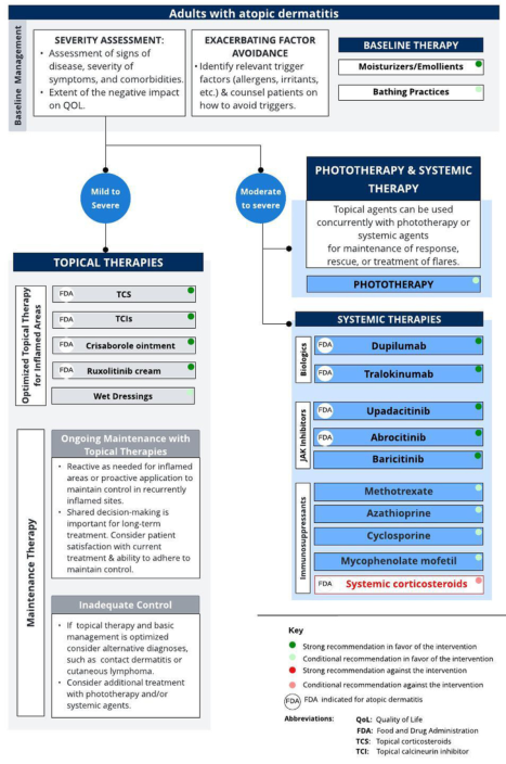
Summary : This flowchart provides a comprehensive management algorithm for adults with atopic dermatitis, detailing baseline management, topical therapies, phototherapy, and systemic therapies, including FDA-approved and recommended treatments, maintenance strategies, and escalation steps for inadequate control.

flowchart:
# Baseline Management :
  • Severity Assessment: Assessment of signs of disease, severity of symptoms, comorbidities, and impact on quality of life (QOL).
  • Exacerbating Factor Avoidance: Identify trigger factors (allergens, irritants, etc.) and counsel patients on avoidance.
  • Baseline Therapy: Moisturizers/Emollients (strong recommendation), Bathing Practices (conditional recommendation).

# Initial Pathways :
  • Mild to Severe: Proceed to Topical Therapies.
  • Moderate to Severe: Proceed to Phototherapy & Systemic Therapy.

# Topical Therapies :
  ## Optimized Topical Therapy for Inflamed Areas :
    • TCS (Topical corticosteroids) (FDA, strong recommendation)
    • TCIs (Topical calcineurin inhibitors) (FDA, strong recommendation)
    • Crisaborole ointment (FDA, strong recommendation)
    • Ruxolitinib cream (FDA, strong recommendation)
    • Wet Dressings (strong recommendation)
  ## Ongoing Maintenance with Topical Therapies :
    • Reactive or proactive application for maintenance.
    • Shared decision-making for long-term treatment.
    • Consider patient satisfaction and adherence.
  ## Inadequate Control :
    • If topical therapy and basic management optimized, consider alternative diagnoses.
    • Consider additional treatment with phototherapy and/or systemic agents.

# Phototherapy & Systemic Therapy :
  • Topical agents can be used concurrently with phototherapy or systemic agents for maintenance, rescue, or flares.

# Phototherapy :
  • No specific agents listed; included as a treatment option for moderate to severe cases.

# Systemic Therapies :
  ## Biologics :
    • Dupilumab (FDA, strong recommendation)
    • Tralokinumab (FDA, strong recommendation)
  ## JAK Inhibitors :
    • Upadacitinib (FDA, strong recommendation)
    • Abrocitinib (FDA, strong recommendation)
    • Baricitinib (strong recommendation)
  ## Immunosuppressants :
    • Methotrexate (strong recommendation)
    • Azathioprine (strong recommendation)
    • Cyclosporine (strong recommendation)
    • Mycophenolate mofetil (strong recommendation)
    • Systemic corticosteroids (FDA, strong recommendation against use)

# Key :
  • Green circle: Strong recommendation in favor.
  • Yellow circle: Conditional recommendation in favor.
  • Red circle: Strong recommendation against.
  • Orange circle: Conditional recommendation against.
  • FDA: Indicated for atopic dermatitis.

# Abbreviations :
  • QOL: Quality of Life
  • FDA: Food and Drug Administration
  • TCS: Topical corticosteroids
  • TCI: Topical calcineurin inhibitor

# Layout :
  • The flowchart is organized from baseline management at the top, splitting into two main pathways (mild to severe and moderate to severe), with further branches into topical, phototherapy, and systemic therapies.
  • Maintenance and escalation steps are included for ongoing management and inadequate control.

# Analysis :
  • The algorithm emphasizes starting with baseline management and topical therapies, escalating to phototherapy and systemic therapies for more severe or refractory cases.
  • Strong recommendations are visually highlighted for first-line agents, with systemic corticosteroids strongly discouraged.
  • FDA-approved options are clearly marked, supporting evidence-based decision-making.
  • The flowchart supports a stepwise, patient-centered approach, integrating maintenance and escalation strategies.

Summary : This flowchart provides a comprehensive management algorithm for adults with atopic dermatitis, detailing baseline management, topical therapies, phototherapy, and systemic therapies, including FDA-approved and recommended treatments, maintenance strategies, and escalation steps for inadequate control. flowchart: # Baseline Management : • Severity Assessment: Assessment of signs of disease, severity of symptoms, comorbidities, and impact on quality of life (QOL). • Exacerbating Factor Avoidance: Identify trigger factors (allergens, irritants, etc.) and counsel patients on avoidance. • Baseline Therapy: Moisturizers/Emollients (strong recommendation), Bathing Practices (conditional recommendation). # Initial Pathways : • Mild to Severe: Proceed to Topical Therapies. • Moderate to Severe: Proceed to Phototherapy & Systemic Therapy. # Topical Therapies : ## Optimized Topical Therapy for Inflamed Areas : • TCS (Topical corticosteroids) (FDA, strong recommendation) • TCIs (Topical calcineurin inhibitors) (FDA, strong recommendation) • Crisaborole ointment (FDA, strong recommendation) • Ruxolitinib cream (FDA, strong recommendation) • Wet Dressings (strong recommendation) ## Ongoing Maintenance with Topical Therapies : • Reactive or proactive application for maintenance. • Shared decision-making for long-term treatment. • Consider patient satisfaction and adherence. ## Inadequate Control : • If topical therapy and basic management optimized, consider alternative diagnoses. • Consider additional treatment with phototherapy and/or systemic agents. # Phototherapy & Systemic Therapy : • Topical agents can be used concurrently with phototherapy or systemic agents for maintenance, rescue, or flares. # Phototherapy : • No specific agents listed; included as a treatment option for moderate to severe cases. # Systemic Therapies : ## Biologics : • Dupilumab (FDA, strong recommendation) • Tralokinumab (FDA, strong recommendation) ## JAK Inhibitors : • Upadacitinib (FDA, strong recommendation) • Abrocitinib (FDA, strong recommendation) • Baricitinib (strong recommendation) ## Immunosuppressants : • Methotrexate (strong recommendation) • Azathioprine (strong recommendation) • Cyclosporine (strong recommendation) • Mycophenolate mofetil (strong recommendation) • Systemic corticosteroids (FDA, strong recommendation against use) # Key : • Green circle: Strong recommendation in favor. • Yellow circle: Conditional recommendation in favor. • Red circle: Strong recommendation against. • Orange circle: Conditional recommendation against. • FDA: Indicated for atopic dermatitis. # Abbreviations : • QOL: Quality of Life • FDA: Food and Drug Administration • TCS: Topical corticosteroids • TCI: Topical calcineurin inhibitor # Layout : • The flowchart is organized from baseline management at the top, splitting into two main pathways (mild to severe and moderate to severe), with further branches into topical, phototherapy, and systemic therapies. • Maintenance and escalation steps are included for ongoing management and inadequate control. # Analysis : • The algorithm emphasizes starting with baseline management and topical therapies, escalating to phototherapy and systemic therapies for more severe or refractory cases. • Strong recommendations are visually highlighted for first-line agents, with systemic corticosteroids strongly discouraged. • FDA-approved options are clearly marked, supporting evidence-based decision-making. • The flowchart supports a stepwise, patient-centered approach, integrating maintenance and escalation strategies.

Now I have all the information I need. Let me compose the comprehensive answer.

Maternal Sepsis: Comprehensive MS OBG Exam Answer


DEFINITION (Key for Viva)

WHO 2017 definition (current gold standard):
"Maternal sepsis is a life-threatening condition defined as organ dysfunction resulting from infection during pregnancy, childbirth, post-abortion, or the postpartum period."
This definition aligns with the adult Sepsis-3 consensus and replaces the older SIRS-based definition.
Septic shock = Sepsis + persisting hypotension requiring vasopressors to maintain MAP ≥65 mmHg + serum lactate >2 mmol/L despite adequate fluid resuscitation. Hospital mortality exceeds 40%.
(RCOG Green-top Guideline No. 64, BJOG 2025; WHO 2017)

INCIDENCE AND IMPORTANCE

ParameterData
Contribution to maternal mortality10-15% of direct maternal deaths globally
Global burden (GLOSS study)70 per 1000 live births (severe maternal infection)
UK incidence (severe sepsis)47 per 100,000 maternities
US incidence49 per 100,000 delivery hospitalizations
Mortality with septic shock>40%
Postpartum cases account for ~47% of maternal bacteremia; antepartum ~17%; intrapartum ~36% (Creasy & Resnik MFM).

AETIOLOGY AND SOURCES

Most common sources (by trimester/period):
PeriodPrimary SourceCommon Organisms
AntepartumUrinary tract (46%), genital (41%)E. coli, Klebsiella, GBS
IntrapartumGenital tract (90%)GBS, E. coli, GAS
PostpartumGenital (54%), urinary (25%)E. coli, GAS, Staphylococcus
Post-abortionUterusAnaerobes, E. coli, Streptococcus
Group A Streptococcus (GAS) - highest risk; rapidly fatal, easily transmitted by healthcare worker hands. Invasive GAS (iGAS) is notifiable.
Other important organisms: MRSA, GBS, E. coli, Klebsiella, Bacteroides, Clostridium. Clinical laboratory identifies organism in only 64% of maternal sepsis cases (UKOSS data).

RISK FACTORS

StrengthFactors
Inconsistent riskObesity, multiple pregnancy, PPROM/PTL, retained products, pregestational diabetes
Modest (OR <2)Nulliparity, African ancestry, age >35, low socioeconomic status
Stronger (OR ≥2)ART (OR~5), cerclage (OR 3.4-9.8), cesarean delivery (OR 2.0-8.1), HIV (OR 3.2-4.2), transfusion (OR 10.9), peripartum hysterectomy (OR 56), chronic renal disease (OR 33), chronic liver disease (OR 55), congestive heart failure (OR 135)
(Creasy & Resnik MFM, Box 71.3)

CLINICAL FEATURES (Recognition - Critical for Exam)

Symptoms suggesting infection:
  • Fever or rigors (temperature >38°C or <36°C)
  • Abdominal/pelvic pain
  • Purulent/offensive vaginal discharge (endometritis, chorioamnionitis)
  • Dysuria, frequency (UTI/pyelonephritis)
  • Productive cough (pneumonia)
  • Rash (GAS, meningococcal)
  • Vomiting, diarrhea
  • No fever does NOT exclude sepsis (immunosuppression, NSAID use can mask temperature)
Warning signs of organ dysfunction / deterioration:
  • Tachycardia >100 bpm
  • Tachypnea >25/min
  • Systolic BP <90 mmHg or MAP <65 mmHg
  • Altered mental status, drowsiness, confusion
  • Reduced urine output <0.5 mL/kg/hr
  • Lactate >2 mmol/L
  • Mottled skin, prolonged capillary refill

SCORING SYSTEMS (High-Yield Viva Tables)

TABLE 1: Obstetrically Modified qSOFA (omqSOFA)

Parameter01
Systolic BP>90 mmHg<90 mmHg
Respiratory rate<25/min>25/min
Altered mental statusAlertNot alert
Score ≥2 = suspect sepsis, escalate care. (SOMANZ guidelines)

TABLE 2: Obstetrically Modified SOFA (omSOFA)

SystemParameterScore 0Score 1Score 2
RespiratoryPaO₂/FiO₂>400300-399<300
CoagulationPlatelets>150,000/μL100-150<100
HepaticBilirubin<20 μmol/L20-32 μmol/L>32 μmol/L
CardiovascularMAP (mmHg)>70<70Vasopressors required
CNSConsciousnessAlertRousable (voice)Rousable (pain)
RenalCreatinine<90 μmol/L90-120 μmol/L>120 μmol/L
(SOMANZ 2017; Creasy & Resnik MFM)
Key note for viva: Standard SOFA/qSOFA are less reliable in pregnancy due to normal physiological changes (lower baseline BP, elevated HR, altered lab norms). Pregnancy-specific modifications must be used.

TABLE 3: Sepsis-3 Definitions (Applied to Obstetrics)

TermDefinition
InfectionSuspected or confirmed pathological process caused by a micro-organism
SepsisOrgan dysfunction (omSOFA ≥2) due to infection in pregnancy/postpartum
Septic shockSepsis + vasopressor requirement to maintain MAP ≥65 mmHg + lactate >2 mmol/L after adequate resuscitation

INVESTIGATIONS

Immediate (within 1 hour):
InvestigationPurpose
Blood cultures x2 (before antibiotics)Identify organism and guide therapy
Serum lactateSeverity assessment; >4 mmol/L = immediate ICU referral
FBCLeukocytosis/leukopenia, thrombocytopenia
CRP, procalcitoninInflammatory markers
Urea, creatinine, LFTsRenal and hepatic dysfunction
Coagulation screen (PT, APTT, D-dimer, fibrinogen)DIC screen
ABGAcid-base, oxygenation
Urine culture and MC&SUTI/pyelonephritis
Vaginal/endocervical swabsGAS, anaerobes
High vaginal swabPostpartum endometritis
Chest X-rayPneumonia
Pelvic/abdominal USS or CTIdentify source (abscess, retained products)
CTGFetal wellbeing in antepartum
Key viva point: Half of UK maternal sepsis deaths had no lactate measured; 67% received antibiotics only on the same day of death (RCOG Green-top 64 data).

MANAGEMENT - THE HOUR-1 BUNDLE (Surviving Sepsis Campaign)

The SSC Hour-1 Bundle (also embedded in RCOG Green-top 64 and SMFM #67):

TABLE 4: Hour-1 Sepsis Bundle (5 Actions Within 1 Hour)

StepAction
1Measure serum lactate
2Obtain blood cultures before antibiotics (do not delay antibiotics for cultures)
3Administer broad-spectrum IV antibiotics
4Give 30 mL/kg IV crystalloid for hypotension or lactate ≥4 mmol/L
5Apply vasopressors if hypotensive during/after fluids to maintain MAP ≥65 mmHg
Re-measure lactate within 1 hour if initial value elevated (>2 mmol/L).

SEPSIS SIX (UK Sepsis Trust - useful for ward-level implementation)

Give 3 to the patient:
  1. IV fluid challenge
  2. IV antibiotics
  3. Oxygen (target SpO₂ >94%)
Take 3 from the patient: 4. Blood cultures 5. FBC and other bloods 6. Lactate level

ANTIBIOTIC THERAPY (Tables for Viva)

Principle: Broad-spectrum empiric IV antibiotics within 1 hour of recognition (GRADE 1C - SMFM; Grade C - RCOG). De-escalate based on culture results.

TABLE 5: Empiric Antibiotic Choices by Source

SourceFirst-lineAlternative/MRSA cover
Genital tract / chorioamnionitisPiperacillin-tazobactam (pip-tazo) 4.5g IV 8-hourlyAdd metronidazole if anaerobes suspected
Urinary tract / pyelonephritisCeftriaxone 1-2g IV ODPiperacillin-tazobactam
Pneumonia (CAP)Co-amoxiclav + clarithromycinCeftriaxone + azithromycin
Suspected GASBenzylpenicillin 1.2g IV 4-hourly + clindamycin 900mg IV 8-hourlyVancomycin (PCN allergy)
MRSA suspectedAdd vancomycin 15-20 mg/kg IVLinezolid
Unknown source / severely illMeropenem or piperacillin-tazobactam ± vancomycinAs per local protocol
Clindamycin is added with penicillin for GAS because it inhibits exotoxin production (anti-toxin effect), reducing tissue destruction.

FLUID RESUSCITATION

  • SMFM 2023: Early IV administration of 1-2 L balanced crystalloid within first 3 hours in hypotension or suspected hypoperfusion (GRADE 1C)
  • RCOG 2025: Immediate 500 mL crystalloid bolus for hypotension or lactate >4 mmol/L; repeat if needed
  • Choice: Balanced crystalloid (Hartmann's/Ringer's lactate or normal saline) - FIRST LINE (GRADE 1B)
  • Avoid: Starches (HES) and gelatin - associated with AKI, worse outcomes (GRADE 1A against)
  • Monitor response with dynamic preload measures (pulse pressure variation, stroke volume variation, passive leg raise test)
  • Use hourly urometer to measure urine output when indicated

VASOPRESSORS AND INOTROPES

AgentRoleNotes
Norepinephrine (noradrenaline)First-line vasopressorMaintain MAP ≥65 mmHg; generally safe in pregnancy
VasopressinAdd-on if norepinephrine doses escalatingMay reduce norepinephrine requirements
DopamineSecond line (if norepinephrine unavailable)Higher arrhythmia risk
PhenylephrineUse with caution in pregnancyCan reduce uterine blood flow
EpinephrineThird-line, refractory shock
Key viva point: Phenylephrine, though a common vasopressor in obstetric anaesthesia, can reduce uteroplacental blood flow and is less preferred than norepinephrine in septic shock.

CORTICOSTEROIDS

  • Consider hydrocortisone 200 mg/day IV (50 mg 6-hourly or continuous infusion) in septic shock not responding to adequate fluids and vasopressors
  • Do not use corticosteroids if adequate fluid resuscitation restores hemodynamic stability
  • Antenatal corticosteroids (betamethasone/dexamethasone) for fetal lung maturity should still be given if indicated, but should not delay sepsis management

SOURCE CONTROL

Critical and time-sensitive:
ConditionSource Control
ChorioamnionitisExpedite delivery (regardless of gestational age)
Endometritis with retained productsSurgical evacuation (ERPC)
Tubo-ovarian abscess / pelvic abscessRadiological drainage or surgical drainage
Septic abortionUterine evacuation
Necrotizing fasciitisUrgent surgical debridement
Urological sourceNephrostomy, ureteric stent
SMFM GRADE 1C: If intrauterine infection is suspected or confirmed, prompt delivery or evacuation of uterine contents is recommended regardless of gestational age.

FETAL CONSIDERATIONS

  • In antepartum sepsis, perform continuous CTG monitoring
  • Maternal fever, tachycardia, and hypotension compromise uteroplacental perfusion causing fetal tachycardia, late decelerations
  • Delivery should be in maternal interest first - do not delay sepsis management for fetal concerns
  • Expedite delivery if: intrauterine infection, fetal compromise, or if delivery would benefit the mother's condition
  • Consider neonatal unit involvement for preterm deliveries
(RCOG Green-top 64: "In a critically ill pregnant woman, birth of the baby can be expedited if it would be beneficial to the woman")

ORGAN SUPPORT AND ICU CARE

Organ SystemManagement
RespiratorySupplemental O₂; target SpO₂ >94%; intubation + mechanical ventilation for ARDS (low tidal volume 6 mL/kg, PEEP, prone positioning)
RenalFluid balance, avoid nephrotoxins; renal replacement therapy (CRRT) if AKI refractory
CoagulationTreat DIC: FFP, cryoprecipitate, platelets; tranexamic acid if indicated
MetabolicTight glycaemic control (target 7-10 mmol/L); avoid hypoglycaemia
NutritionalEnteral nutrition within 48 hours where possible
DVT prophylaxisLMWH (when coagulation permits), TED stockings
Stress ulcerPPI/H2 blocker in ventilated patients
Threshold for ICU transfer: Lactate >4 mmol/L, vasopressor requirement, respiratory failure, refractory septic shock. Involve critical care and infectious disease specialists early.

INFECTION CONTROL

  • GAS and MRSA: easily transmitted by healthcare workers - strict hand hygiene, contact precautions
  • Invasive GAS (iGAS): notifiable disease - inform infection control team + UKHSA
  • CPE and VRE carriers: isolate per local protocols
  • Post-caesarean sepsis prevention (FIGO 2025): antibiotics 30-60 minutes before surgery, vaginal + skin antiseptic prep, glove change after placental delivery

PREVENTION

StrategyEvidence
Prophylactic antibiotics at LSCSCefazolin IV before skin incision (reduces SSI by >50%)
GBS screening & intrapartum prophylaxisReduces neonatal + maternal GBS sepsis
Hand hygiene (5 moments)Most effective prevention measure
Antiseptic vaginal prep (chlorhexidine)Reduces post-caesarean endometritis
Early recognition protocols + sepsis screeningPerformance improvement programs recommended (SMFM GRADE 1B)
Influenza vaccinationReduces viral sepsis in pregnancy

SPECIAL SITUATIONS

Post-Abortal Sepsis

  • Suspect after any termination or miscarriage with fever, uterine tenderness, foul discharge
  • Common organisms: anaerobes, E. coli, streptococcus, Clostridium perfringens
  • Management: IV antibiotics (cover anaerobes - add metronidazole) + surgical evacuation

Group A Streptococcal Sepsis

  • Can be rapidly fatal within hours of onset
  • Door-to-antibiotic time is critical
  • Benzylpenicillin + clindamycin combination (anti-toxin effect)
  • Isolate patient; notify public health

Sepsis Following Influenza

  • Influenza in pregnancy: higher ARDS rate (9.7% vs 5.4% in non-pregnant), higher ECMO requirement
  • Early oseltamivir + bacterial superinfection cover

OUTCOMES

  • Long-term: >50% of sepsis survivors have some cognitive impairment; physical, emotional sequelae common
  • SMFM Best Practice: Offer comprehensive ongoing support for sepsis survivors and their families
  • Perinatal: Antepartum sepsis associated with preterm birth, fetal compromise, IUGR, stillbirth

QUICK VIVA TABLE SUMMARY

TABLE 6: Key "Numbers" for Viva

ParameterValue
MAP target with vasopressors≥65 mmHg
Lactate: immediate escalation/ICU≥4 mmol/L
Lactate: abnormal (monitor)>2 mmol/L
Antibiotic target timeWithin 1 hour
IV crystalloid initial bolus (RCOG)500 mL (repeat if needed)
IV crystalloid (SMFM)1-2 L within 3 hours
Fluid for lactate ≥4 or hypotension (SSC)30 mL/kg crystalloid
Urine output target≥0.5 mL/kg/hr
SpO₂ target>94%
Blood glucose target (ICU)7-10 mmol/L
Hydrocortisone dose (refractory shock)200 mg/day (50 mg 6-hourly)

TABLE 7: Guideline Sources (Examiner May Ask)

GuidelineBodyYear
Green-top Guideline No. 64 (Identification and Management of Maternal Sepsis)RCOG/BJOG2025
SMFM Consult Series #67: Maternal SepsisSMFM (endorsed by ACOG)2023, reaffirmed 2025
Surviving Sepsis Campaign Hour-1 BundleSSC (ESICM/SCCM)Updated 2018+
WHO Maternal Sepsis DefinitionWHO2017
SOMANZ Sepsis in Pregnancy GuidelinesSOMANZ2017
Preventing Post-Caesarean Sepsis (FIGO review)FIGO/IJGO2025
APT-Sepsis Trial (new WHO data on prevention/management)WHO/HRP2025

TABLE 8: Differential Diagnoses Causing Maternal Collapse / Mimicking Sepsis

ConditionDistinguishing Feature
Pulmonary embolismSudden dyspnea, pleuritic pain, D-dimer raised, CTPA
Amniotic fluid embolismSudden collapse during/after delivery, DIC, bronchospasm
EclampsiaSeizures, proteinuria, hypertension
Haemorrhage (concealed)Bleeding source, pallor without fever
Acute liver failureJaundice, coagulopathy, encephalopathy
Acute pancreatitisEpigastric pain, raised amylase/lipase
AnaphylaxisUrticaria, bronchospasm, known allergen exposure

TABLE 9: Multidisciplinary Team in Maternal Sepsis

SpecialistRole
ObstetricianSource identification, delivery decision, uterine source control
Intensivist / Critical careICU management, mechanical ventilation, vasopressors
Infectious disease specialistAntibiotic optimization, ID source
MicrobiologistCulture interpretation, antibiotic de-escalation
AnaesthetistAirway management, invasive monitoring
HaematologistDIC management
NeonatologistFetal/neonatal welfare if antepartum
RadiologistUSS/CT for source, interventional drainage

ESSAY STRUCTURE OUTLINE (For MS OBG Long Question)

Introduction: Define, state WHO 2017 definition, epidemiological importance (10-15% maternal deaths), global GLOSS data.
Body:
  1. Aetiology and sources (genital > urinary tract, by period)
  2. Risk factors (table)
  3. Clinical features and recognition (symptoms, warning signs)
  4. Scoring tools (omqSOFA, omSOFA - tables)
  5. Investigations
  6. Management - Hour-1 Bundle, antibiotics, fluids, vasopressors, source control (detailed)
  7. ICU care and organ support
  8. Fetal considerations and delivery
  9. Prevention
  10. Special situations (GAS, post-abortal)
Conclusion: Maternal sepsis is a time-critical emergency requiring immediate MDT response, early antibiotics, source control, and ICU support. Adherence to Hour-1 bundle and institution-specific protocols improves survival.

Sources:
This is a shared conversation. Sign in to Orris to start your own chat.