
Sarcoidosis
sarcoidosis diagnosis management
sarcoidosis histology non-caseating granuloma lung
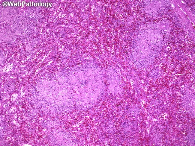
This is a photomicrograph of a splenic parenchyma section from a splenectomy specimen, prepared for light microscopy and stained with hematoxylin and eosin. The specimen shows multiple subcentimeter nodules within the splenic parenchyma, with well-formed, non-caseating granulomas composed of tightly arranged epithelioid macrophages and multinucleated giant cells surrounded by a lymphocytic rim. The granulomas lack central necrosis, which, along with histiocytic microarchitecture, is highly suggestive of granulomatous inflammation due to Toxoplasma gondii infection. Serologic tests confirmed toxoplasmosis in this patient, correlating with disseminated disease or focal splenic involvement associated with lymphadenopathy and splenomegaly. In the spleen, toxoplasmosis can produce discrete nodules or focal granulomatous lesions that may mimic lymphoma or other granulomatous diseases on imaging or gross examination. Key differential diagnoses include sarcoidosis, fungal infections such as histoplasmosis, mycobacterial infections, and other parasitic infestations; clinical correlation with serology and immune status is essential. This image is relevant for teaching granulomatous splenitis due to toxoplasmosis, assessing granuloma morphology, distinguishing non-caseating from caseating granulomas, and correlating histology with systemic infection. Appropriate clinical scenarios include immunocompromised patients with fever, cytopenias, or organomegaly and routine pathology review in splenectomy specimens. This image exemplifies infectious granulomatous disease companion to serology and morphology in clinical practice.
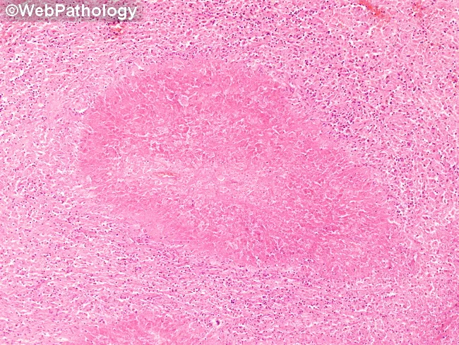
This is a low-magnification brightfield histology image of a tissue section stained with Hematoxylin and Eosin (H&E). The specimen shows granulomatous inflammation with aggregates of epithelioid macrophages forming a rounded granuloma, often with multinucleated giant cells, surrounded by a lymphocytic cuff. The central focus is well circumscribed within native parenchyma, suggesting a chronic, organized immune response to a persistent antigen. Necrosis is not clearly evident at this magnification, though subtle caseation cannot be excluded. Morphology is compatible with tuberculoid or non-caseating granulomas, and infectious versus noninfectious etiologies must be distinguished with ancillary studies. This pattern can occur in lymph nodes or solid organs and prompts a differential that includes tuberculous lymphadenitis, sarcoidosis, fungal granulomatous infections (Histoplasma, Coccidioides), and foreign-body reaction. Clinically, recognition of granulomas guides testing: acid-fast bacilli staining (Ziehl-Neelsen), fungal stains (GMS/PAS), cultures, PCR panels, and radiologic correlation. The image is educational for medical trainees, illustrating granuloma architecture, macrophage differentiation, and the spectrum of chronic inflammatory responses. Educational context.
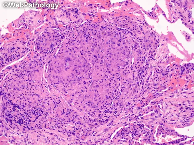
Pulmonary tissue from a wedge resection stained with Hematoxylin and Eosin, analyzed by brightfield light microscopy. The specimen shows robust granulomatous inflammation within the lung parenchyma consistent with a granulomatous infectious process. Nodular aggregates of epithelioid histiocytes are surrounded by a rim of lymphocytes and scattered plasma cells; multinucleated Langhans-type giant cells are present, producing a palisading appearance. The granulomas appear well circumscribed within the interstitium and may encroach upon adjacent alveolar spaces. Central necrosis is variably present and not conspicuously predominant in this field, a feature compatible with coccidioidal granulomatous reaction (coccidioidomycosis) that may display BCG-like granulomas in tissue specimens. The background alveolar architecture is preserved in portions, with mild interstitial inflammation and occasional eosinophils. Although fungal organisms are not definitively visualized in this image, the histology is compatible with fungal infection of the lung in the clinical context of pulmonary coccidioidomycosis, potentially associated with chronic granulomatous response. This image emphasizes characteristic granuloma morphology useful for differential diagnosis from mycobacterial tuberculosis or sarcoidosis, and provides histopathologic confirmation to guide clinical management, including antifungal therapy decisions and serology correlation. It is suitable for education on pulmonary granulomatous disease and wedge resection pathology.
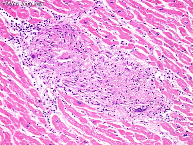
Histology, light microscopy of cardiac tissue, reveals classic non-caseating granulomatous inflammation characteristic of cardiac sarcoidosis within the full thickness of the myocardium. The predominant architectural feature is well-demarcated granulomas composed of epithelioid histiocytes with multinucleated giant cells, embedded in a mononuclear lymphocytic infiltrate. Rare eosinophils may be present, but necrosis is minimal or absent. Granulomas are distributed along lymphatic channels and are frequently associated with the cardiac conduction system tracts, explaining potential electrical disturbances. The surrounding myocytes show preserved morphology with only focal interstitial fibrosis in many areas, reflecting chronicity and prior inflammatory injury. This histologic pattern differentiates sarcoid granulomas from other granulomatous myocarditis such as tuberculosis or hypersensitivity reactions, where caseation or different inflammatory profiles may occur. The tissue shows extensive lymphatic involvement and perivascular localization, contributing to impaired conduction and remodeling. The specimen, stained with Hematoxylin and Eosin, highlights the pink cytoplasm of myocardium and the dark nuclei of inflammatory cells, facilitating recognition of granulomatous foci, giant cells, and fibrotic change. Clinically, these findings support a diagnosis of cardiac sarcoidosis and carry implications for prognosis, arrhythmia risk, and indications for immunosuppressive therapy and device management. This description enhances searchability for cardiac pathology, granulomatous myocarditis, and sarcoid research applications.
Sarcoidosis
Definition
- Goldman-Cecil Medicine, p. 978
Epidemiology
| Feature | Details |
|---|---|
| Age | Peaks 20-39 years; develops before age 50 in most |
| Sex | Women more often affected across all racial groups |
| Race (USA) | Black Americans: 35.5/100,000; White Americans: 10.9/100,000 (3.5x higher) |
| Highest global incidence | Northern Europe: 5-40/100,000/year |
| Family risk | 5x increased risk in first-degree relatives; 80-fold increase in monozygotic twins |
| Worst outcomes | Black women in the US (highest lifetime risk 2.7%); Black patients have later onset, more chronic/fatal disease |
Pathobiology
- Tightly organized collections of macrophages and macrophage-derived epithelioid cells
- Encircled by lymphocytes
- Contain multinucleated giant cells (fused epithelioid cells)
- Non-caseating (no central necrosis) - a key distinguishing feature
- HLA-mediated antigen processing by macrophages
- T-cell activation → oligoclonal expansion of CD4+ (Th1) lymphocytes
- CD4+ T cells produce IL-2 and IFN-γ
- TNF-α from macrophages organizes inflammatory cells into granulomas
- Th17 and T-regulatory (Treg) cells modulate granuloma formation and resolution
- Treg activity explains the immune paradox: anergy to tuberculin despite active inflammation
- B cells also play a role (polyclonal hypergammaglobulinemia, response to anti-B-cell therapy)
-
Airborne antigens (lungs are most common site)
-
Mycobacteria and propionibacteria (proposed, not confirmed)
-
Environmental exposures: mold, musty odors, agricultural work, pesticide industries (OR ~1.5 each)
-
Genetic risk: HLA-D and other MHC genes on chromosome 6p
-
Goldman-Cecil Medicine, p. 979
Clinical Manifestations
General Symptoms
- 30-50% are asymptomatic at diagnosis (found incidentally on CXR)
- Common: night sweats, weight loss, fatigue
- Fatigue is often persistent and related to inflammation, small fiber neuropathy, depression, or corticosteroid use
Löfgren Syndrome (Acute Presentation)
- Erythema nodosum (predominantly in women)
- Bilateral hilar adenopathy on CXR
- Polyarthritis (predominantly in men)
- May also include fever and uveitis
- Carries a favorable prognosis with spontaneous resolution
Respiratory System (>90% of patients)
- Most common symptoms: dry cough and dyspnea; chest pain is vague and nonspecific
- Upper respiratory tract (2-6%): nasal mucosal involvement, anosmia, septal erosion, stridor, dysphonia
- Bilateral hilar adenopathy is the most common radiographic finding
| Stage | Finding | Spontaneous Remission |
|---|---|---|
| 0 | Normal | - |
| I | Bilateral hilar adenopathy (BHL) alone | ~60-80% |
| II | BHL + pulmonary infiltrates | ~40-70% |
| III | Pulmonary infiltrates without BHL | ~10-20% |
| IV | Pulmonary fibrosis | Rare |
Skin (25-35% of patients)
- Lupus pernio: violaceous indurated plaques on the nose, cheeks, lips - associated with chronic disease, pulmonary fibrosis, and upper airway involvement
- Red-brown non-scaly papules/plaques on face, especially around nose and mouth
- Erythema nodosum (10%, usually in Löfgren syndrome)
- Lesions can form within scars and tattoos
Eyes
- Uveitis is the most common ocular manifestation; can precede diagnosis by decades
- Any part of the eye and adnexa can be involved
Cardiac Sarcoidosis (25-30% in USA; up to 60% in Japan by MRI/PET)
- Often clinically silent
- Can cause: complete heart block, ventricular arrhythmias, sudden cardiac death, cardiomyopathy
- MRI and PET are the preferred imaging modalities for detection
Musculoskeletal
- Arthritis/arthralgias: 4-38% of patients
- Acute type: part of Löfgren syndrome (knees and ankles; self-limiting)
- Chronic type (<5%): synovitis, polyarthritis/oligoarthritis, dactylitis; granulomas on synovial biopsy
- Lytic or sclerotic bone lesions (3-13%): "punched-out" lesions of phalanges
- Myopathy (3%)
Neurologic (Neurosarcoidosis)
- Cranial nerve palsies (especially CN VII - facial nerve)
- Requires high-dose corticosteroids (80-100 mg prednisone/day)
Renal
- Hypercalcemia (from granulomatous 1α-hydroxylase activity → excess 1,25-OH vitamin D) → nephrolithiasis, nephrocalcinosis
- Granulomatous interstitial nephritis
Liver/GI
- Hepatic granulomas are common but usually subclinical
- Rarely causes portal hypertension
Diagnosis
- Compatible clinical and radiographic findings
- Histologic evidence of non-caseating granulomas (from accessible site - skin, lymph node, bronchial mucosa, transbronchial biopsy)
- Exclusion of other granulomatous diseases (tuberculosis, fungal infection, foreign body reaction, malignancy)
| Test | Finding / Purpose |
|---|---|
| Chest X-ray | BHL (Stage I-II most common) |
| HRCT chest | Perilymphatic micronodules, upper-lobe predominance |
| Serum ACE (angiotensin-converting enzyme) | Elevated in ~60% - useful for monitoring, not diagnostic |
| Serum calcium / 24-hr urine calcium | Hypercalciuria/hypercalcemia |
| LFTs, renal function | Organ involvement screening |
| ECG | Arrhythmias, conduction block (cardiac sarcoidosis) |
| Ophthalmologic exam | Uveitis |
| PFTs | Restrictive pattern ± obstructive (endobronchial) |
| BAL | Increased CD4+:CD8+ ratio >3.5 (supportive) |
| MRI/PET | Cardiac and neurosarcoidosis |
| Tissue biopsy | Gold standard |
- Goldman-Cecil Medicine, pp. 980-984
Treatment
When to Treat
- Progressive decline in pulmonary function
- Significant symptoms impeding daily life
- Serious extrapulmonary involvement (cardiac, neurologic, ocular, hypercalcemia)
First-Line: Corticosteroids
- Oral prednisone 20-40 mg/day for 3 months (initial therapy for pulmonary disease)
- Assess response at 3 months (symptoms, FVC, imaging)
- If response: taper to 10-15 mg/day for 6-9 months, then taper off
- Recurrence after stopping: 30-80% of patients → monitor for 1-2 years
- Neurosarcoidosis requires high-dose prednisone (80-100 mg/day)
- Hypercalcemia: responds to prednisone 15-20 mg/day within days
Steroid-Sparing Agents (second-line or refractory disease)
| Drug | Role |
|---|---|
| Methotrexate | First-choice steroid-sparer; significant steroid-sparing effect |
| Azathioprine | Alternative steroid-sparer |
| Hydroxychloroquine | Skin and hypercalcemia |
| Infliximab (anti-TNF) | Refractory cases; superior for lupus pernio; neurosarcoidosis |
| Adalimumab | Anti-TNF alternative |
| Roflumilast | PDE-4 inhibitor; small trial showed reduced exacerbations in fibrotic sarcoidosis |
| Repository corticotropin | Prednisone-sparing; improves lung function/PET parameters |
Special Considerations
- Inhaled corticosteroids: only for bronchial hyperreactivity or persistent cough
- Pulmonary rehabilitation: recommended for pulmonary fibrosis (Stage IV)
- Skin lesions: intralesional corticosteroid injections (2-4 week intervals); systemic steroids for widespread disease
- Corticosteroids should be continued ≥1 month after adding immunosuppressives
- Allow 3-6 months of cytotoxic therapy to assess response
Prognosis
- Stage I: 60-80% spontaneous remission
- Löfgren syndrome: generally favorable
- Black patients: more likely to have chronic, severe, fatal disease
- Lupus pernio and angiolupoid sarcoidosis: markers of chronic disease
- Stage IV (fibrosis): irreversible; lung transplant may be considered
- Leading causes of sarcoidosis death: respiratory failure, cardiac arrhythmias, neurosarcoidosis
- Isolated cardiac sarcoidosis (2025 systematic review, PMID 41193496) carries a high risk of sudden cardiac death and requires particular vigilance
Summary Box
| Feature | Key Point |
|---|---|
| Hallmark lesion | Non-caseating granuloma |
| Most common organ | Lung (>90%) |
| Most common radiographic finding | Bilateral hilar adenopathy |
| Acute syndrome | Löfgren (erythema nodosum + BHL + arthritis) |
| Skin marker of chronic disease | Lupus pernio |
| Immune paradox | Anergy despite active granulomatous inflammation |
| Hypercalcemia mechanism | Granulomatous 1α-hydroxylase → excess 1,25-OH Vit D |
| First-line treatment | Oral prednisone |
| Best steroid-sparer | Methotrexate or azathioprine |
| Refractory/lupus pernio | Infliximab |