
treatment protocol to follow with drugs and dosage in detail for haemprotozoal disease in dogs and cats with sign symptoms
haemoprotozoa dogs cats treatment protocol drugs dosage 2024 veterinary
https://www.abcdcatsvets.org/guideline-for-babesiosis
https://www.merckvetmanual.com/generalized-conditions/ricket…
haemoprotozoa dogs cats treatment review
blood smear Babesia dog intraerythrocytic protozoa parasite
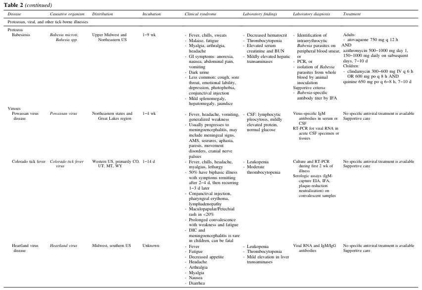
Table 2 (continued) <table><thead><tr><th>Disease</th><th>Causative organism</th><th>Distribution</th><th>Incubation</th><th>Clinical syndrome</th><th>Laboratory findings</th><th>Laboratory diagnosis</th><th>Treatment</th></tr></thead><tbody><tr><td colspan="8">Protozoan, viral, and other tick-borne illnesses</td></tr><tr><td>Protozoa<br>Babesiosis</td><td>Babesia microti, Babesia spp.</td><td>Upper Midwest and Northeastern US</td><td>1–9 wk</td><td>- Fever, chills, sweats<br>- Malaise, fatigue<br>- Myalgia, arthralgia, headache<br>- GI symptoms: anorexia, nausea, abdominal pain, vomiting<br>- Dark urine<br>- Less common: cough, sore throat, emotional lability, depression, photophobia, conjunctival injection<br>- Mild splenomegaly, hepatomegaly, jaundice</td><td>- Decreased hematocrit<br>- Thrombocytopenia<br>- Elevated serum creatinine and BUN<br>- Mildly elevated hepatic transaminases</td><td>- Identification of intraerythrocytic Babesia parasites on peripheral blood smear, or<br>- PCR, or<br>- Isolation of Babesia parasites from whole blood by animal inoculation<br>Supportive criteria:<br>- Babesia-specific antibody titer by IFA</td><td>Adults:<br>- atovaquone 750 mg q 12 h AND<br>azithromycin 500–1000 mg day 1, 150–1000 mg daily on subsequent days, 7–10 d<br>Children:<br>- clindamycin 300–600 mg IV q 6 h OR 600 mg po q 8 h AND<br>quinine 650 mg po q 6–8 h, 7–10 d</td></tr><tr><td>Viruses<br>Powassan virus disease</td><td>Powassan virus</td><td>Northeastern states and Great Lakes region</td><td>1–4 wk</td><td>- Fever, headache, vomiting, generalized weakness<br>- Usually progresses to meningoencephalitis, may include meningeal signs, AMS, seizures, aphasia, paresis, movement disorders, cranial nerve palsies</td><td>- CSF: lymphocytic pleocytosis, mildly elevated protein, normal glucose</td><td>Virus-specific IgM antibodies in serum or CSF<br>RT-PCR for viral RNA in acute CSF specimen or tissues</td><td>No specific antiviral treatment is available<br>Supportive care</td></tr><tr><td>Colorado tick fever</td><td>Colorado tick fever virus</td><td>Western US, primarily CO, UT, MT, WY</td><td>1–14 d</td><td>- Fever, chills, headache, myalgias, lethargy<br>- 50% have biphasic illness with symptoms remitting after 2–4 d, then recurring 1–3 d later<br>- Conjunctival injection, pharyngeal erythema, lymphadenopathy<br>- Maculopapular/Petechial rash in <20%<br>- Prolonged convalescence with weakness and fatigue<br>- DIC and meningoencephalitis is rare in children, can be fatal</td><td>- Leukopenia<br>- Moderate thrombocytopenia</td><td>Culture and RT-PCR during first 2 wk of illness<br>Serologic assays (IgM-capture EIA, IFA, plaque-reduction neutralization) on convalescent samples</td><td>No specific antiviral treatment is available<br>Supportive care</td></tr><tr><td>Heartland virus disease</td><td>Heartland virus</td><td>Midwest, southern US</td><td>Unknown</td><td>- Fever<br>- Fatigue<br>- Decreased appetite<br>- Headache<br>- Arthralgia<br>- Myalgia<br>- Nausea<br>- Diarrhea</td><td>- Leukopenia<br>- Thrombocytopenia<br>- Mild elevation in liver transaminases</td><td>Viral RNA and IgM/IgG antibodies</td><td>No specific antiviral treatment is available<br>Supportive care</td></tr></tbody></table>
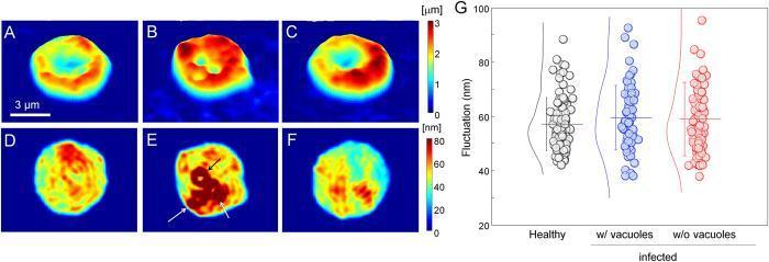
This composite image presents topographical and mechanical analyses of red blood cells (RBCs) infected with Babesia microti. Panels A-C show two-dimensional topographical height profiles. A healthy RBC (A) displays a characteristic biconcave discocyte shape with a central dimple. An infected Bm-RBC with parasite vacuoles (B) shows a loss of this central dimple and an irregular height profile, while an infected Bm-RBC without visible vacuoles (C) largely maintains the donut shape. Panels D-F depict dynamic membrane fluctuations measured by root-mean-square (RMS) height displacement. In panel E, arrows highlight high-value ring-shaped fluctuations surrounding the parasite vacuoles. The color scales indicate height (0-3 μm) and fluctuation (0-80 nm). Panel G is a scatter plot comparing membrane fluctuations (nm) among healthy RBCs, infected RBCs with vacuoles, and infected RBCs without vacuoles. The plot demonstrates that despite structural changes, the mechanical deformability of the host membrane remains statistically comparable across all three groups, a feature that distinguishes Babesia infection from Plasmodium-induced stiffening.
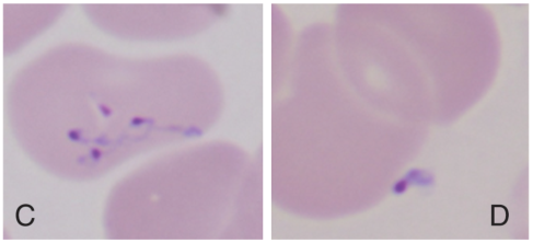
Summary : This figure shows two microscopy images (labeled C and D) of stained blood cells, highlighting the presence of small, dark-stained structures within or near the cells. photo: Scene Overview : • Two side-by-side light microscopy images of blood smears, labeled "C" (left) and "D" (right). • Main subjects are red blood cells with small, dark purple/blue-stained inclusions or organisms visible in the cytoplasm or adjacent to the cells. • Background is pale pink, typical of Giemsa or Wright’s stain. • The images are slightly out of focus, but the inclusions are clearly visible. Technical Details : • No visible scale bar or magnification indicator. • Staining method appears to be a standard hematological stain (e.g., Giemsa/Wright). • No on-image UI elements or additional text besides the labels "C" and "D" in the lower left and right corners, respectively. Spatial Relationships : • In image C, multiple inclusions are present within a single red blood cell, some connected by fine thread-like structures. • In image D, a single inclusion is seen near the edge of a red blood cell. • Both images focus on the central area of the field, with cells partially overlapping. Analysis : • The images likely compare two different morphological forms or stages of an intraerythrocytic organism (e.g., a blood parasite such as Plasmodium or Babesia). • Image C shows a more complex or segmented form, while image D shows a simpler, possibly single-nucleated form. • The visual differences suggest a comparison of developmental stages, species, or treatment effects on the organisms within red blood cells.
Haemoprotozal Diseases in Dogs and Cats: Complete Treatment Protocol
1. BABESIOSIS
Causative Agents
- Dogs: Babesia canis (large Babesia - most common in Europe/Africa), Babesia gibsoni (small Babesia - SE Asia, USA), Babesia vulpes/B. microti-like (very small)
- Cats: Babesia felis (South Africa), Babesia vogeli, Babesia clade VI
Signs and Symptoms
| System | Clinical Signs |
|---|---|
| General | Fever (39.5-41°C), lethargy, anorexia, weakness |
| Haematological | Haemolytic anaemia, pale/icteric mucous membranes, thrombocytopaenia |
| Urinary | Haemoglobinuria ("red/brown urine"), bilirubinuria |
| GI | Vomiting, diarrhoea, hepatosplenomegaly |
| Neurological | Ataxia, seizures, "cerebral babesiosis" (B. canis) |
| Severe/Complicated | Acute kidney injury, ARDS, DIC, immune-mediated haemolytic anaemia (IMHA) |
Treatment Protocol - DOGS
| Drug | Dose | Route | Interval | Duration | Notes |
|---|---|---|---|---|---|
| Imidocarb dipropionate (drug of choice) | 5-6 mg/kg | IM or SC | 2 weeks apart (2 injections) | 2 doses | Pre-treat with atropine (0.05 mg/kg SC) to prevent cholinergic side effects; effective for B. canis; less effective for B. gibsoni |
| Atovaquone + Azithromycin (for B. gibsoni/B. vulpes) | 13.5 mg/kg + 10 mg/kg | PO with fatty meal + PO | q8h + q24h | 10 days | Most effective for clearing parasitaemia (83% PCR-negative); expensive but best for B. gibsoni |
| Diminazene aceturate (not available in USA) | 3.5 mg/kg (B. canis) / 5-7.5 mg/kg (B. gibsoni) | IM | Single dose / repeat q2 weeks x2-3 | 1-3 doses | Available in Asia, Africa, Europe; narrow therapeutic window |
| Phenamidine isethionate | 15-16 mg/kg | SC | 24h apart | 2 doses | Alternative to imidocarb in some regions |
Treatment Protocol - CATS
| Drug | Dose | Route | Interval | Duration | Notes |
|---|---|---|---|---|---|
| Primaquine phosphate (drug of choice - B. felis) | 0.5 mg/kg | PO or IM | q72h x 1-3 doses, then weekly x 3 weeks | 3-4 weeks total | Narrow therapeutic window - NEVER exceed 1 mg/kg (lethal); causes vomiting PO; monitor CBC and biochem weekly |
| Imidocarb dipropionate (B. vogeli, clade VI) | 5 mg/kg | IM or SC | 2 weeks apart | 2 doses | Effective for non-B. felis species |
| Diminazene aceturate (B. vogeli, clade VI) | 2-3 mg/kg | IM | Single dose | 1 dose | Alternative for non-B. felis |
Warning for cats: Most canine anti-Babesia drugs are ineffective or toxic in cats with B. felis. Primaquine is the only reliably effective drug for B. felis sensu stricto. Do not use buparvaquone, enrofloxacin, or danofloxacin - no anti-Babesia effect demonstrated.
Supportive Care (Both Species)
- IV fluid therapy for dehydration/haemoglobinuria (Hartmann's solution or 0.9% NaCl)
- Blood transfusion if PCV < 15% (dogs) or < 12% (cats)
- Prednisolone 1-2 mg/kg/day PO if IMHA component confirmed
- Monitor renal parameters, bilirubin, coagulation profile
2. EHRLICHIOSIS AND ANAPLASMOSIS
Causative Agents
- Ehrlichia canis (Canine Monocytic Ehrlichiosis - CME) - most important; tick-borne (Rhipicephalus sanguineus)
- Anaplasma phagocytophilum - dogs and cats; Ixodes ticks
- Anaplasma platys (Canine Cyclic Thrombocytopaenia)
- Ehrlichia ewingii - dogs only
Signs and Symptoms
| Phase | Signs |
|---|---|
| Acute (1-4 weeks) | High fever, lethargy, lymphadenopathy, splenomegaly, thrombocytopaenia, epistaxis, petechiae |
| Sub-clinical | Mild thrombocytopaenia, patient appears clinically well |
| Chronic (severe) | Pancytopaenia, bone marrow aplasia, bleeding diathesis, weight loss, ocular lesions (uveitis, hyphaema), neurological signs (ataxia, seizures), polyarthritis (A. phagocytophilum) |
Treatment Protocol - DOGS AND CATS
| Drug | Dose | Route | Interval | Duration | Notes |
|---|---|---|---|---|---|
| Doxycycline (drug of choice) | 10 mg/kg | PO or IV | q24h (or 5 mg/kg q12h) | Minimum 4 weeks (28 days) | Rapid clinical improvement within 24-48h. Avoid in puppies < 6 months (tooth staining). Give with food. |
| Minocycline | 5-12.5 mg/kg | PO | q12h | 4 weeks | Alternative tetracycline |
| Rifampicin/Rifampin | 10-20 mg/kg | PO | q12h | 3-4 weeks | May help in experimental ehrlichiosis; not reliably curative |
| Chloramphenicol | 15-25 mg/kg | PO | q8h | 4 weeks | Used in dogs < 6 months (avoid doxycycline); monitor for bone marrow suppression |
Note: Clinical improvement expected within 24-48 hours of starting doxycycline. If no improvement in 48 hours, reconsider diagnosis. Cats with anaplasmosis respond equally well to doxycycline 10 mg/kg PO q24h x 28 days. (ESCCAP Guideline 2024)
Supportive Care
- Blood/platelet transfusion for severe haemorrhage
- Avoid glucocorticoids unless concurrent IMHA is confirmed (may worsen infection)
- Treat secondary infections if present
3. HEPATOZOONOSIS
Causative Agents
- Dogs: Hepatozoon canis (tick ingestion - Rhipicephalus spp.) and Hepatozoon americanum (USA - Amblyomma maculatum)
- Cats: Hepatozoon felis (usually subclinical)
Signs and Symptoms
| Species/Organism | Clinical Signs |
|---|---|
| H. canis (mild-moderate) | Lethargy, weight loss, lymphadenopathy, anaemia, mild fever |
| H. americanum (severe) | Marked pain on palpation, muscle wasting, periosteal bone proliferation (pathognomonic), severe cachexia, purulent ocular discharge, cyclic fever |
| Cats (H. felis) | Usually subclinical; mild anaemia occasionally |
Treatment Protocol - DOGS
| Drug | Dose | Route | Interval | Duration |
|---|---|---|---|---|
| Imidocarb dipropionate | 5 mg/kg | IM or SC | 2 weeks apart | 2-4 doses |
| Doxycycline | 10 mg/kg | PO | q24h | 3-4 weeks |
| Toltrazuril | 5-10 mg/kg | PO | q24h | 5-7 days |
| Drug | Dose | Route | Interval | Duration |
|---|---|---|---|---|
| Trimethoprim-sulfadiazine | 15 mg/kg | PO | q12h | 14 days (induction) |
| Pyrimethamine | 0.25 mg/kg | PO | q24h | 14 days (induction) |
| Clindamycin | 10 mg/kg | PO | q8h | 14 days (induction) |
| Maintenance: Decoquinate | 10-20 mg/kg | PO mixed with food | q12h | Lifelong (prevents relapse) |
Decoquinate (Deccox) maintenance is critical for H. americanum - discontinuation leads to relapse in most dogs.
4. CYTAUXZOONOSIS (CATS ONLY)
Causative Agent
Signs and Symptoms
- Rapid onset: high fever (40-41.5°C), profound lethargy, anorexia
- Pale/icteric mucous membranes
- Dyspnoea, tachypnoea
- Tachycardia, hypothermia (terminal)
- Icterus, hepatosplenomegaly
- Death within 1-5 days if untreated
Treatment Protocol - CATS
| Drug | Dose | Route | Interval | Duration | Notes |
|---|---|---|---|---|---|
| Atovaquone + Azithromycin (preferred combination) | 15 mg/kg + 10 mg/kg | PO with fatty meal + PO | q8h + q24h | 10 days | Best survival rates (60% vs 25% with supportive only); give together |
| Imidocarb dipropionate (adjunct) | 2 mg/kg | IM | Once | - | Some protocols combine with atovaquone |
| Heparin | 200-300 IU/kg | SC | q6-8h | During hospitalisation | For prevention/treatment of thromboembolic disease |
Supportive Care (Intensive - essential for survival)
- IV fluid therapy (avoid over-hydration; monitor for pulmonary oedema)
- Nutritional support (nasogastric tube if not eating)
- NSAIDs - Meloxicam 0.1 mg/cat SC/PO q24h for fever/pain
- Anti-nausea: Maropitant 1 mg/kg SC q24h
- Blood transfusion if PCV < 12%
- Broad-spectrum antibiotics for secondary infections
5. LEISHMANIOSIS (DOGS - Leishmania infantum)
Signs and Symptoms
- Skin lesions: alopecia, scaling, hyperkeratosis (nose/footpads), nodules, ulcers
- Lymphadenopathy (generalised)
- Weight loss and muscle atrophy (cachexia)
- Epistaxis, polyuria/polydipsia (renal involvement)
- Ocular lesions (uveitis, keratoconjunctivitis)
- Splenomegaly and hepatomegaly
- Lameness (polyarthritis)
Treatment Protocol - DOGS
| Drug | Dose | Route | Interval | Duration | Notes |
|---|---|---|---|---|---|
| Meglumine antimoniate | 50 mg/kg | SC | BID (q12h) | 4-6 weeks | First-line; combine with allopurinol |
| Allopurinol | 10 mg/kg | PO | BID (q12h) | 6-18 months | Maintenance/secondary prevention; continues after antimoniate stopped |
| Miltefosine | 2 mg/kg | PO with food | q24h | 28 days | Oral alternative to antimoniate; teratogenic - use with caution |
| Meglumine antimoniate + Allopurinol | See above | Combined | - | Combined protocol | Most effective combination; standard of care in endemic areas |
Leishmaniosis is not curable - treatment achieves clinical remission and reduces parasite burden. Lifelong monitoring with serology and PCR is recommended. (ESCCAP 2024)
6. HAEMOPLASMOSIS (Mycoplasma haemofelis / Haemobartonellosis)
Causative Agents
- Cats: Mycoplasma haemofelis (most pathogenic), 'Candidatus M. haemominutum', 'Candidatus M. turicensis'
- Dogs: Mycoplasma haemocanis (mainly in splenectomised/immunosuppressed dogs)
Signs and Symptoms
- Acute haemolytic anaemia: pale/white mucous membranes, tachycardia, tachypnoea
- Lethargy, weakness, collapse
- Icterus (jaundice)
- Splenomegaly
- Cyclic fever
- Anorexia, weight loss
- Dogs: often subclinical unless immunocompromised or splenectomised
Treatment Protocol - CATS AND DOGS
| Drug | Dose | Route | Interval | Duration | Notes |
|---|---|---|---|---|---|
| Doxycycline (drug of choice) | 5-10 mg/kg | PO or IV | q12h-q24h | Minimum 4 weeks (some recommend 8 weeks for cats) | Reduces parasitaemia, improves clinical signs; may not fully eliminate organism; give with food/water to prevent oesophageal stricture in cats |
| Marbofloxacin (alternative) | 2-2.5 mg/kg | PO | q24h | 3-4 weeks | Fluoroquinolone alternative |
| Pradofloxacin | 5 mg/kg | PO | q24h | 3-4 weeks | Licensed for cats in some countries |
| Enrofloxacin | 5 mg/kg | PO | q24h | 3-4 weeks | Caution in cats - risk of retinal toxicity at higher doses; use with care |
Supportive Care
- Prednisolone 1-2 mg/kg PO q24h if concurrent IMHA (titrate down over 4-8 weeks)
- Blood transfusion if PCV < 12% cats or < 15% dogs
- Syringe/assisted feeding if anorexic
- Treat underlying immunosuppression (e.g., FIV/FeLV in cats)
7. TRYPANOSOMOSIS
Causative Agents
- Trypanosoma cruzi (Chagas disease) - dogs, cats; Americas
- Trypanosoma brucei (Sleeping sickness) - dogs; Africa (rare in pets)
- Trypanosoma evansi (Surra) - dogs; Asia, Africa, South America
Signs and Symptoms
| Phase (T. cruzi) | Signs |
|---|---|
| Acute | Fever, lymphadenopathy, oedema, myocarditis, sudden death (young dogs) |
| Indeterminate | Subclinical |
| Chronic | Dilated cardiomyopathy, arrhythmias, CHF signs: ascites, pleural effusion, exercise intolerance |
Treatment Protocol - DOGS (T. cruzi)
| Drug | Dose | Route | Interval | Duration | Notes |
|---|---|---|---|---|---|
| Benznidazole (drug of choice) | 5-7 mg/kg | PO | q12h | 60 days | Most effective in acute phase; poor efficacy in chronic phase; monitor for GI/neurological side effects |
| Nifurtimox | 2-7 mg/kg | PO | q8h | 3-4 months | Alternative; not widely available; causes GI side effects |
Note: Treatment efficacy for T. cruzi is limited and largely ineffective in the chronic cardiac phase. Cardiac management (antiarrhythmics, ACE inhibitors) becomes the focus.
| Drug | Dose | Route | Interval |
|---|---|---|---|
| Diminazene aceturate | 3.5-7 mg/kg | IM | Single dose, repeat if needed |
| Isometamidium chloride | 0.5-1 mg/kg | IM | Single dose |
Summary Reference Table: Key Drugs at a Glance
| Drug | Disease | Dogs Dose | Cat Dose | Route |
|---|---|---|---|---|
| Imidocarb dipropionate | Babesiosis, Hepatozoonosis, Ehrlichiosis | 5-6 mg/kg x2 (2 wks apart) | 5 mg/kg x2 | IM/SC |
| Atovaquone + Azithromycin | B. gibsoni, Cytauxzoonosis | 13.5 + 10 mg/kg | 15 + 10 mg/kg | PO q8h + q24h x10d |
| Primaquine phosphate | Babesiosis (B. felis) | - | 0.5 mg/kg q72h x3 then weekly x3 | PO/IM |
| Diminazene aceturate | Babesiosis, Trypanosomosis | 3.5-7.5 mg/kg | 2-3 mg/kg | IM |
| Doxycycline | Ehrlichiosis, Anaplasmosis, Haemoplasmosis | 10 mg/kg q24h x28d | 5-10 mg/kg q12-24h x28d+ | PO/IV |
| Meglumine antimoniate | Leishmaniosis | 50 mg/kg BID x4-6 wks | Limited data | SC |
| Allopurinol | Leishmaniosis (maintenance) | 10 mg/kg BID x6-18 months | 10-15 mg/kg BID | PO |
| Clindamycin + TMP-SDZ + Pyrimethamine | H. americanum | See TABS protocol above | - | PO |
| Benznidazole | T. cruzi | 5-7 mg/kg q12h x60d | Same | PO |
General Principles of Management
- Diagnosis first - blood smear (Giemsa stain), PCR, serology, CBC, biochemistry before starting treatment
- Supportive care is as important as antiprotozoal therapy - especially fluids, blood transfusions, and nutritional support
- Vector control (tick/flea prevention) is mandatory to prevent reinfection and transmission to humans (zoonotic risk for Babesia, Leishmania, T. cruzi)
- Recheck PCR/serology at 1-3 months post-treatment to assess response
- Co-infections are common (e.g., Ehrlichia + Babesia) - screen for multiple pathogens in tick-endemic areas
- Pre-treat with atropine (0.05 mg/kg SC) before every imidocarb injection to prevent cholinergic crisis
Generate a clinical decision tree for diagnosing co-infections in dogs and cats
A detailed veterinary clinical decision tree diagram for diagnosing haemoprotozal co-infections in dogs and cats. The flowchart starts at the top with "Febrile/Ill Dog or Cat with Tick/Vector Exposure History" and branches down through multiple decision nodes. Include the following flow: Node 1 (Start): "Febrile/Anaemic/Thrombocytopenic Dog or Cat - Tick or Vector Exposure?" → YES branch continues down → NO branch: "Consider non-vector-borne differentials" Node 2: "Perform Initial Diagnostics: CBC, Blood Smear (Giemsa), Biochemistry, Urinalysis" Node 3: Splits into DOG and CAT pathways side by side DOG pathway nodes: - "Anaemia + Thrombocytopaenia + Intraerythrocytic organisms on smear?" → Babesia canis/gibsoni - "Thrombocytopaenia + Morulae in monocytes/neutrophils?" → Ehrlichia/Anaplasma - "Severe myalgia + periosteal bone reaction?" → Hepatozoon americanum - "Lymphadenopathy + skin lesions + renal involvement?" → Leishmania CAT pathway nodes: - "Severe haemolytic anaemia + intraerythrocytic organisms?" → Babesia felis / Mycoplasma haemofelis - "Mild anaemia + thrombocytopaenia + tick exposure?" → Anaplasma phagocytophilum - "Acute fever + dyspnoea + rapid deterioration?" → Cytauxzoon felis CO-INFECTION DETECTION node at the bottom: "If poor response to single-agent therapy after 48h → Suspect Co-infection → Run PCR Panel for all pathogens + Serology" Final boxes: "Tailor combination therapy" with arrows pointing back up Use a clean medical infographic style with a white background, color-coded boxes (blue for dogs, orange for cats, red for co-infection node, green for diagnostic steps, yellow for treatment), clear arrows, and professional veterinary clinical typography. Include a legend at the bottom.
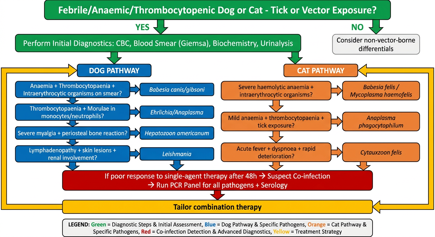
Clinical Decision Tree: Diagnosing Haemoprotozal Co-infections in Dogs and Cats
STEP 1 - Entry Criteria
- Fever (>39.5°C)
- Lethargy + anorexia
- Pale/icteric mucous membranes
- Unexplained anaemia or thrombocytopaenia
- Tick/flea/sandfly/triatomine bug exposure history
STEP 2 - Initial Diagnostics (All Cases)
- CBC - look for anaemia (PCV), thrombocytopaenia, leucopaenia/leucocytosis
- Blood smear (Giemsa stain) - intraerythrocytic organisms, morulae, RBC morphology
- Serum biochemistry - ALT, ALP, creatinine, BUN, bilirubin, total protein
- Urinalysis - haemoglobinuria, bilirubinuria, proteinuria (leishmania)
- Tick burden assessment - species ID if possible (guides likely pathogens)
STEP 3A - DOG Pathway
| Finding on Initial Screen | Suspect | Confirm With |
|---|---|---|
| Haemolytic anaemia + intraerythrocytic piriform/ring organisms + thrombocytopaenia | Babesia canis / B. gibsoni | PCR (differentiates species), IFA serology |
| Thrombocytopaenia + morulae in monocytes/lymphocytes + fever | Ehrlichia canis (CME) | PCR, IFA serology ≥1:80 |
| Thrombocytopaenia + morulae in neutrophils + joint pain | Anaplasma phagocytophilum | PCR, IFA serology |
| Severe muscle pain + hyperaesthesia + periosteal bone proliferation on X-ray | Hepatozoon americanum | Muscle biopsy (cysts), PCR |
| Lymphadenopathy + progressive weight loss + skin scaling + proteinuria | Leishmania infantum | Cytology, PCR, IFAT serology ≥1:160 |
| Pale mucous membranes + epiphora + splenomegaly - splenectomised or immunosuppressed | Mycoplasma haemocanis | PCR (blood smear often negative) |
STEP 3B - CAT Pathway
| Finding on Initial Screen | Suspect | Confirm With |
|---|---|---|
| Severe haemolytic anaemia (PCV < 15%) + intraerythrocytic organisms + S. Africa / endemic region | Babesia felis | PCR, blood smear |
| Moderate anaemia + organisms on RBC surface/within cells + FIV/FeLV positive | Mycoplasma haemofelis | PCR (most sensitive; smear often missed) |
| Acute: high fever + dyspnoea + rapid collapse + S-central USA | Cytauxzoon felis | Blood smear (piroplasms in RBCs + large schizonts in macrophages), PCR |
| Thrombocytopaenia + fever + joint swelling | Anaplasma phagocytophilum | PCR, serology |
| Subclinical anaemia + mild thrombocytopaenia (incidental) | Babesia vogeli / Hepatozoon felis | PCR (serology unreliable) |
STEP 4 - Co-infection Detection Gate
- Poor or incomplete response to single-agent therapy after 48-72 hours
- Initial CBC shows multiple-lineage cytopaenias (anaemia + thrombocytopaenia + neutropaenia)
- Blood smear shows organisms of different morphology simultaneously
- Patient is from a highly tick-endemic area (Mediterranean, SE Asia, USA South)
- Concurrent systemic signs not explained by one diagnosis (e.g., renal failure + haemolysis + joint pain)
STEP 5 - Common Co-infection Combinations
| Co-infection Pair | Region | Clinical Clue |
|---|---|---|
| Ehrlichia canis + Babesia canis | Global (tropics/subtropics) | Severe pancytopaenia + haemolysis; poor response to doxycycline alone |
| E. canis + Leishmania infantum | Mediterranean, S. America | Thrombocytopaenia + skin lesions + chronic wasting |
| Babesia gibsoni + Ehrlichia/Anaplasma | SE Asia, USA | Refractory anaemia + thrombocytopaenia |
| M. haemofelis + FIV/FeLV | Worldwide (cats) | Non-regenerative anaemia, immunosuppression |
| C. felis + M. haemofelis | USA South (cats) | Acute collapse + haemolysis in cat from endemic area |
| Babesia + Hepatozoon (dogs) | Africa, Middle East | Fever + myalgia + anaemia |
STEP 6 - Treatment Tailoring for Co-infections
| Co-infection | Combined Treatment |
|---|---|
| E. canis + B. canis | Doxycycline 10 mg/kg q24h x 28d + Imidocarb dipropionate 6 mg/kg IM x 2 doses |
| Ehrlichia + Leishmania | Doxycycline + Meglumine antimoniate + Allopurinol |
| B. gibsoni + Ehrlichia | Atovaquone + Azithromycin (10d) + Doxycycline (28d) |
| M. haemofelis + FIV/FeLV | Doxycycline + Prednisolone (if IMHA) + manage retroviral disease |
| C. felis + M. haemofelis (cats) | Atovaquone + Azithromycin + Doxycycline + intensive supportive care |
Key Rule of Thumb
"In tick-endemic areas, never assume a single pathogen. If the patient does not improve in 48 hours, broaden the diagnostic net with PCR and broaden the treatment to cover the most likely co-infecting organisms."