
Fundoscopy signs of myopia
Reading File
Quick answer: Fundoscopy in myopia (especially high myopia) shows a characteristic set of changes including a temporal crescent/staphyloma, tigroid/tessellated fundus, lattice degeneration, Fuchs' spot, lacquer cracks, and posterior staphyloma. The optic disc often appears tilted with peripapillary atrophy.
Searching sources...
Finding Sources
Finding Sources
Finding Sources
Finding Sources
Good results. Now let me read the detailed sections from Kanski's and Wills Eye Manual on degenerative myopia signs.
Reading File
Reading File
Excellent content retrieved. Let me also get the rest of the Kanski section and fetch the fundus image from Wills Eye Manual.
Reading File
Loading Image
I now have comprehensive textbook content. Let me fetch a couple of the key Kanski fundus images to enrich the response.
Loading Image
Loading Image
I have comprehensive textbook data and clinical images. Here is the full answer:
Fundoscopy Signs of Myopia
These signs are most prominent in high myopia (>-6D) and degenerative/pathological myopia, where axial elongation drives mechanical stretching and atrophy of ocular layers.
Optic Disc Signs
1. Tilted (Oblique) Disc
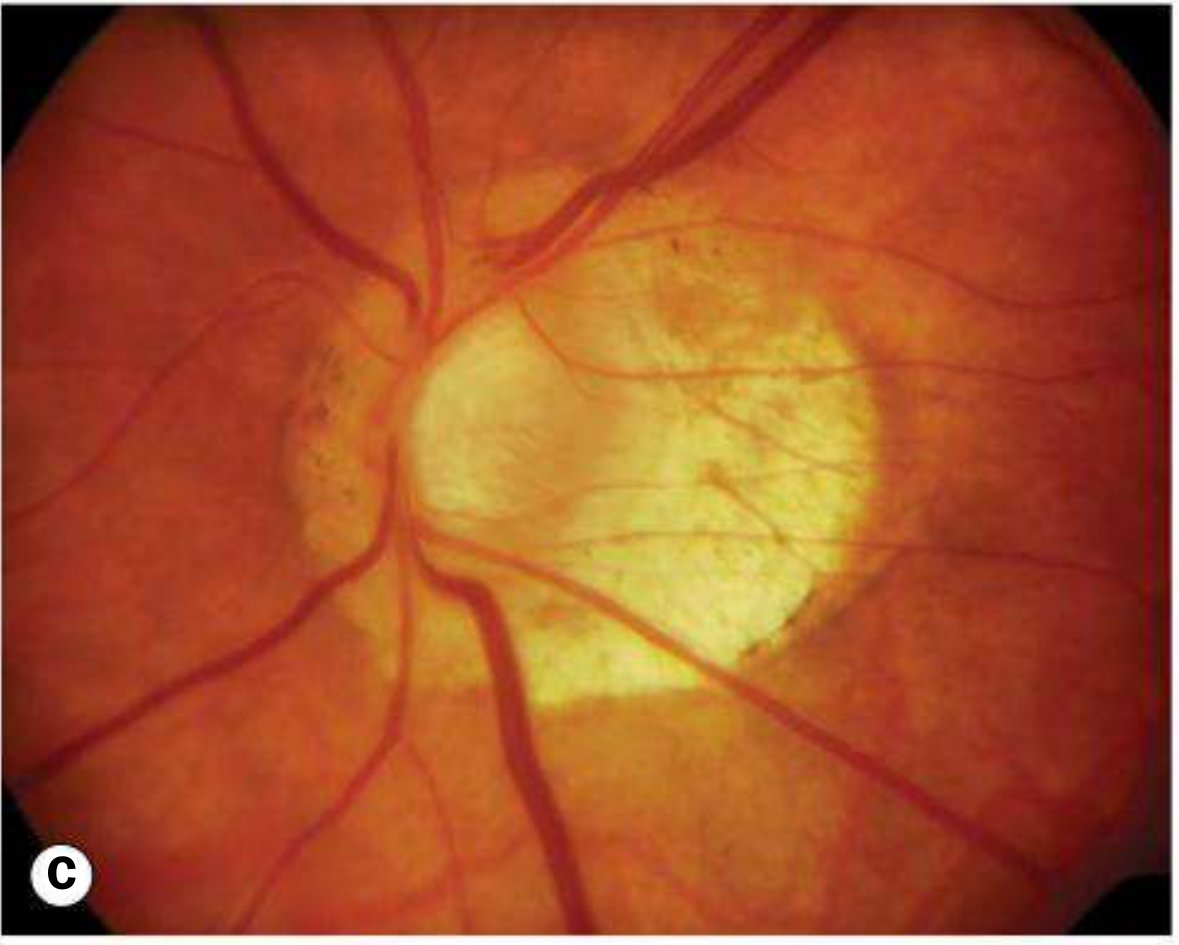
The optic disc appears obliquely inserted, often with vertical elongation. This is one of the most recognizable disc features of myopia. Retinal vessels may enter through the nasal part of the optic cup ("situs inversus").
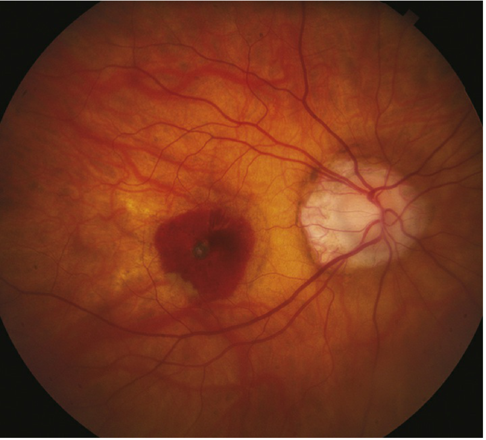
2. Myopic / Temporal Crescent (Conus)
A crescent-shaped area of white sclera or exposed choroidal vessels is seen immediately adjacent to the disc (most commonly temporal). It is separated from normal-appearing fundus by a hyperpigmented border line. This is due to peripapillary RPE/choriocapillaris thinning or absence.
3. Temporal Optic Disc Pallor
The temporal rim of the disc appears pale due to underlying chorioretinal atrophy.
4. Acquired Optic Disc Pit
Occurs as the peripapillary region expands with globe elongation; clinically appears as a small excavation within the disc.
(Image below shows a highly myopic fundus with macular hemorrhage and temporal crescent)
Retinal / Choroidal Background Signs
5. Tessellated (Tigroid) Fundus
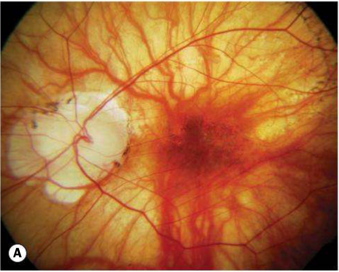
The fundus has a pale, tessellated/tigroid appearance due to diffuse attenuation of the RPE, making the underlying large choroidal vessels visible. This gives the retina a "striped" or "tiled" look.
6. Posterior Staphyloma
A focal ectasia (outpouching) of the posterior sclera at the peripapillary or macular region, seen in approximately one-third of eyes with pathological myopia. It results from focal scleral thinning and expansion. Associations include macular hole and dome-shaped macula.
(Image below shows a fundus with prominent temporal crescent, tessellated background and subretinal haemorrhage in degenerative myopia)
Macular Signs
7. Lacquer Cracks
Fine, irregular, yellowish lines criss-crossing the posterior pole, representing ruptures through the RPE-Bruch membrane-choriocapillaris complex. Seen in ~5% of highly myopic eyes. They are a precursor to subretinal hemorrhage and myopic neovascularization (MNV).
8. Fuchs Spot (Foster-Fuchs Spot)
A dark, hyperpigmented spot at the macula. It represents a pigmented scar that follows resolution of subretinal hemorrhage or regressed myopic choroidal neovascularization (MNV). It signals prior macular damage and is associated with significant central visual loss.
9. Subretinal "Coin" Haemorrhages
Small, round subretinal hemorrhages at the macula can occur from lacquer cracks even in the absence of MNV.
10. Focal Chorioretinal Atrophy
Patchy areas where both choroid and RPE are absent, revealing underlying sclera. Large geographic atrophic patches are a key cause of legal blindness in degenerative myopia.
11. Macular Retinoschisis (Foveoschisis)
Splitting within the retinal layers at the macula, often seen in association with posterior staphyloma due to vitreous traction. Can progress to macular retinal detachment without macular hole formation.
Peripheral Signs
12. Lattice Degeneration
Peripheral retinal thinning with crisscrossing white lines (sclerosed vessels) and pigmentation, predisposing to retinal tears and detachment. Much more common in myopia.
13. Peripheral Retinal Thinning / Atrophic Holes
Areas of thinned peripheral retina with risk of rhegmatogenous retinal detachment (RRD). RRD risk is significantly elevated in high myopia.
Other Associated Signs
14. Peripapillary Intrachoroidal Cavitation (ICC)
Formerly called "peripapillary detachment of pathological myopia." Appears as a small yellowish-orange area typically inferior to the disc. It can produce visual field defects that mimic glaucoma on testing.
15. Vitreous Changes
Premature posterior vitreous detachment (PVD) is common; the vitreous is often liquefied and may show fibrillar degeneration.
Summary Table
| Sign | Location | Notes |
|---|---|---|
| Tilted disc | Optic disc | Oblique insertion; vessels enter nasally |
| Temporal/myopic crescent | Peripapillary | Exposed sclera/choroid temporal to disc |
| Tessellated (tigroid) fundus | Background | RPE attenuation, choroidal vessels visible |
| Posterior staphyloma | Posterior pole | Scleral ectasia; ~1/3 of pathological myopia |
| Lacquer cracks | Posterior pole | Yellow lines; Bruch membrane rupture |
| Fuchs spot | Macula | Dark pigmented scar; prior MNV/haemorrhage |
| Subretinal coin haemorrhages | Macula | From lacquer cracks; no MNV |
| Focal chorioretinal atrophy | Posterior pole | Patchy RPE/choroid loss |
| Macular retinoschisis | Macula | Retinal layer splitting with staphyloma |
| Lattice degeneration | Periphery | Risk of RRD |
| Peripapillary intrachoroidal cavitation | Peripapillary | Mimics glaucoma VF defect |
(Image below: tilted optic disc with large peripapillary atrophy/crescent in degenerative myopia)
Sources: Kanski's Clinical Ophthalmology, 10th ed. (Ch. 14 - Degenerative Myopia); The Wills Eye Manual, Section 11.22 - Pathologic/Degenerative Myopia.
This is a shared conversation. Sign in to Orris to start your own chat.