
Pleomorphic edenoma
pleomorphic adenoma parotid gland histology
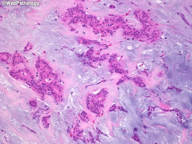
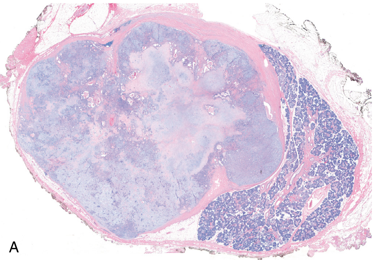
Pleomorphic adenoma histology, parotid salivary gland, examined by bright-field microscopy on Hematoxylin and Eosin stained sections. Imaging modality: Light microscopy; technique: Hematoxylin and Eosin staining. The sample reveals a classic biphasic neoplasm comprising epithelial ductal structures and myoepithelial cells embedded in an abundant stromal backdrop. Epithelial components form cords, ducts, and small island formations lined by cuboidal to columnar cells with eosinophilic cytoplasm and hyperchromatic nuclei. Intermixed myoepithelial cells display plasmacytoid or spindle morphologies, often surrounding epithelial elements. The stroma is a prominent feature, ranging from basophilic, myxoid to more eosinophilic, hyalinized areas. The myxoid matrix is rich in glycosaminoglycans and imparts a loose, gelatinous appearance, while hyalinized zones contribute dense eosinophilic bands. Overall architecture is often well circumscribed with minimal pleomorphism and low mitotic activity, consistent with benign behavior. Clinical significance includes differentiation from malignant salivary tumors; prognosis is favorable with complete surgical excision. Differential considerations include mucoepidermoid carcinoma, adenoid cystic carcinoma, basal cell adenoma, and Warthin tumor. This image is useful for educational purposes in pathology, otolaryngology, and head-and-neck surgery, illustrating quintessential pleomorphic adenoma features: epithelial and myoepithelial proliferation in a chondromyxoid stroma, with duct-like structures and a variable stromal composition. This histologic pattern aids diagnosis and education.
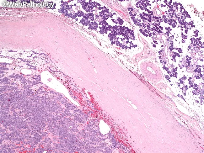
Pleomorphic adenoma of a major salivary gland (parotid) is depicted on hematoxylin and eosin stained histology. The lesion is a well circumscribed, encapsulated neoplasm: a thick fibrous capsule delineates tumor from adjacent normal parotid tissue seen in the upper right, with the mass projecting toward the lower left. The tumor shows biphasic histology with epithelial elements forming duct-like structures and nests, embedded in a variably myxoid to chondromyxoid stroma. Epithelial cells range from cuboidal to columnar, often arranged in sheets and tubules, while myoepithelial cells display plasmacytoid to spindle morphologies, contributing to a heterogeneous, hyalinized background. The stroma may contain cartilaginous or osseous-like foci in places; overall architecture remains orderly rather than infiltrative. Capsule integrity is a key diagnostic cue; the capsule here is thick and typically prevents invasion, although small capsular breaches may explain rare recurrences if not completely excised. No significant necrosis or high mitotic activity is observed. Clinically, this benign mixed tumor presents as a slow-growing, painless parotid mass with preserved facial nerve function. Differential diagnoses include basal cell adenoma and Warthin tumor; mucoepidermoid carcinoma must be considered when ductal differentiation or invasion is suspected. The image highlights the classic pleomorphic adenoma biology: myxoid/chondroid stroma with epithelial/myoepithelial components within a robust capsule.
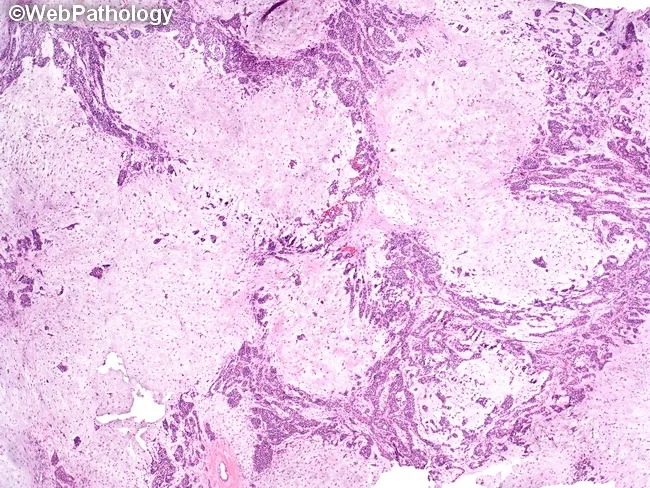
Histology of a pleomorphic adenoma involving the parotid gland. This light-microscopy image derives from a hematoxylin and eosin–stained paraffin section. It demonstrates a biphasic neoplasm with epithelial ductal–acinar elements embedded in a myxoid to chondromyxoid stroma. Epithelial components arrange as trabeculae, tubules, and duct-like acini, often lined by a two-layered cast: inner cuboidal to columnar luminal cells and outer basal/myoepithelial cells with plasmacytoid or spindle morphology. The surrounding stroma is variably myxoid, sometimes chondroid, producing a lobulated architecture with intimately interwoven cellular and stromal zones. Clefts and interspersed myxoid pools may be evident; ossification or calcifications are occasional. The lesion is typically well circumscribed within the parotid, showing no frank invasion of adjacent parenchyma on histology. Mitotic activity is rare. The overall appearance—myxoid/chondromyxoid matrix with epithelial tubules and ducts—supports pleomorphic adenoma; however, histologic overlap with other salivary gland neoplasms necessitates clinical correlation and, when indicated, ancillary immunohistochemistry (e.g., cytokeratin, S-100, GFAP) or molecular testing. Clinically, pleomorphic adenoma presents as a slow-growing, painless parotid mass in adults, with a favorable prognosis after complete surgical excision, though recurrence and potential for malignant transformation exist if spillage or capsular rupture occurs.
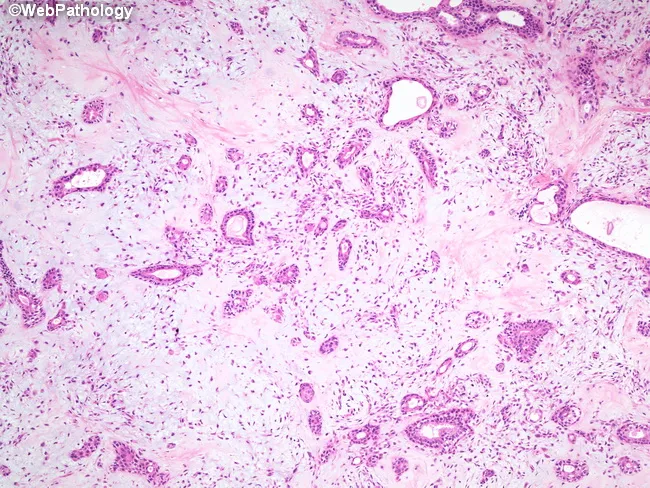
Low-power brightfield histology of a salivary gland tumor stained with hematoxylin and eosin reveals the classical pleomorphic adenoma pattern. The lesion is encapsulated within the parotid region, presenting as a benign, slow-growing mass. The epithelial component forms duct-like and acinar structures lined by a single layer of cuboidal to columnar cells, while an outer layer of neoplastic myoepithelial cells displays plasmacytoid, spindled, or stellate morphologies. The stroma is variably myxoid to chondroid, producing a heterogeneous, often cartilaginous appearance and imparting the 'pleomorphic' descriptor. Cellular atypia and mitotic activity are absent or minimal, consistent with benign behavior. The lesion demonstrates biphasic architecture with a mosaic of epithelial nests, cords, and tubules embedded in an abundant extracellular matrix rich in mucopolysaccharides and chondroid material; occasional hyalinization may be seen. Clinically, this histology corresponds to a pleomorphic adenoma, the most common benign salivary gland neoplasm, characteristically presenting in adults with a painless, mobile mass. Differential diagnoses include mucoepidermoid carcinoma, adenoid cystic carcinoma, and basal cell adenoma; however, the combination of ductal epithelial cells with myoepithelial myxochondroid stroma supports benign pleomorphic adenoma. The diagnostic significance lies in confirming a benign, surgically curable process requiring complete excision to minimize recurrence. Correlation with clinical exam advised today.
Pleomorphic Adenoma (Benign Mixed Tumor)
Overview
- ~60% of all salivary gland tumors
- ~65% of all parotid gland tumors
- ~40% of intraoral minor salivary gland tumors
- Annual incidence: 2.4-3.05 per 100,000
Epidemiology
- Occurs at all ages; most common between the 3rd and 6th decade (average age ~45 years)
- Slightly more common in women
- Risk factors: radiation exposure, long duration of untreated tumor
Location
| Site | Frequency |
|---|---|
| Parotid gland | >80% (mostly superficial lobe) |
| Submandibular gland | Less common |
| Minor salivary glands | ~10% of all salivary tumors |
| Hard palate | Most common intraoral site |
Clinical Features
- Painless, slow-growing, well-defined, solitary, mobile mass
- Gradual progression over many years; can reach enormous size if untreated
- Sudden increase in size or facial nerve palsy suggests malignant transformation
- Firmness varies: soft (myxoid tumors) to hard (chondroid/collagenous tumors)
- Occasional synchronous or metachronous presentation
Pathology
Gross Appearance
- Well-demarcated, rounded/ovoid, nodular, firm mass
- Rarely exceeds 6 cm
- Cut surface: gray-white with myxoid and blue translucent chondroid areas
- May show cystic change, calcification, or ossification
- Variable capsule: thick and fibrotic OR attenuated and incomplete
- Pericapsular nodules (pseudopods) may project beyond the main mass - the key reason enucleation leads to recurrence
Microscopic Appearance
- Epithelial component: ductal cells forming ducts, acini, tubules, strands, or sheets; cuboidal to columnar cells
- Myoepithelial component: plasmacytoid, spindle, stellate, or clear cells surrounding ductal structures
- Stroma: chondromyxoid matrix (myxoid + chondroid + hyalinized areas); may contain true cartilage, bone, squamous islands
Immunohistochemistry
| Cell type | Markers |
|---|---|
| Luminal (ductal) cells | CK7 (strong, diffuse) |
| Myoepithelial cells | p63, S-100, SOX10, SMA (smooth muscle actin) |
Molecular Pathology
- Most cases involve chromosomal rearrangements causing overexpression of PLAG1 - a transcription factor that activates growth factor receptor signaling pathways
- Cases lacking PLAG1 overexpression often carry HMGA2 gene mutations (encodes a DNA-binding protein)
- All tumor elements - including the mesenchymal-looking stroma - are thought to be of myoepithelial or ductal reserve cell (stem cell) origin
Imaging
- Ultrasound: first-line; well-defined, hypoechoic, lobulated mass
- CT/MRI: useful for deep lobe or parapharyngeal extension; calcifications within the mass strongly suggest PA
- FNAC: useful pre-operatively; however, the histologic heterogeneity can make diagnosis from limited samples challenging
Treatment
- Need for definitive histology
- Continued growth if untreated
- Risk of malignant transformation
- Superficial parotidectomy (or total parotidectomy if deep lobe involved) with preservation of the facial nerve
- Excision should include a cuff of normal tissue to encompass pericapsular pseudopods
- Enucleation alone is contraindicated - recurrence rates approach 25% (vs. ~4% with formal parotidectomy) due to capsular pseudopods being left behind
- Intraoral/minor salivary gland PAs: complete transoral excision
- CO2 laser can be used for intraoral lesions
Recurrence
- After parotidectomy: ~4%
- After enucleation: ~25%
- Recurrences are typically multifocal (seeding effect)
- Risk factors for recurrence: capsular rupture/spillage, enucleation, incomplete excision, pericapsular nodules
Malignant Transformation: Carcinoma Ex Pleomorphic Adenoma
| Duration | Risk of malignant transformation |
|---|---|
| < 5 years | ~2% |
| > 15 years | ~10% |
| Overall | ~6% |
- Older patient age
- Male sex
- Long duration of tumor
- Multiple recurrences
- Deep lobe parotid location
Differential Diagnosis
| Tumor | Key distinguishing features |
|---|---|
| Warthin tumor | Older males, smokers; bilateral in 10%; oncocytic cells + lymphoid stroma |
| Basal cell adenoma | Uniform basaloid cells; no myxoid stroma; well-encapsulated |
| Mucoepidermoid carcinoma | Mixed mucous + epidermoid + intermediate cells; no chondromyxoid matrix |
| Adenoid cystic carcinoma | Cribriform "Swiss cheese" pattern; perineural invasion; aggressive |
| Myoepithelioma | Pure myoepithelial cells; no ductal elements; no chondroid stroma |
Key Points to Remember
- Most common salivary gland tumor overall
- Parotid (superficial lobe) is the most common site
- Biphasic: epithelial + myoepithelial cells in chondromyxoid stroma
- PLAG1 and HMGA2 are the key molecular drivers
- Enucleation = high recurrence; always do formal excision with cuff
- Duration increases malignant transformation risk
- Calcification on imaging = strong clue to PA diagnosis
Parotid gland
parotid gland anatomy facial nerve branches
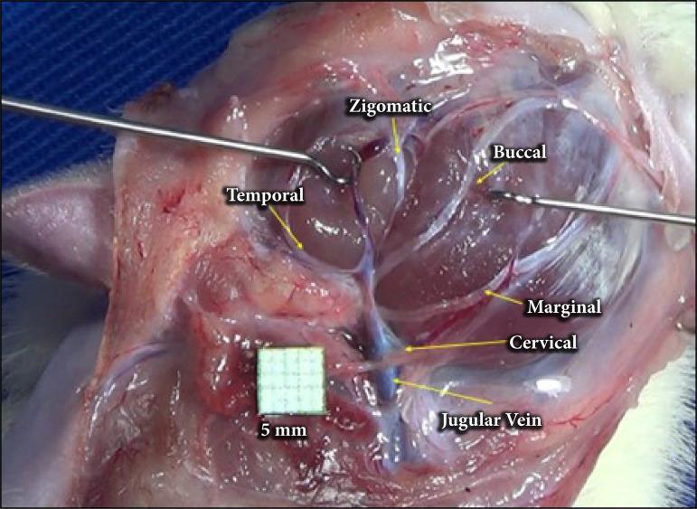
A clinical intraoperative photograph showing the anatomical arrangement of the facial nerve branches in a surgical dissection, following parotidectomy. The image provides a lateral view of the cervicofacial region where the parotid gland has been removed to expose the extratemporal facial nerve plexus. Labeled branches include the temporal branch extending superiorly, the zygomatic and buccal branches traveling toward the mid-face, and the marginal mandibular and cervical branches descending inferiorly. The cervical branch is depicted in close proximity to the jugular vein, which is visible in the lower surgical field. Surgical instruments (retractors) are positioned to maintain exposure of the deep tissues. A 5mm grid scale is included for anatomical measurement. This visual serves as an educational guide for surgical anatomy, identifying key landmarks for nerve preservation during parotid gland surgery or facial nerve decompression.
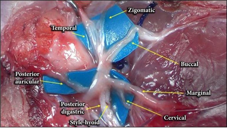
This clinical photograph displays a high-magnification surgical dissection of the facial nerve (Cranial Nerve VII) and its extratemporal branches. The image highlights the anatomical arrangement after the parotid gland has been removed. Blue surgical background material is placed beneath the nerves to enhance visual contrast. Visible and labeled branches radiating from the main trunk include the temporal, zygomatic, buccal, marginal mandibular, and cervical branches. Additionally, the posterior auricular branch is visible posteriorly, while the posterior digastric and stylohyoid branches are shown inferiorly. The branches appear as glistening, whitish-cord structures overlying surrounding muscle and connective tissue. This educational visual demonstrates the typical branching patterns and spatial relationships essential for parotidectomy procedures, maxillofacial surgery, and head and neck anatomy education. The labels indicate the specific distribution of motor innervation to the muscles of facial expression.
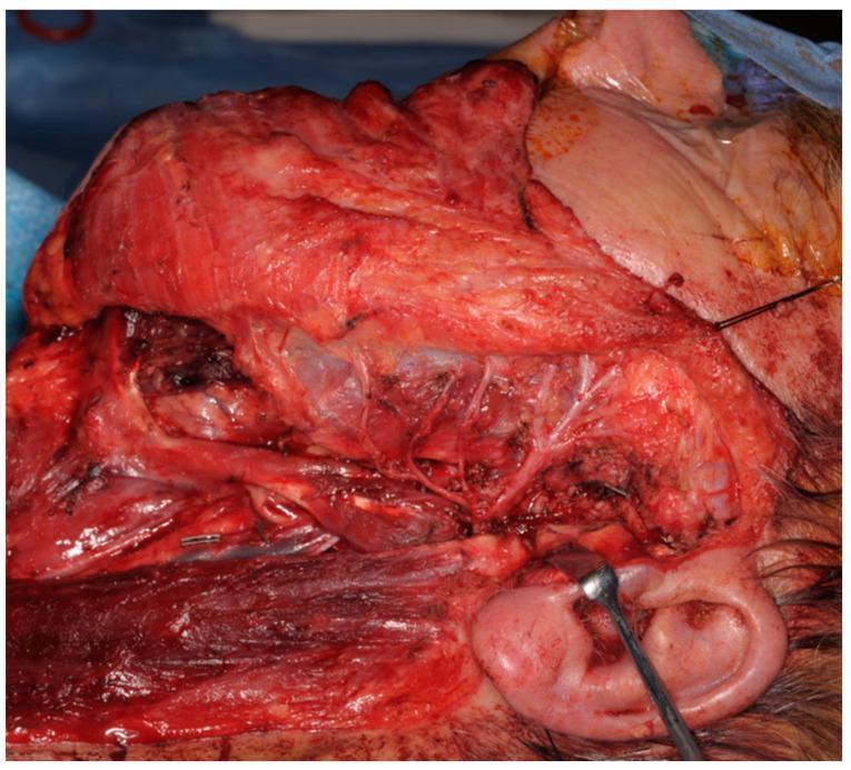
This clinical intra-operative photograph displays a large-scale dissection of the parotid and facial region during a total parotidectomy. The surgical field reveals an anatomical display of the facial nerve (Cranial Nerve VII) and its distal branching network preserved and traversing the surgical void created by the resection of the parotid gland. Key structures visible include the masseter muscle superiorly, the sternocleidomastoid muscle at the inferior margin, and the external ear at the lower right, where a surgical retractor is positioned for tissue displacement. Multiple fine nerve branches are seen branching across the exposed muscle bed and subcutaneous fat. The image demonstrates a 'Type 0 defect,' characterized by an intact facial nerve following tumor resection. This visual serves to educate on the intricate neurovascular anatomy of the head and neck, specifically highlighting surgical landmarks necessary for nerve preservation during oncological resections in otorhinolaryngology and maxillofacial surgery.
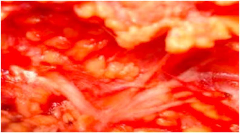
This intraoperative clinical photograph displays an anatomical dissection of the facial nerve (Cranial Nerve VII) within the parotid region. The image illustrates a 'Type II' branching pattern characterized by a single facial nerve trunk (FNT) that divides directly into multiple terminal branches. Notably, this variant lacks the typical primary bifurcation into temporofacial and cervicofacial divisions. The nerve branches appear as thin, pale, string-like structures radiating through the reddish, fleshy tissue of the surgical field. The surrounding area shows exposed glandular and soft tissue relevant to a parotidectomy procedure. This visual serves as an educational reference for surgical anatomy, highlighting normal anatomical variations of the extracranial facial nerve that are critical for surgeons to identify during parotid gland surgery to avoid nerve injury and post-operative facial paralysis.
Parotid Gland
Embryology
- The parotid is ectodermal in origin (unlike the submandibular and sublingual glands, which are endodermal)
- Develops in the 6th week of gestation when epithelial buds invaginate from the oral mucosa into the surrounding mesenchyme
- Uniquely, the parotid has delayed encapsulation - the facial nerve, vessels, and lymphatics all become embedded within the gland before the capsule finally fuses around them
- This explains why: (1) lymph nodes are found inside the gland, (2) the facial nerve runs through its substance
Gross Anatomy
Position and Borders
- Largest of the three paired major salivary glands; purely serous gland
- Situated anterior and inferior to the external ear, wedged between the ramus of the mandible, base of skull, and mastoid process
| Border | Structure |
|---|---|
| Anterior | Masseter muscle |
| Superior | Zygomatic arch |
| Posterior | Tragal cartilage + sternocleidomastoid muscle |
| Deep | Pretyloid compartment of parapharyngeal space |
| Inferior | "Tail" between mandibular ramus and SCM, overlying the digastric |
Lobes
- The facial nerve divides the gland into superficial and deep lobes (anatomically - no true fascial plane between them)
- 80% of parenchyma is in the superficial lobe (lateral to facial nerve)
- Deep lobe is the retromandibular component with minimal functional tissue
- Covered by the parotidomasseteric fascia (SMAS is closely approximated to this capsule)
Accessory Parotid Gland
- Found along the Stensen duct in 21-61% of individuals (autopsy data)
- Lies on the masseter muscle anterior to the duct, with a secondary duct joining Stensen's duct
- ~1% of parotid neoplasms arise in the accessory gland
Stensen's Duct (Parotid Duct)
- Exits the anterior edge of the gland midway between the zygomatic arch and corner of the mouth
- Crosses the masseter muscle horizontally, then turns deep at its medial border
- Pierces the buccinator muscle to enter the oral cavity
- Opens opposite the upper second molar tooth
Key Structures Within the Parotid
1. Facial Nerve (CN VII) - Most Important
- Exits the skull via the stylomastoid foramen (posterolateral to the styloid process, anteromedial to the mastoid process)
- Enters the parotid and forms the pes anserinus (goose's foot) as it divides into upper and lower trunks
- 5 terminal branch groups emerge from the gland borders:
| Branch | Exits from | Supplies |
|---|---|---|
| Temporal | Upper border | Frontalis, orbicularis oculi (upper) |
| Zygomatic | Upper-anterior border | Orbicularis oculi (lower) |
| Buccal | Anterior border | Buccinator, upper lip muscles |
| Marginal mandibular | Lower border | Lower lip muscles |
| Cervical | Lower border | Platysma |
- Tympanomastoid suture line - main trunk lies ~6-8 mm deep to its inferior end
- Posterior belly of digastric - marks the depth of the nerve
- Tragal pointer - main trunk lies ~1 cm deep and slightly inferior to its tip
- Retrograde identification - tracing a distal branch back to the trunk
⚠️ In children under 2 years, the facial nerve trunk and divisions are more superficial - higher surgical risk.
2. Retromandibular Vein
- Formed by the union of maxillary + superficial temporal veins within the gland
- Lies deep to the facial nerve (useful retrograde landmark during surgery)
- Drains via the posterior facial vein to the external jugular vein
- Can give off an anterior branch joining the internal jugular vein
3. External Carotid Artery
- Courses medial to the parotid gland
- Gives off the posterior auricular artery before dividing into its two terminal branches:
- Maxillary artery - passes deep, horizontally behind the mandible
- Superficial temporal artery - ascends, gives off the transverse facial artery, and exits the superior border
Innervation
Parasympathetic (Secretomotor)
- Pathway: Inferior salivatory nucleus → CN IX (glossopharyngeal) → Jacobson's nerve (tympanic branch) → tympanic plexus (middle ear) → lesser petrosal nerve → exits via foramen ovale → otic ganglion (synapse) → postganglionic fibers join the auriculotemporal nerve → parotid gland
- Neurotransmitter: ACh - acts on muscarinic receptors causing acinar activity, ductal transport, vasodilatation, and myoepithelial cell contraction
Sympathetic
- From the superior cervical ganglion (via perivascular plexus)
- Causes vasoconstriction and secretion of thick, mucoid saliva
Sensory
- Auriculotemporal nerve (branch of V3) - also carries the postganglionic parasympathetics
- Great auricular nerve (C2, C3) - supplies overlying skin; has anterior and posterior branches; posterior branch may sometimes be preserved during parotidectomy to reduce auricular numbness
Lymphatics
- Unique among salivary glands in containing ~20 intraglandular lymph nodes (in addition to periglandular nodes) because lymphatics develop within the gland after the capsule fuses
- Most nodes lie in the superficial (preauricular) lobe lateral to the masseter
- Clinical significance: lymph node metastases from skin (scalp, face, ear), parotid tumors, and Stensen's duct carcinomas can present as intraparotid masses
Parotid Diseases - Overview
Inflammatory / Infective
| Condition | Key Features |
|---|---|
| Acute bacterial parotitis | Staphylococcal, elderly/dehydrated patients; tender swelling, pus at Stensen's duct orifice |
| Viral parotitis (mumps) | Paramyxovirus; bilateral; common in children |
| Chronic recurrent parotitis | Recurrent swelling ± calculi |
| Parotid calculi (sialolithiasis) | Less common than submandibular; postprandial swelling |
Tumors
- ~80% of parotid neoplasms arise in the superficial lobe
- ~75% are benign, ~25% malignant (1 in 4 parotid tumors is malignant)
- Features suggesting malignancy: rapid growth, pain, cervical lymphadenopathy, skin/deep fixation, facial nerve palsy (44% of parotid cancers cause pain; 25% cause facial palsy)
| Tumor | Type |
|---|---|
| Pleomorphic adenoma | Most common benign |
| Warthin's tumor | 2nd most common benign; smokers, older males |
| Mucoepidermoid carcinoma | Most common malignant |
| Adenoid cystic carcinoma | Perineural invasion; late metastases |
| Acinic cell carcinoma | Low-grade |
| Carcinoma ex pleomorphic adenoma | Aggressive |
Parotidectomy and Complications
Surgical Procedures
- Superficial parotidectomy: adequate for most superficial lobe tumors (with appropriate cuff of normal tissue)
- Total conservative parotidectomy: for deep lobe tumors; both lobes removed with facial nerve preservation
- Extracapsular dissection: growing evidence-base as an alternative for selected small tumors
Complications
| Complication | Details |
|---|---|
| Temporary facial weakness | Neuropraxia; resolves in 4-6 weeks; most common |
| Permanent facial palsy | ~1-2% risk; degeneration injuries take 6-12+ months to recover |
| Frey's syndrome (gustatory sweating) | Gustatory sweating/flushing over the parotid skin during eating; due to aberrant reinnervation of sweat glands by auriculotemporal parasympathetic fibers (ACh acts on both sweat glands and salivary receptors); treated with botulinum toxin or interposition flap |
| Great auricular nerve numbness | Unavoidable; skin over angle of mandible and lower 2/3 of pinna; reduces over 12 months; posterior branch preservation may help |
| Cosmetic hollow | Loss of bulk behind the ramus; can be mitigated with fat transfer |
| Sialocele / salivary fistula | Saliva collection or leakage from remnant acinar tissue |
Parapharyngeal Space Relationship
- The deep parotid lobe's deep border abuts the pretyloid compartment of the parapharyngeal space
- Deep lobe tumors can extend into this space as a dumbbell tumor presenting as a paratonsillar bulge
- The poststyloid compartment (behind the styloid process) contains the carotid sheath and cranial nerves IX-XII - separate from the parotid
Summary: Key Facts to Remember
- Ectodermal origin, delayed encapsulation - explains intraglandular nodes and facial nerve course
- Purely serous gland (unlike submandibular = mixed; sublingual = mostly mucous)
- Facial nerve divides gland into lobes - 80% superficial
- Structures within: Facial nerve (most superficial) → Retromandibular vein → External carotid artery (deepest)
- Parasympathetic supply: CN IX → lesser petrosal nerve → otic ganglion → auriculotemporal nerve
- Stensen's duct opens at upper 2nd molar
- Frey's syndrome = aberrant reinnervation of sweat glands by auriculotemporal parasympathetics post-parotidectomy
- ~25% of parotid tumors are malignant; facial nerve involvement = malignant until proven otherwise