
Grattage test and auspitz sign
- Grattage test (Scratch test): Scraping a psoriatic plaque with a glass slide or fingernail reveals a characteristic sequence - first a candle-wax scaling, then a last transparent membrane (Bulkeley's membrane), then pinpoint bleeding.
- Auspitz sign: The pinpoint bleeding that appears after removing the last thin membrane - caused by dermal papillae being close to the surface with dilated capillaries.
Auspitz sign psoriasis pinpoint bleeding grattage test
Grattage Test and Auspitz Sign in Psoriasis
Grattage Test (Scratch/Curettage Test)
| Step | Finding | Basis |
|---|---|---|
| 1st | Candle-wax (silvery-white) scales crumble off like scrapings from a candle | Dry, loosely attached parakeratotic stratum corneum |
| 2nd | Last shiny membrane (Bulkeley's or Bulkley's membrane) - a thin, transparent, glistening layer | Thinned suprapapillary epidermis |
| 3rd | Pinpoint bleeding (Auspitz sign) - multiple tiny bleeding points on a red, moist base | Dilated, tortuous capillaries in elongated dermal papillae, exposed after membrane removal |
Auspitz Sign
- In psoriasis, the capillaries in the dermal papillae become elongated, tortuous, and dilated - they extend upward into club-shaped elongated papillae
- The suprapapillary epidermal plate overlying these papillae is markedly thinned
- Scraping removes this thin plate and traumatizes the exposed dilated capillaries directly below, causing pinpoint bleeding
"If the superficial silvery white (micaceous) scales are removed, then a wet surface is seen with characteristic pinpoint bleeding. This finding, called Auspitz sign, is the clinical reflection of elongated vessels in the dermal papillae together with thinning of the suprapapillary epidermis."
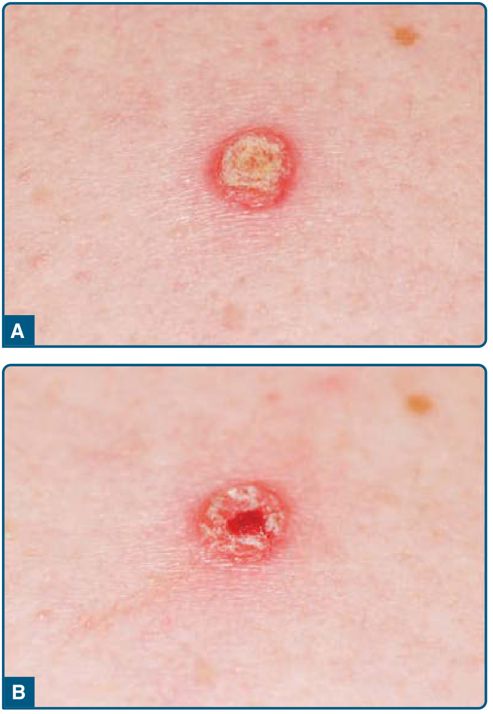
Specificity of Auspitz Sign
"Removal of scales in psoriasis may disclose bleeding points (Auspitz sign). This sign is common but lacks great specificity."
- Darier's disease (keratosis follicularis)
- Some cases of actinic keratosis
Histopathological Correlation
"These vessels bleed when the scale is removed, giving rise to multiple punctate bleeding points (Auspitz sign). Neutrophils form small aggregates within both the spongiotic superficial epidermis (spongiform pustule of Kogoj) and the parakeratotic stratum corneum (Munro microabscesses)."
Koebners sign
Koebner phenomenon psoriasis trauma skin lesion
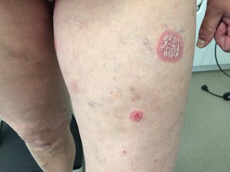
This clinical photograph displays a human anterior proximal thigh exhibiting classic psoriatic lesions as part of the Koebner phenomenon. The primary feature is a large, well-demarcated, circular-to-oval erythematous plaque. This lesion is characterized by a raised red border and a central surface covered in thick, silvery-white micaceous scales. Disseminated across the surrounding skin are smaller, satellite erythematous papules and plaques of varying diameters, some appearing as pinpoint lesions. The background skin shows signs of mild solar damage, visible superficial vasculature (telangiectasia), and minor blemishes. This visual presentation demonstrates an isomorphic response, where localized skin trauma—specifically insulin injection sites in this clinical context—triggers the development of psoriatic plaques in a patient with a history of psoriasis. This is typical of a disease flare-up following the withdrawal of systemic therapy like methotrexate. The image is a primary educational resource for dermatological morphology, illustrating plaque psoriasis and the Koebner phenomenon.
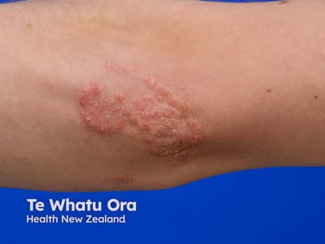
Imaging modality: Clinical photography. This is a high-resolution, close-up surface photograph of an in vivo elbow lesion, focusing on the antecubital region. Primary subject: a chronic plaque psoriasis plaque with a proximal Koebner line after scratching. Anatomical context: cutaneous skin of the elbow (integumentary system; extensor surface). Visual features: well-defined erythematous plaque with adherent silvery scale, thickened hyperkeratosis, slight fissuring at the margins, and a linear, proximal, scratch-induced lesion corresponding to koebnerization. Notable findings: localized psoriasis morphology with coalescent plaques and intact surrounding skin; no ulceration; mild perifollicular changes may be absent. Clinical significance: koebner phenomenon is a diagnostic and prognostic indicator in psoriasis; trauma-induced lesions reflect disease activity and help distinguish psoriasis from other dermatoses. Differential considerations: eczema/atopic dermatitis, tinea corporis, lichen simplex chronicus; clinical correlation: assess for additional plaques at other sites (scalp, knees, elbows), screen for nail/arthritis involvement, tailor therapy accordingly. Potential clinical use cases: educational dermatology resources, patient education on avoiding skin trauma, documentation for treatment response and research into Koebnerization patterns. The image serves as a reference for lesion morphology classification, aids in teaching koebnerization mechanisms, and supports standardized annotation for image-based psoriasis databases. Clinicians may compare longitudinally to monitor therapeutic response and disease activity.
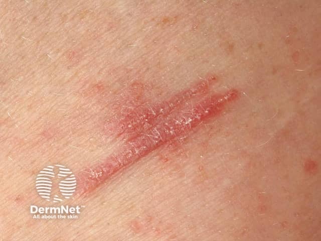
Clinical photograph demonstrating Koebner phenomenon in lichen planus. Modality: in vivo color image acquired with standard clinical photography using visible light; perspective: close‑up, surface view of the skin, showing a linear trajectory along a history of trauma. Anatomical context: integumentary system; skin (cutaneous tissue); epidermis with superficial dermis involvement; location not specified on the body; region described as a linear band on a trunk/limb surface; laterality not indicated. Visual features: a linear, plaque‑like lesion composed of erythematous to mildly violaceous linear band, with slight desquamation and a glossy surface; margins are relatively well defined; surrounding skin shows additional punctate papules and subtle erythema. The lesion aligns with a scratch or trauma line, illustrating the Koebner response in lichen planus. Pathophysiology: lichenoid inflammation triggered by cutaneous trauma leads to linearization of LP lesions; hallmark features may include Wickham’s striae not visible in this image. Diagnostic significance: supports a clinical diagnosis of lichen planus with Koebnerization; may help differentiate from linear psoriasis or lichen striatus in ambiguous cases and prompt correlation with history and biopsy if needed. Clinical context: management favors trauma avoidance, topical steroids, calcineurin inhibitors, and close monitoring for mucocutaneous involvement; educate patient about triggering events.
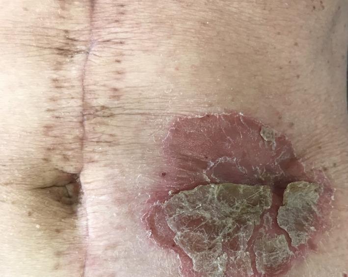
This clinical photograph displays a primary dermatological lesion on the human abdomen, specifically a well-demarcated peristomal plaque. The lesion is a large, circular, erythematous (reddish) plaque characterized by sharply defined borders that distinguish it from the healthy surrounding skin. The surface exhibits significant desquamation, with thick, micaceous (silvery), and yellowish-brown scales, many of which are flaky and partially peeling. The plaque is situated on the left abdominal wall adjacent to a stoma site. Medially to the lesion, a vertical surgical midline incision scar is visible, showing post-operative hyperpigmentation and healing markers. The surrounding abdominal skin shows signs of chronological aging or sun damage, including fine rhytids and scattered lentigines. This image illustrates the Koebner phenomenon or peristomal psoriasis, emphasizing the typical morphology of psoriatic lesions in areas of chronic mechanical irritation or localized skin trauma.
Koebner Phenomenon (Isomorphic Response)
Definition
- Named after Heinrich Koebner, who first described it in psoriasis in 1877
- The word "isomorphic" means the new lesions are identical in morphology to the patient's existing disease lesions
Key Features in Psoriasis
- The Koebner reaction usually occurs 7 to 14 days after injury
- It is an all-or-none phenomenon - if psoriasis occurs at one site of injury, it will occur at all sites of injury
- Between 25% to 75% of patients with psoriasis may develop trauma-related Koebner phenomenon at some point during their disease
- It occurs more frequently during flares/active disease
Triggering trauma types:
- Scratches, incisions, surgical scars, burns
- Phototherapy burns (overdose of UV)
- Needle prick sites, tattoos
- Friction, pressure
- Viral exanthema (pityriasis rosea trigger)
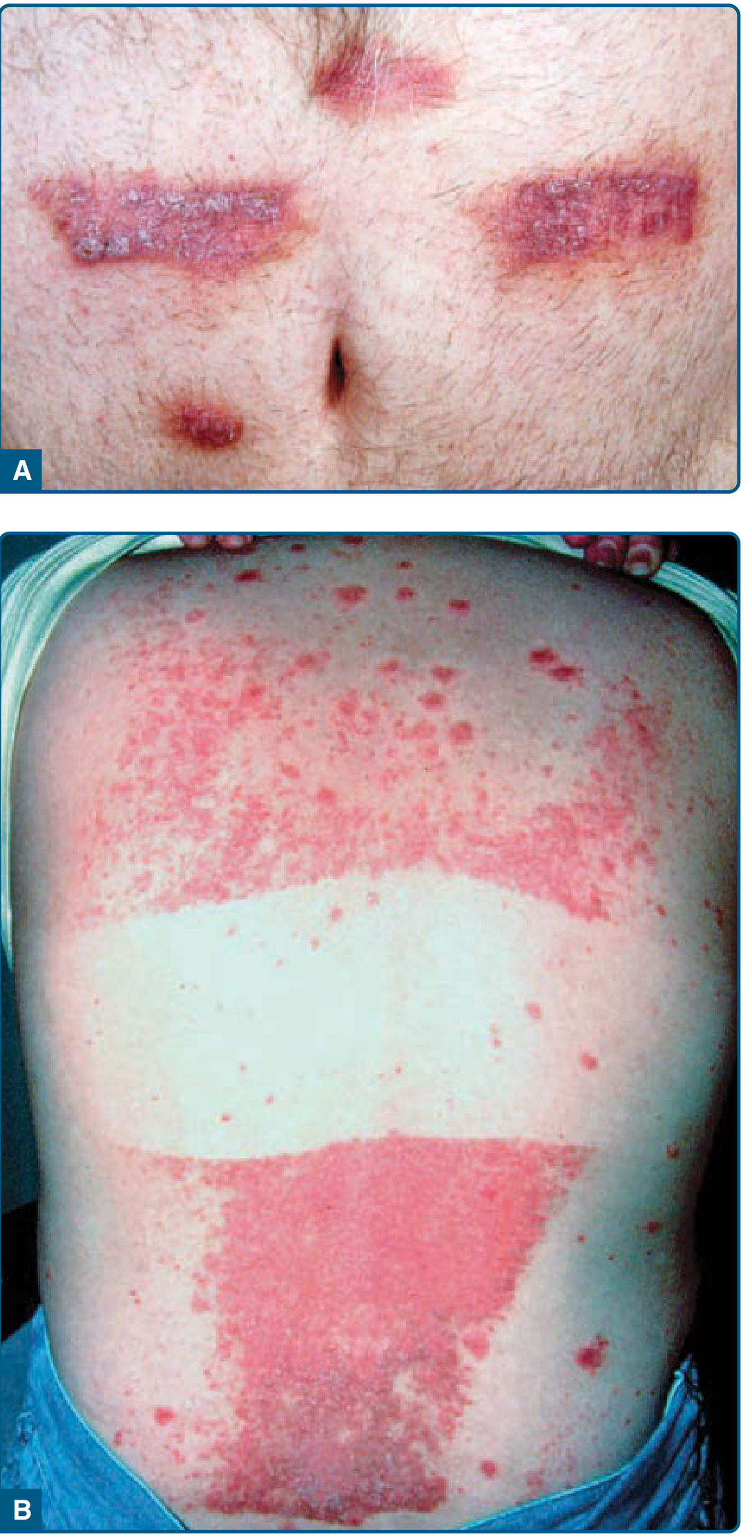
Diseases That Show the Koebner Phenomenon
| Category | Diseases |
|---|---|
| Most classic | Psoriasis, lichen planus, vitiligo |
| Others | Lichen nitidus, pityriasis rubra pilaris, reactive perforating collagenosis, cutaneous small vessel vasculitis, Still disease |
Clinical significance
- Diagnostic clue - helps confirm psoriasis or lichen planus when lesions appear in a linear pattern following trauma
- Marker of disease activity - its presence indicates an active phase; absent in quiescent disease
- Prognostic indicator - Koebnerization tends to predict more widespread or aggressive disease
- Practical implication - patients must be advised to avoid skin trauma (scratching, tattoos, piercings, surgery during active disease) to prevent new lesion formation
Related Phenomena - Important Distinctions
| Phenomenon | Description | Example |
|---|---|---|
| True Koebner (isomorphic response) | New lesions of the same disease at trauma site | Psoriasis plaques at surgical scar |
| Pseudo-Koebner (autoinoculation) | Infection spread to traumatized skin | Warts, molluscum at scratch sites |
| Wolf isotopic response | A second, different skin disease appearing at site of a prior, healed skin disease | Granuloma annulare at healed herpes zoster site |
| Pathergy | Exaggerated inflammatory response to trivial injury (papule/pustule at needle prick) | Classic in Behcet's disease |
| Reverse Koebner | Clearance of existing lesions at a trauma site | Psoriasis clearing at site of UV burn |
Do we suture category 3 dog bite
Suturing of Category 3 Dog Bites
Short Answer
WHO Category 3 - Recap
| Category | Exposure | PEP Required |
|---|---|---|
| I | Touch/feed animal, licks on intact skin | None |
| II | Nibbling uncovered skin, minor scratches without bleeding | Vaccine + local wound care |
| III | Single/multiple transdermal bites or scratches, licks on broken skin, mucous membrane contact with saliva, bat contact | Vaccine + RIG + local wound care |
Why NOT Suture Immediately
"Bite wounds should not be immediately sutured to prevent additional trauma which may help spread the virus into deeper tissues. If suturing is necessary, it should be done 24-48 hours later, applying minimum possible stitches, under the cover of rabies immunoglobulin locally."
- Rabies virus spread - Suturing compresses and drives virus deeper into tissues along nerve endings
- Infection risk - Bite wounds (especially punctures) are heavily contaminated; primary closure traps bacteria and promotes anaerobic infection
Exceptions - When Suturing IS Permitted
| Situation | Approach |
|---|---|
| Facial wounds | Can suture primarily (face has excellent blood supply, low infection rate; cosmesis important) - with copious irrigation + debridement + antibiotics |
| Life-threatening bleeding | Haemostatic sutures permissible |
| Deferred closure | Reassess at 48-72 h; if no infection signs, delayed primary closure |
| All other locations | Leave open or use loose approximation with adhesive strips only |
"Bite wounds of the face and scalp from any species that are less than 6 hours old may be sutured after appropriate wound preparation... Puncture wounds, wounds more than 12 hours old, and wounds infected at presentation should not be sutured."
"Primary wound closure is not recommended for animal bite wounds, with the exception of those to the face... bite wounds should not be sealed with a tissue adhesive, no matter their age or appearance."
Complete Management Protocol for Category 3 Dog Bite
1. Local Wound Care (FIRST and most important)
- Wash immediately with soap and water for ≥15 minutes under running tap (reduces rabies risk by up to 80%)
- Irrigate under pressure with saline/sterile water
- Apply virucidal agent: povidone-iodine, alcohol (40-70%), or 0.01% aqueous iodine
- For puncture wounds: use a catheter for deep irrigation
- DO NOT suture immediately
2. Rabies Post-Exposure Prophylaxis (PEP)
- Rabies Immunoglobulin (RIG) - infiltrate as much as possible around the wound; remainder given IM at a distant site
- Rabies vaccine - full course (Day 0, 3, 7, 14 ± 28 by Essen regimen)
3. Other
- Anti-tetanus prophylaxis
- Prophylactic antibiotics - amoxicillin-clavulanate (drug of choice for dog bites) - especially for hand/foot wounds, deep wounds, immunocompromised
- Observe the dog for 10 days (if available)
Which antibiotic should i prescribe for category 3 dog bite with laceration
Antibiotic for Category 3 Dog Bite with Laceration
First-Line: Amoxicillin-Clavulanate (Co-amoxiclav / Augmentin)
Dosing
| Patient | Dose | Duration |
|---|---|---|
| Adult | Amoxicillin-clavulanate 875/125 mg orally twice daily | 5 days (prophylaxis) / 5-7 days (established infection) |
| Child | 25-45 mg/kg/day (amoxicillin component) in 2-3 divided doses | 5 days |
Why Amoxicillin-Clavulanate?
| Organism | Notes |
|---|---|
| Pasteurella multocida / P. canis | Most important - requires beta-lactam coverage |
| Capnocytophaga canimorsus | Can cause fatal sepsis in asplenic/immunocompromised patients |
| Staphylococcus aureus | Skin flora |
| Streptococcus spp. | Skin and oral flora |
| Oral anaerobes | Deep punctures/lacerations |
"Treatment failure of Pasteurella infections caused by dog and cat bites using monotherapy with erythromycin, clindamycin, penicillinase-resistant penicillins, and first-generation cephalosporins has been described." - Rosen's Emergency Medicine
Penicillin-Allergic Patients
| Allergy type | Alternative |
|---|---|
| Non-severe allergy | Doxycycline (good Pasteurella coverage; avoid in children <8 yrs) |
| Penicillin-allergic adult | Ciprofloxacin + Metronidazole OR Moxifloxacin (monotherapy, covers anaerobes) OR Clindamycin + TMP-SMX |
| Penicillin-allergic child | TMP-SMX + Clindamycin (TMP-SMX covers Pasteurella, MRSA; clindamycin covers anaerobes + streptococci) |
| Can tolerate cephalosporins | Ceftriaxone IV + clindamycin (inpatient setting) |
Note: Erythromycin, first-gen cephalosporins (cephalexin), and clindamycin alone are NOT adequate for dog bites - they lack Pasteurella coverage.
When to Give Antibiotics for Dog Bites?
- ✅ Lacerations (your case)
- ✅ Hand or foot wounds
- ✅ Deep puncture wounds
- ✅ Wounds involving tendon, joint, bone, or cartilage
- ✅ Delayed presentation (>6 hours)
- ✅ Immunocompromised / asplenic / advanced liver disease
- ✅ Wounds that are primarily closed
- ✅ Facial wounds in children
If Wound is Already Infected (vs. Prophylaxis)
| Severity | Treatment |
|---|---|
| Mild local infection | Oral amoxicillin-clavulanate, 5-7 days |
| Spreading cellulitis / systemic signs | IV ampicillin-sulbactam (Unasyn) or piperacillin-tazobactam |
| Penicillin-allergic + severe | IV clindamycin + ciprofloxacin OR carbapenem |
Complete Management Summary for Your Patient
- Wound care - irrigate copiously with saline, debride, povidone-iodine
- Do NOT suture primarily (or defer 24-48 h if facial)
- Rabies PEP - RIG infiltrated at wound site + rabies vaccine series
- Anti-tetanus prophylaxis
- Antibiotic - Amoxicillin-clavulanate 875/125 mg BD x 5 days (oral)
- Review at 48-72 hours for wound check and consider delayed closure if clean
What about levofloxacin
Levofloxacin for Category 3 Dog Bite
Short Answer
Why Levofloxacin Falls Short Alone
"Fluoroquinolones, including moxifloxacin and levofloxacin, are very active in vitro against P. multocida and other Pasteurella spp., although published experience in humans is limited."
Accepted Role of Levofloxacin in Bite Wounds
| Situation | Preferred | If Severe Penicillin Allergy |
|---|---|---|
| Animal bite, no surgery, risk factors present | Amoxicillin-clavulanate (PO) | (Doxycycline OR levofloxacin) + metronidazole |
| Animal bite requiring surgical intervention | Ampicillin-sulbactam (IV) | (Doxycycline or levofloxacin) + metronidazole |
"The combination of an antibiotic active against gram-positive cocci and anaerobes (such as clindamycin or metronidazole) with trimethoprim-sulfamethoxazole or a fluoroquinolone, which is active against many of the other potential pathogens, would appear reasonable."
"A fluoroquinolone, such as ciprofloxacin or moxifloxacin, or clindamycin combined with trimethoprim-sulfamethoxazole may be used in penicillin-allergic patients."
Comparison: Fluoroquinolones in Bite Wounds
| Fluoroquinolone | Pasteurella | Anaerobes | Verdict for Dog Bite |
|---|---|---|---|
| Ciprofloxacin | Excellent | Poor | Use + metronidazole |
| Levofloxacin | Excellent | Poor-moderate | Use + metronidazole |
| Moxifloxacin | Excellent | Good | Can use as monotherapy (preferred FQ if using one alone) |
Key point: Moxifloxacin is the preferred fluoroquinolone for bite wounds because its anaerobic coverage means it can be used as a single agent, unlike levofloxacin or ciprofloxacin.
Practical Summary
| Scenario | Best Choice |
|---|---|
| No penicillin allergy | Amoxicillin-clavulanate (first-line) |
| Penicillin-allergic, oral | Moxifloxacin alone OR levofloxacin + metronidazole OR doxycycline + metronidazole |
| Penicillin-allergic, IV (admitted) | Clindamycin + ciprofloxacin OR carbapenem |
| Penicillin-allergic child | TMP-SMX + clindamycin |
What about fdc cefixime and ofloxacin
FDC Cefixime + Ofloxacin for Category 3 Dog Bite
Bottom Line Up Front
- Poor anaerobic coverage - neither cefixime nor ofloxacin covers oral anaerobes reliably
- Poor gram-positive (Staphylococcus aureus) coverage - cefixime (3rd-gen cephalosporin) has reduced activity against S. aureus
Breaking Down Each Component
Cefixime (3rd-generation oral cephalosporin)
| Property | Detail |
|---|---|
| Gram-negative coverage | Good - covers Pasteurella, H. influenzae, Enterobacteriaceae |
| Pasteurella coverage | Yes - listed as effective (Red Book 2021) |
| S. aureus coverage | Poor - 3rd-gen cephalosporins have reduced gram-positive activity vs 1st-gen |
| Anaerobic coverage | None |
| MRSA | No |
"Third generation: cefixime... as for second generation but also active against Pseudomonas aeruginosa" - with less gram-positive coverage than earlier generations.
Ofloxacin (2nd-generation fluoroquinolone)
| Property | Detail |
|---|---|
| Gram-negative coverage | Good |
| Pasteurella coverage | Yes |
| Anaerobic coverage | Poor to variable |
| Gram-positive (S. aureus) | Moderate |
"Second-generation quinolones (e.g. ciprofloxacin, ofloxacin, norfloxacin) have good coverage of gram-negative rods and some gram-positive coverage... Third-generation quinolones (e.g. levofloxacin) are broader... Fourth-generation (e.g. moxifloxacin) [add] anaerobic coverage."
"Many fluoroquinolones (e.g. ciprofloxacin, levofloxacin, ofloxacin) display reasonable activity against anaerobes [but] other antibiotics are preferred."
Coverage Gap Analysis for Dog Bite Pathogens
| Pathogen | Needed? | Cefixime | Ofloxacin | FDC Together |
|---|---|---|---|---|
| Pasteurella multocida/canis | ✅ | ✅ Good | ✅ Good | ✅ |
| S. aureus | ✅ | ❌ Poor | ⚠️ Moderate | ⚠️ Borderline |
| Capnocytophaga canimorsus | ✅ | ✅ | ✅ | ✅ |
| Oral anaerobes (Fusobacterium, Bacteroides, Prevotella) | ✅ | ❌ None | ❌ Poor | ❌ Gap |
| Streptococci | ✅ | ⚠️ Reduced | ✅ | ✅ |
| MRSA | If suspected | ❌ | ❌ | ❌ |
Why This FDC is Popular in India (But Not for Bites)
- Typhoid / enteric fever
- Complicated UTIs
- RTIs
- Gonorrhea
What Should You Do Instead?
| Situation | Choice |
|---|---|
| Standard (no penicillin allergy) | Amoxicillin-clavulanate 625 mg TDS x 5-7 days |
| Penicillin-allergic | Doxycycline 100 mg BD + Metronidazole 400 mg TDS |
| Want a fluoroquinolone | Moxifloxacin 400 mg OD (has anaerobic coverage, unlike ofloxacin/levofloxacin) OR Levofloxacin + Metronidazole |
| If using cefixime + ofloxacin | Must add Metronidazole to plug the anaerobic gap - but even then, S. aureus coverage remains suboptimal |
A person is having category three dog bite. Now he wants to eat meat. Is it allowed for him to eat the meat?
WHO rabies PEP dietary restrictions meat allowed after dog bite guidelines
Can a Category 3 Dog Bite Patient Eat Meat?
Yes - absolutely allowed. There is no dietary restriction.
What the Evidence Says
"Ingestion of raw meat or other tissues from animals infected with rabies is not a source of human infection."
- Severe egg allergy (for chick embryo-based vaccines only)
- Previous severe reaction to vaccine components
Why This Myth Exists
- Meat (especially chicken/mutton)
- Spicy food
- Sour food
- Alcohol (this one actually has some basis - see below)
The Alcohol Question
What You Should Tell Your Patient
| Concern | Medical Advice |
|---|---|
| Eating meat | ✅ Fully allowed |
| Spicy food | ✅ Allowed |
| Sour/citrus food | ✅ Allowed |
| Heavy alcohol | ⚠️ Avoid during vaccine course (may blunt immune response) |
| Smoking | No specific restriction |
- Complete the full rabies vaccine course (do NOT skip doses)
- Ensure RIG was given at the wound site on Day 0
- Keep the wound clean
- Take the prescribed antibiotics (amoxicillin-clavulanate)
- Anti-tetanus prophylaxis
Should we suture this
Should We Suture a Category 3 Dog Bite Laceration?
The Core Principle: It Depends on Location, Timing, and Wound Type
Decision Table
| Factor | Suture? |
|---|---|
| Face or scalp + < 6 hours old + simple laceration | ✅ YES - suture (primary closure) |
| Trunk / limbs + uncomplicated laceration + < 6 hours | ✅ Probably safe to suture |
| Hand or foot | ❌ NO - high infection risk, rarely suture |
| Puncture wound (any location) | ❌ NO - never suture punctures |
| Wound > 12 hours old | ❌ NO - delayed presentation, too contaminated |
| Signs of infection at presentation | ❌ NO - treat infection first |
| Crush injury / devitalized tissue | ❌ NO - debride and leave open |
| Immunocompromised patient | ❌ NO - avoid primary closure |
| Deep wound near joint/tendon | ❌ NO - leave open, surgical referral |
What the Textbooks Say
"Bite wounds of the face and scalp from any species that are less than 6 hours old may be sutured after appropriate wound preparation. It is probably safe to suture most other uncomplicated dog bite wounds, although bites of the hands and feet are at high risk for infection and should rarely be sutured. Puncture wounds, wounds more than 12 hours old, and wounds infected at presentation should not be sutured."
- Animal = dog
- Location = face or scalp
- Wound = simple, appropriate for single-layer closure, no devitalized tissue
- No underlying fracture
- Host = no systemic immunocompromising conditions
"A skilled practitioner can primarily close the wound... with a risk of post-repair wound infection of 5-15%... we recommend antibiotic prophylaxis with amoxicillin-clavulanic acid if wound closure is desired."
Alternatives to Primary Suturing
| Option | When to Use |
|---|---|
| Leave open + non-adherent dressing | Deep, contaminated, high-risk wounds |
| Adhesive strips (steri-strips) | Loosely approximate edges for cosmesis without sealing |
| Delayed primary closure (DPC) | Reassess at 48-72 hours - if no infection signs, close then |
| Secondary intention healing | Small, cosmetically unimportant wounds |
Important: Suturing + Category 3 = Mandatory RIG First
"If suturing is necessary, it should be done 24-48 hours later, applying minimum possible stitches, under the cover of rabies immunoglobulin locally."
- Give RIG infiltrated around and into the wound FIRST on Day 0
- Do not suture in a way that would prevent RIG infiltration at the wound site
- Suturing should never delay or compromise RIG administration
Quick Summary for Your Patient
- Face → Suture (after irrigation, debridement, RIG) with antibiotics cover
- Hands/feet → Do NOT suture
- Puncture wound → Do NOT suture
- Trunk/limb laceration → Can suture if < 6 h, clean, uncomplicated; otherwise DPC at 48-72 h
- All cases → RIG + vaccine + antibiotics (amoxicillin-clavulanate) regardless of suturing decision

What about this Should we suture this This is category three dig bite
Wound Assessment
- Location: Forearm or lower leg (extremity - NOT face/hand/foot)
- Type: Linear laceration, ~3-5 cm long
- Depth: Full-thickness - through epidermis and dermis, subcutaneous tissue visible
- Edges: Gaping, relatively clean-cut, well-demarcated
- Contamination: No gross dirt/debris visible; some loose hairs near wound edges
- Devitalized tissue: None visible - wound bed looks viable and vascular
- Signs of infection: None - no pus, no spreading erythema, no swelling
Should You Suture This? Conditionally YES - with strict conditions
Factors Favoring Suturing:
- ✅ Clean laceration with viable edges - no devitalized tissue
- ✅ Wound is gaping and full-thickness - edges won't approximate on their own
- ✅ No signs of infection
- ✅ Extremity (not hand/foot - those are high risk)
- ✅ Not a puncture wound
Factors Requiring Caution:
- ⚠️ Category 3 dog bite - contaminated with polymicrobial oral flora
- ⚠️ Location is extremity - moderate infection risk
- ⚠️ RIG must be infiltrated into this wound first before any closure
Recommended Approach
Step 1 - Right Now (Day 0)
- Thorough irrigation - copious saline under pressure for 10-15 min
- Povidone-iodine application
- Debride any hair/debris from wound edges
- Infiltrate RIG into and around the wound - this is mandatory and must NOT be compromised by early suturing
- Start rabies vaccine (Day 0)
- DO NOT suture yet - leave open with a non-adherent dressing
Step 2 - At 48-72 Hours (Delayed Primary Closure)
- Re-examine the wound
- If no signs of infection (no redness, no swelling, no pus, no warmth) → Suture now (delayed primary closure)
- If signs of infection appear → treat infection, leave open, allow secondary healing
Why Delay?
- Allows RIG to work in the wound tissue
- Allows monitoring for early infection
- Infection rate of sutured dog bite wounds = 5-15% vs ~5% for unsutured - the delay reduces this risk significantly
If Suturing is Done (Primary or Delayed)
- Use minimum number of sutures - do not over-tighten
- Use interrupted sutures only - allows drainage
- Avoid deep/subcutaneous buried sutures if possible - additional foreign material increases infection risk
- Cover with antibiotics: Amoxicillin-clavulanate 625 mg TDS x 5-7 days
- Review in 48 hours post-closure
Summary
| Option | Recommended? |
|---|---|
| Immediate primary suturing today | ⚠️ Not ideal - give RIG first, then reassess |
| Loose approximation with steri-strips | ✅ Acceptable today for wound edges |
| Delayed primary closure at 48-72 h | ✅ Best option for this wound |
| Leave open entirely | Acceptable if DPC not feasible |
How is ofloxacin for uti
Ofloxacin for UTI
Pharmacological Profile Relevant to UTI
- Mechanism: Inhibits DNA gyrase (topoisomerase II) and topoisomerase IV
- Spectrum: Excellent gram-negative coverage (E. coli, Klebsiella, Proteus, Pseudomonas with some activity), moderate gram-positive
- Urinary excretion: ~70-80% excreted unchanged in urine - achieves very high urinary concentrations
- Bioavailability: Near 100% oral absorption - IV and oral doses are equivalent
- Dosing: 200-400 mg orally every 12 hours (twice daily - unlike levofloxacin which is once daily)
- Prostate penetration: Excellent - reaches therapeutic concentrations in prostatic secretions
Efficacy by UTI Type
1. Uncomplicated Cystitis (Women)
"Fluoroquinolones remain active against most E. coli strains causing uncomplicated cystitis, although resistance is increasing in most locations and use for cystitis is discouraged."
"The FDA has stated that the risks of systemic fluoroquinolone antibacterial drugs outweigh their benefits for uncomplicated cystitis."
| Drug | Dose | Duration |
|---|---|---|
| Nitrofurantoin (first-line) | 100 mg BD | 5 days |
| TMP-SMX (if resistance <20% locally) | 160/800 mg BD | 3 days |
| Fosfomycin | 3g single dose | 1 day |
| Fluoroquinolones (incl. ofloxacin) | 200 mg BD | 3 days |
2. Acute Pyelonephritis
"Fluoroquinolones [dose varies by agent], women: 7 days, men: 7 days - first-line for pyelonephritis."
- Ofloxacin 200-400 mg BD x 7-14 days for outpatient pyelonephritis
- Must confirm local E. coli fluoroquinolone resistance <10-20% before empiric use
- If resistance >10% locally, give a single dose of IV ceftriaxone first, then oral fluoroquinolone
3. Complicated UTI / Catheter-Associated UTI
- Fluoroquinolones (including ofloxacin) are appropriate options
- Culture and sensitivity guided whenever possible
- Duration: 7-14 days
4. Prostatitis - Ofloxacin's BEST indication in urology
"Ofloxacin 300 mg twice daily - first-line treatment for chronic bacterial prostatitis."
Ofloxacin vs. Levofloxacin for UTI
| Feature | Ofloxacin | Levofloxacin |
|---|---|---|
| Relationship | Racemate | S-isomer of ofloxacin (more active) |
| Dosing | 200-400 mg BD | 250-500 mg OD |
| UTI efficacy | Equivalent | Equivalent |
| Convenience | Less (twice daily) | Better (once daily) |
| Cost | Often cheaper | Usually more expensive |
| FDA status for UTI | Listed as option | Listed as option |
Key Cautions with Ofloxacin/Fluoroquinolones for UTI
| Concern | Detail |
|---|---|
| Rising resistance | E. coli FQ resistance >30-40% in parts of India - always check local antibiogram |
| FDA black box warning | Tendinopathy, tendon rupture (especially Achilles), peripheral neuropathy, CNS effects |
| Avoid in | Pregnancy, breastfeeding, children <18 years (unless no alternative) |
| Not for cystitis in uncomplicated cases | FDA/IDSA discourage routine use - preserve for more serious infections |
| Drug interactions | NSAIDs (seizure risk), antacids reduce absorption (separate by 2h) |
Practical Summary
| UTI Type | Role of Ofloxacin | Preferred Agent Instead |
|---|---|---|
| Uncomplicated cystitis | Not recommended (FDA/IDSA) | Nitrofurantoin, fosfomycin, TMP-SMX |
| Pyelonephritis | ✅ First-line (if FQ resistance <20%) | Ciprofloxacin or levofloxacin preferred over ofloxacin (OD dosing) |
| Complicated UTI | ✅ Appropriate | Culture-guided |
| Prostatitis | ✅ Excellent - first-line | Ofloxacin 300 mg BD x 4-6 weeks |
| UTI in men | ✅ Reasonable option | 7 days minimum |