
Crescent shaped incision - used during CLP - clinical photo
crescent shaped incision cleft lip palate repair clinical photo surgical technique
Rose-Thompson crescent incision cleft lip repair technique clinical photo
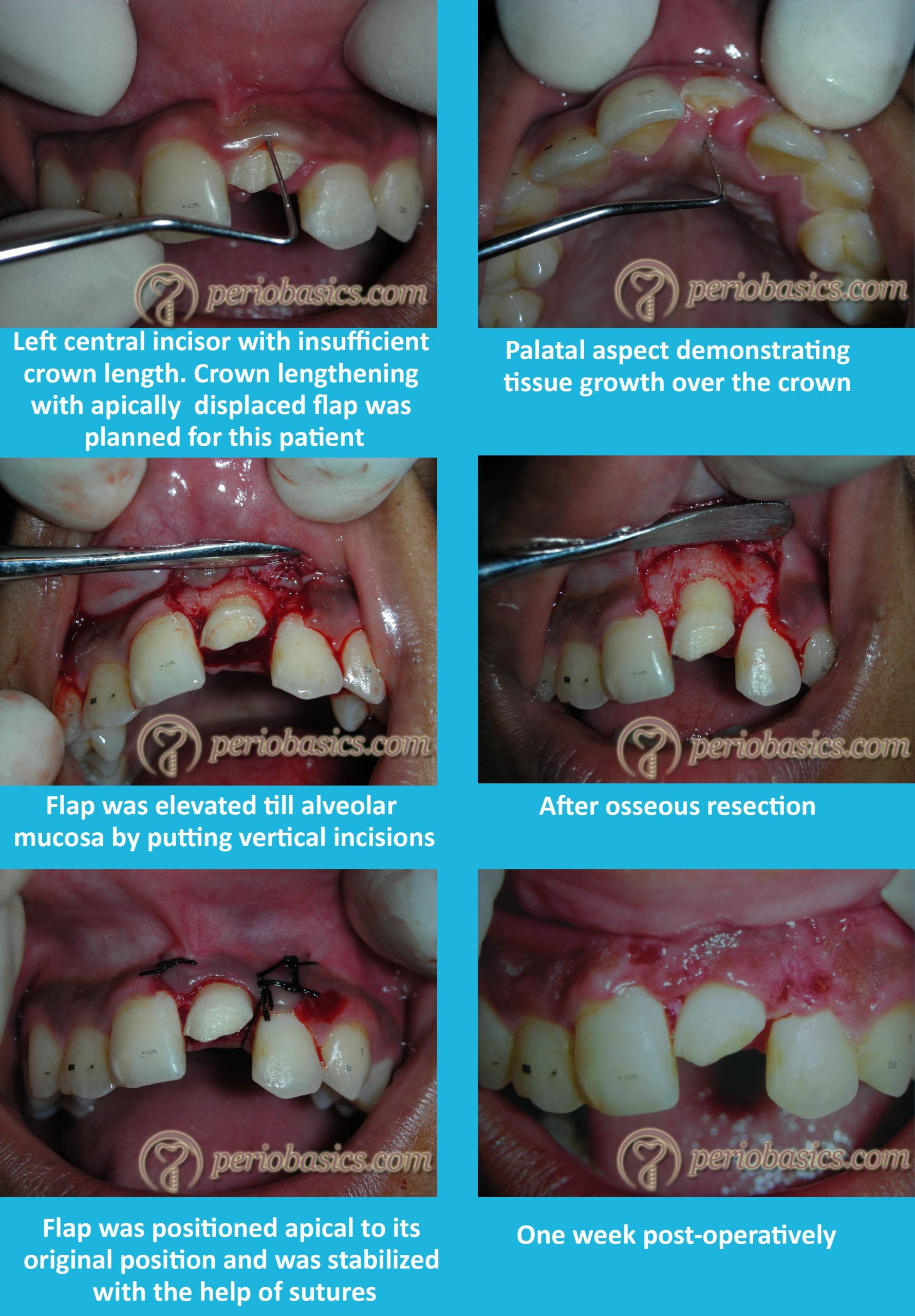
Crescent shaped incision - used during Crown lengthening procedure - clinical photo
crescent shaped incision crown lengthening procedure periodontal clinical photo
semilunar crescent incision crown lengthening periodontal surgery apically repositioned flap clinical photograph
"crescent incision" OR "semilunar incision" crown lengthening gingiva clinical photograph periodontics
Crescent / Semilunar Incision in Crown Lengthening
What it is
- The convexity of the crescent faces coronally (toward the gingival margin)
- The incision is placed entirely within attached keratinized gingiva, apical to the free gingival margin
- It is used when no bone recontouring is needed and adequate attached gingiva is present
- The crescent of tissue between the incision and the gingival crest is excised, effectively shifting the gingival margin apically and exposing more clinical crown
Clinical Photographs

Key Technique Points
| Feature | Detail |
|---|---|
| Incision shape | Convex crescent/half-moon, apex pointing apically |
| Location | Within attached keratinized gingiva, 2-3 mm apical to the free gingival margin |
| Indication | Esthetic crown lengthening (altered passive eruption, "gummy smile"), adequate bone-to-margin distance (≥3 mm), no osseous surgery needed |
| Advantage | Preserves keratinized tissue, no releasing incisions needed, predictable healing |
| Limitation | Cannot address bone; requires sufficient band of attached gingiva (≥5 mm ideally) |
| Also called | Semilunar incision, inverse scalloped incision |
Context in Crown Lengthening Techniques
- Gingivectomy - excision of excess gingival tissue from the coronal aspect (for cases with ≥3 mm bone-to-margin distance and no need for bone work)
- Crescent/semilunar incision - apical excision of a crescent of attached gingiva to shift the margin apically without raising a full flap
- Apically repositioned flap (with/without osseous resection) - full-thickness flap reflected and sutured apically; required when bone reshaping is needed
Double papilla flap clinical photo
double papilla flap periodontal root coverage clinical photo procedure
double papilla flap technique Cohen Ross 1968 root coverage intraoperative clinical photograph periodontics
https://europepmc.org/article/pmc/pmc4295462
double papilla flap pre-op intraoperative suturing post-op clinical images gingival recession
Double Papilla Flap (Cohen & Ross, 1968)
Technique Overview
Clinical Photograph - Suturing Stage

Surgical Steps (Illustrated Diagram)

- (A-C) Measuring the recession and marking the MGJ
- (D-F) Incisions on mesial and distal papillae with horizontal and vertical cuts
- (G-I) Raising the two pedicles and root planing/conditioning
- (J-L) Joining the two papillae at the midline with sutures over the recession
Indications, Steps & Key Points
| Feature | Detail |
|---|---|
| Introduced by | Cohen & Ross, 1968 |
| Classification | Pedicle flap (lateral sliding type) |
| Indication | Isolated gingival recession (Miller Class I & II), narrow recession with adequate adjacent papilla height |
| Advantage | Dual blood supply (from both sides), no donor site morbidity, retains keratinized tissue |
| Disadvantage | Risk of recession at donor papilla sites, limited to isolated defects, not for wide recession |
Step-by-Step Procedure
- Recipient site preparation - De-epithelialize a margin around the recession; root plane and condition with citric acid or EDTA
- Papilla flap design - Horizontal incision at base of each adjacent interdental papilla + vertical releasing incisions
- Flap elevation - Raise both papillae as full-thickness pedicle flaps
- Mobilization - Slide both flaps toward the midline of the recession
- Suturing - Join the two flaps together at the midline with interrupted sutures, and anchor to the periosteum apically
- Periodontal dressing - Coe-Pak placed for 1 week