
Ameoba and ameobiasis
Reading File
Now let me search the library for comprehensive coverage on amoeba and amoebiasis.
Finding Sources
Searching PubMed
"Entamoeba histolytica" AND "amoebiasis"
Good results. Now let me read the most comprehensive sources in parallel - the gastroenterology textbook, the microbiology/lab textbook, Bailey & Love (surgery), and the preventive medicine textbook.
Reading File
Reading File
Reading File
Reading File
Good content flowing in. Now let me read more of the Park's PSM textbook (epidemiological determinants, host, environment) and the gastroenterology textbook (clinical features, diagnosis, treatment) in parallel.
Reading File
Reading File
Excellent content. Now let me get the treatment section and also read more of the Park's PSM for clinical features and control/prevention.
Reading File
Reading File
The second read jumped to Giardia content. Let me get the clinical features + treatment from the gastroenterology book (Yamada's) and also read the PSM content on clinical features and prevention/control.
Finding Sources
Reading File
Now let me get the pathogenesis section and treatment/management from the Yamada's gastroenterology book, plus the clinical features from the gastroenterology section.
Finding Sources
Finding Sources
I now have excellent material. Let me also fetch the key histology image of E. histolytica for the visual.
Loading Image
Loading Image
I now have comprehensive material to write a thorough response. Let me compile everything.
Amoeba and Amoebiasis
The Parasite: Entamoeba histolytica
Amoebiasis is caused by Entamoeba histolytica, a pathogenic protozoan. It is important to distinguish it from two morphologically identical but genetically distinct species:
- E. dispar - a harmless commensal (most common in stool-positive individuals)
- E. moshkovskii - nonpathogenic but recently associated with diarrhoea in some studies
Only E. histolytica causes invasive amoebiasis.
Life Cycle
The parasite exists in two forms:
| Form | Size | Features | Role |
|---|---|---|---|
| Trophozoite (vegetative) | 20-40 µm | Motile, pseudopodia, single nucleus; may contain ingested RBCs | Tissue invasion; NOT infective outside body |
| Cyst (infective stage) | 10-15 µm | Quadrinucleate (4 nuclei); chromatoid bars with rounded ends | Transmission; resistant to environment |
Life cycle steps:
- Cysts shed in faeces by infected humans (up to 1.5 × 10⁷ cysts/day from a carrier)
- Ingestion of cysts in contaminated food/water
- Excystation in small intestine - each cyst produces 8 trophozoites
- Trophozoites colonize the large intestine
- May invade colonic mucosa → disseminate via portal blood → liver/other organs
- Trophozoites re-encyst → passed in stool → continue cycle
Man is the only reservoir of infection. Trophozoites are rapidly destroyed outside the body and by gastric acid - they play no role in transmission.
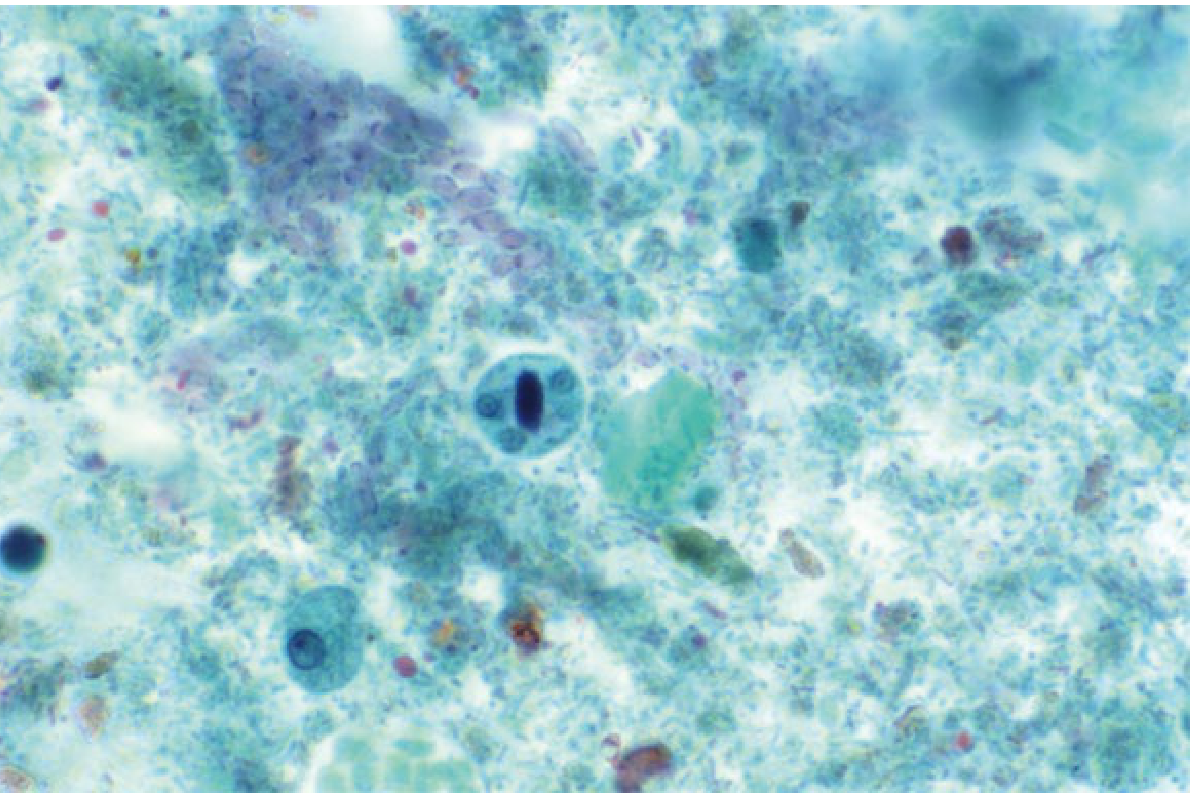
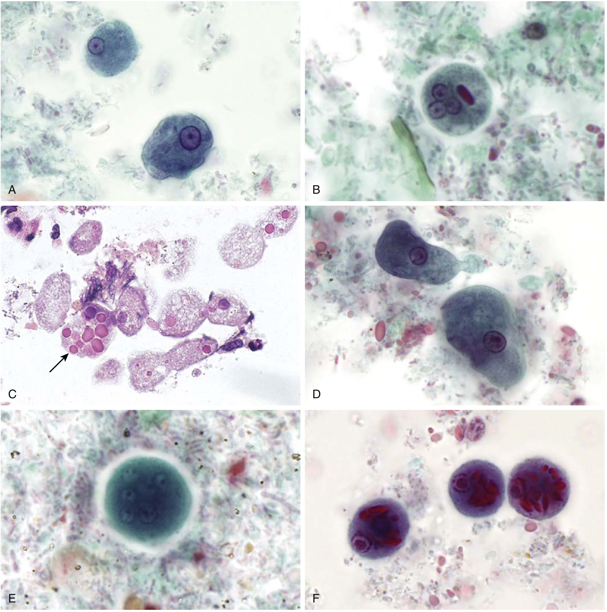
Epidemiology
| Fact | Data |
|---|---|
| Global infected | ~500 million with Entamoeba spp. (all three species combined) |
| Invasive disease | ~50 million develop colitis or extraintestinal abscesses |
| Annual deaths | ~50,000 |
| India prevalence | ~15% (range 3.6-47.4% by region) |
| High-burden regions | China, SE Asia, W Asia, Latin America (especially Mexico), sub-Saharan Africa |
Among parasitic diseases, only cryptosporidiosis, malaria, and schistosomiasis have similar or higher mortality than amoebiasis.
High-risk groups in developed countries: Recent travellers/immigrants, institutionalized/imprisoned persons, men who have sex with men (MSM).
Important epidemiological facts:
- 90% of infections are caused by E. dispar (non-invasive) - symptomatic disease occurs in <10% of E. histolytica infections
- Amebic liver abscess (ALA) is 3-10 times more common in men, though colonic disease is equal between sexes
- Children (especially neonates), pregnant and postpartum women have increased risk of severe disease
- Risk factors for severity: malignancy, malnutrition, corticosteroids
Agent Factors: Zymodemes
E. histolytica can be differentiated into at least 18 zymodemes (populations differing in electrophoretic mobility of enzymes). At least 7 are pathogenic and 11 are non-pathogenic. Pathogenic strains belong to specific zymodemes, but the reason a particular zymodeme can invade is not yet fully understood.
Mode of Transmission
- Faecal-oral route (main):
- Contaminated water (cysts survive for days-weeks; NOT killed by chlorination at routine doses)
- Raw/uncooked vegetables irrigated with sewage water
- Food handlers (cysts found on hands and under fingernails)
- Direct hand-to-mouth
-
Sexual transmission: Oral-rectal contact (especially among MSM)
-
Vectors: Flies, cockroaches, and rodents can mechanically carry cysts to food
Incubation period: 2 to 4 weeks (may be longer)
Period of communicability: As long as cysts are excreted - potentially years if unrecognized/untreated.
Cyst resistance:
- Survive days-weeks in water, faeces, sewage, and soil with moisture and low temperature
- Resistant to chlorine at routine water purification concentrations
- Killed by: drying, heating to ~55°C, or freezing
Pathogenesis
Within the colon, excystation releases trophozoites that may simply colonize without invading. Invasion involves:
- Contact-dependent killing of host cells via galactose/N-acetyl-D-galactosamine (Gal/GalNAc) lectin attachment to colonic epithelium
- Release of amebapores (pore-forming proteins that lyse cell membranes)
- Cysteine proteases that degrade extracellular matrix
- Trophozoites cause flask-shaped ulcers in the submucosa - narrow neck at mucosa with wide base below - most commonly in cecum, rectosigmoid, and ascending colon
- Portal bloodstream invasion → liver (most common extraintestinal site)
Clinical Features
A. Intestinal Amoebiasis
1. Asymptomatic carrier - the most common outcome (~90%)
2. Amebic dysentery (acute)
- Bloody diarrhoea with mucus
- Abdominal pain and cramping
- Trophozoite invasion of mucosa → ulceration → risk of perforation and peritonitis
- Classically: mucus- and blood-stained stools (no pus - helps distinguish from shigellosis, where pus cells are prominent)
3. Amebic colitis (subacute/chronic)
- Milder than dysentery; may mimic inflammatory bowel disease (ulcerative colitis, Crohn's)
- Non-bloody diarrhoea, constipation, abdominal cramping, weight loss
- Flask-shaped submucosal ulcers that expand
- Segments affected: cecum > rectosigmoid > ascending colon
4. Ameboma
- Granulomatous mass in the intestine from chronic amoebic infection
- Produces a "napkin ring" lesion - may be misdiagnosed as carcinoma
5. Fulminant/toxic megacolon - rare but life-threatening
B. Extraintestinal Amoebiasis
1. Amebic Liver Abscess (ALA) - most common extraintestinal form (~5% of intestinal cases)
- Right upper quadrant pain + fever (most consistent finding)
- Usually a single, large abscess in the right lobe
- Anchovy sauce/chocolate paste pus (liquefied hepatic tissue)
- Stool O&P negative in >50% of ALA cases at time of presentation
- Men affected 3-10x more often than women
- Onset ~12 weeks after exposure
- ALP and aminotransferases elevated
- Can rupture into pleura, pericardium, or peritoneum
2. Pulmonary amoebiasis - by contiguous spread from liver abscess across diaphragm or haematogenous spread
3. Cerebral amoebiasis - rare; haematogenous spread; distinguish from free-living amoeba
4. Cutaneous amoebiasis - skin involvement; perianal/genital
Diagnosis
Stool Examination
- Microscopy (fresh stool) - demonstration of trophozoites containing RBCs is diagnostic
- Must examine immediately (cooling destroys trophozoites)
- Look in fresh mucus passed per rectum
- Absence of pus cells in stool: helpful differential from shigellosis
- Note: cannot distinguish E. histolytica from E. dispar by morphology alone
Serology
- Often negative in intestinal amoebiasis but positive in extraintestinal disease
- Positive serology provides a clue to extraintestinal amoebiasis
- Indirect Haemagglutination (IHA) - regarded as the most sensitive serological test
- Counter Immunoelectrophoresis (CIE) and ELISA - newer techniques
- TechLab E. histolytica II antigen detection - can specifically identify E. histolytica (vs E. dispar)
Imaging (for ALA)
- Ultrasound: peripherally located, ovoid abscess; well-circumscribed border; homogeneous hypoechoic centre
- CT scan or MRI for complex cases
Stool Antigen Tests
- Species-specific monoclonal antibody-based antigen tests can differentiate E. histolytica from E. dispar
Treatment
Symptomatic Intestinal Amoebiasis / Amebic Colitis
| Drug | Dose | Notes |
|---|---|---|
| Metronidazole (first-line) | 30 mg/kg/day in 3 divided doses after meals for 8-10 days | Clinical response within 48 h confirms diagnosis |
| Tinidazole (alternative) | Single-dose regimen effective | Longer-acting nitroimidazole |
| Ornidazole | Single-dose regimen | Effective in developing countries |
Nitroimidazoles act against trophozoites (tissue forms) but do NOT eliminate luminal cysts.
Luminal Agent (always add after nitroimidazole)
- Paromomycin (aminoglycoside) - acts on organisms in the intestinal lumen; eliminates cysts; first-line for asymptomatic carriers
- Also used to prevent further transmission after metronidazole course
Amebic Liver Abscess
| Approach | Detail |
|---|---|
| Metronidazole IV/oral | 750 mg q8h for 7-10 days - drug of choice; >90% respond within 72 h |
| Chloroquine | Used in combination for liver abscess in some regimens |
| Luminal agent | Add paromomycin to eradicate bowel cysts (72% of ALA patients have bowel infection) |
| Aspiration | Indicated only if: ruling out pyogenic abscess, no response in 3-5 days, threat of imminent rupture, left-lobe abscess threatening pericardium |
| Surgery | Reserved for bowel perforation, pericardial rupture |
Asymptomatic carriers: In endemic areas, consensus is generally not to treat unless the person is a food handler or travelling to a non-endemic area.
Mortality: <1% with early diagnosis and treatment for uncomplicated ALA.
Prevention and Control
Primary Prevention
- Sanitation - safe disposal of human excreta; handwashing after defecation and before eating
- Water supply - sand filtration and boiling are more effective than chlorination (cysts are chlorine-resistant); protect from faecal contamination
- Food hygiene:
- Disinfect raw vegetables/fruits with 5-10% acetic acid solution or full-strength vinegar
- Thorough washing with detergents in running water removes cysts
- Periodic examination, treatment and education of food handlers
- Health education
Secondary Prevention
- Early diagnosis via stool microscopy and serology
- Prompt treatment of cases
- Trace and treat contacts
Key Differentiating Facts (High-Yield)
| Feature | E. histolytica | E. dispar |
|---|---|---|
| Pathogenic | Yes | No (commensal) |
| Morphology | Identical | Identical |
| Distinction | Contains ingested RBCs in trophozoite | No RBC ingestion |
| Serology | Positive in invasive disease | Negative |
| Prevalence | ~10% of Entamoeba infections | ~90% |
| Feature | Amebic dysentery | Bacillary (Shigella) dysentery |
|---|---|---|
| Pus cells in stool | Absent | Present |
| Macroscopic | Blood + mucus | Blood + mucus + pus |
| Ulcer shape | Flask-shaped | Superficial |
| Onset | Gradual | Acute |
| Response to metronidazole | Yes | No |
Recent Literature
Two recent systematic reviews add current context:
- A 2025 meta-analysis from India (PMID 39979548) provides updated data on epidemiology, risk factors, and clinical characteristics of ALA specifically in the Indian subcontinent.
- A 2026 systematic review and meta-analysis on E. histolytica intestinal infection in Malaysia (PMID 42126905) highlights ongoing high burden in SE Asia.
Sources: Yamada's Textbook of Gastroenterology 7e | Henry's Clinical Diagnosis and Management by Laboratory Methods | Park's Textbook of Preventive and Social Medicine | Harrison's Principles of Internal Medicine | Bailey and Love's Short Practice of Surgery 28e
This is a shared conversation. Sign in to Orris to start your own chat.