
How to calculate cardiac axis in degree simplify
The cardiac axis in degrees is calculated by looking at leads I and aVF (and sometimes lead II) on the ECG. Here is the simplified method:
Searching sources...
Reading File
Searching Images
cardiac axis calculation ECG leads hexaxial diagram
Finding Sources
Searching Images
hexaxial reference system ECG axis degrees leads
Finding Sources
Searching the Web
calculate cardiac axis in degrees ECG simplified method leads I aVF
Reading File
Reading File
Loading Image
Loading Image
Here is a complete, simplified guide to calculating cardiac axis in degrees.
Cardiac Axis: How to Calculate It (Simplified)
The Hexaxial Reference System
Each limb lead points in a specific direction (in degrees) on the frontal plane. The diagram below shows each lead's angle:
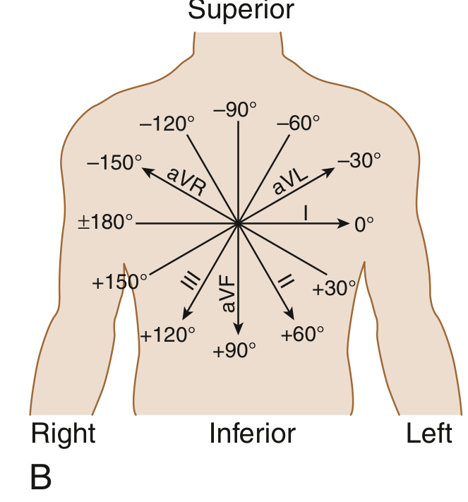
| Lead | Degree |
|---|---|
| Lead I | 0° |
| Lead II | +60° |
| Lead aVF | +90° |
| Lead III | +120° |
| aVL | -30° |
| aVR | -150° |
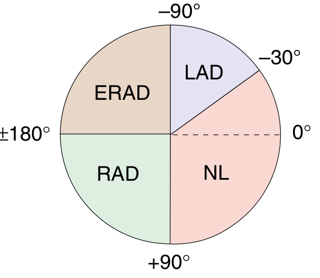
Normal axis: -30° to +90°
Method 1: Quick Quadrant Method (Lead I + aVF)
This is the fastest clinical method. Just check whether Lead I and aVF are positive (upward) or negative (downward):
| Lead I | aVF | Axis |
|---|---|---|
| Positive (+) | Positive (+) | Normal (0° to +90°) |
| Positive (+) | Negative (-) | Left Axis Deviation (LAD) (-30° to -90°) |
| Negative (-) | Positive (+) | Right Axis Deviation (RAD) (+90° to +180°) |
| Negative (-) | Negative (-) | Extreme/NW Axis (-90° to ±180°) |
Method 2: 3-Lead Method (More Accurate - Leads I, II, aVF)
Add Lead II to distinguish true LAD from borderline:
- If Lead I and Lead II are both positive → Normal axis
- If Lead I is positive but Lead II is negative → LAD
- If Lead I is negative, check aVF:
- aVF positive → RAD
- aVF negative → Extreme axis deviation
Rule of thumb: "If Lead I is positive, look at Lead II. If Lead I is negative, look at aVF."
Method 3: Precise Degree Calculation (Tangent Formula)
To get the exact degree, use the net QRS amplitude (R wave - S wave in mm):
Formula:
tan(θ) = aVF amplitude ÷ Lead I amplitude θ = arctan(aVF / Lead I)
Steps:
- In Lead I: calculate net deflection = R height - S depth (in mm)
- In aVF: calculate net deflection = R height - S depth (in mm)
- Apply: θ = tan⁻¹(aVF value ÷ Lead I value)
Worked example:
- Lead I: R=7, S=2 → net = +5 mm
- aVF: R=5, S=0 → net = +5 mm
- θ = tan⁻¹(5/5) = tan⁻¹(1) = 45° → Normal axis ✓
If both values are positive → axis is in the normal quadrant (0° to +90°), the answer stands.
If one or both are negative → you may need to add 90° to the result and confirm using the quadrant method.
Axis Deviation Ranges (Summary)
| Axis | Range | Lead I | aVF |
|---|---|---|---|
| Normal | -30° to +90° | + | + |
| LAD (Left) | -30° to -90° | + | - |
| RAD (Right) | +90° to +180° | - | + |
| Extreme (NW) | -90° to ±180° | - | - |
Subtypes of LAD: Moderate = -30° to -45°; Marked = -45° to -90°
Subtypes of RAD: Moderate = +90° to +120°; Marked = +120° to +180°
Common Causes
| Deviation | Common Causes |
|---|---|
| LAD | Left ventricular hypertrophy (HTN), left anterior fascicular block, inferior MI |
| RAD | Right ventricular hypertrophy (pulmonary HTN, PE), left posterior fascicular block, dextrocardia |
| Extreme/NW | Ventricular tachycardia, severe emphysema, hyperkalemia |
Note: Mild RAD is normal in children, adolescents, and young adults. - Goldman-Cecil Medicine
Key takeaway: For clinical practice, the 3-lead method (I + II + aVF) is fast and reliable. For a precise number in degrees, use the arctan formula with Leads I and aVF net amplitudes.
This is a shared conversation. Sign in to Orris to start your own chat.