
Sialadenitis
sialadenitis
sialadenitis salivary gland inflammation histology parotid
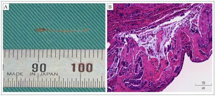
This composite image illustrates the macroscopic and microscopic features of an eosinophilic salivary plug discharged from the parotid gland. Panel A is a clinical photograph showing a macroscopic view of a white, elongated, and translucent fibrous mass, approximately 13 mm in length, against a metric ruler. This mass represents an 'allergic plug' typical of eosinophilic sialadenitis (Kussmaul disease). Panel B is a photomicrograph of the same tissue stained with hematoxylin and eosin (H&E) at high magnification (scale bar 150 µm). The histology reveals a dense inflammatory exudate dominated by eosinophils, characterized by their bright eosinophilic (pink) cytoplasm and bilobed nuclei. These cells are embedded within a network of amorphous eosinophilic fibrin. Additionally, fragments of degenerated ductal epithelial cells are visible, indicating the obstructive nature of the plug within the salivary duct system. The combination of findings is characteristic of chronic obstructive sialadenitis with an allergic or eosinophilic etiology, often seen in patients with comorbid asthma or sinusitis.
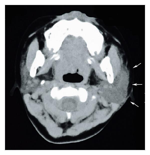
This diagnostic image is an axial computed tomography (CT) scan of the neck at the level of the mandible, demonstrating clinical findings of sialadenitis. The primary focus is the left parotid gland, which is significantly enlarged and swollen compared to the contralateral side. The left parotid exhibits a heterogeneous parenchymal density and ill-defined margins, indicated by white arrows, which suggest active inflammation and adjacent soft tissue edema. In contrast, the right parotid gland appears relatively normal in size with more uniform density. Key anatomical structures visible include the mandibular rami, the base of the tongue, the oropharynx, and the cervical vertebrae. The image serves as an educational example of inflammatory pathology of the salivary glands, highlighting the use of cross-sectional imaging to assess glandular swelling, tissue plane blurring, and potential complications such as abscess formation in cases of acute sialadenitis or mumps-related parotitis.
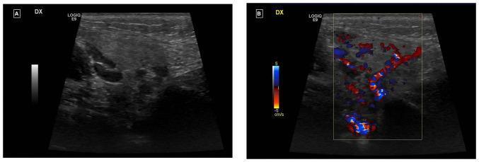
This comparative ultrasound study displays the right parotid gland in two modalities: (A) B-mode grayscale and (B) Color Doppler. Panel A shows an enlarged salivary gland characterized by a heterogeneous, predominantly hyperechoic parenchymal texture with scattered small, ill-defined hypoechoic areas. No discrete focal masses, abscesses, or obstructing sialoliths are visible. Panel B demonstrates an Ecocolor-Doppler overlay within a central region of interest. The image reveals a diffuse and prominent increase in intraparenchymal vascularity, indicated by dense red and blue color signals throughout the gland. This hypervascularity, in the absence of obstructive features, is clinically consistent with acute sialadenitis or an inflammatory process of the salivary gland. The Doppler velocity scale is set between -5 and 5 cm/s, highlighting low-velocity flow typical of glandular inflammation and reactive adenitis.
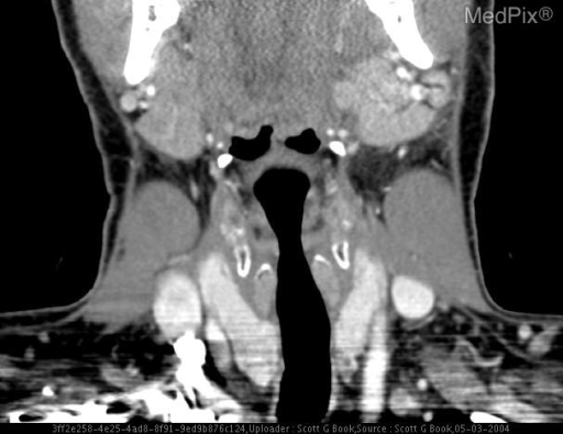
**Imaging Modality:** Contrast-enhanced Computed Tomography (CT), Coronal reformatted view. **Anatomical Region:** Head and neck, specifically focusing on the salivary glands and suprahyoid/infrahyoid neck spaces. **Observed Pathology:** The image demonstrates bilateral enlargement of the parotid glands, consistent with sialadenitis. The glands appear hyperdense and mildly heterogeneous compared to typical fatty parotid parenchyma. There is associated soft tissue swelling in the submandibular and cervical regions. **Characteristic Visual Features:** - **Bilateral Parotid Gland Involvement:** Symmetric enlargement and increased enhancement of the parotid glands. - **Differentiating Features:** The absence of discrete, rim-enhancing fluid collections suggests diffuse inflammatory change (sialadenitis) rather than a mature abscess. - **Secondary Findings:** Mild inflammatory stranding is visible in the overlying subcutaneous fat. The airway remains patent and midline. **Clinical Context:** Findings are highly suggestive of acute infectious sialadenitis, frequently caused by viral pathogens (such as mumps/myxovirus) or bilateral bacterial infection. The imaging helps differentiate diffuse inflammation from obstructive sialolithiasis or localized neoplastic processes.
Sialadenitis
Classification
| Type | Gland Most Affected | Key Feature |
|---|---|---|
| Acute suppurative (bacterial) | Parotid > submandibular | Retrograde bacterial infection |
| Chronic non-specific | Submandibular > parotid | Recurrent episodes, obstruction |
| Viral (mumps) | Bilateral parotid | Paramyxovirus, self-limited |
| Autoimmune (Sjögren) | Major + minor glands | Lymphocytic destruction |
| Granulomatous | Parotid (esp. TB/sarcoid) | Intraglandular lymph node |
| Radioiodine-induced | Bilateral parotid | Post-I-131 therapy |
1. Acute Suppurative (Bacterial) Sialadenitis
Pathophysiology
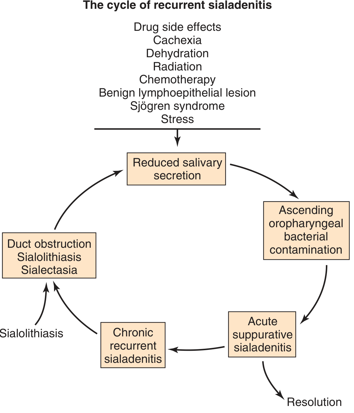
Predisposing Factors
- Dehydration (most important) - postoperative patients (esp. major abdominal/hip surgery), elderly
- Drugs reducing salivary flow - anticholinergics, antihistamines, phenothiazines
- Systemic disease - diabetes mellitus, hypothyroidism, renal failure, Sjögren syndrome
- Sialolithiasis and ductal obstruction (more often causes chronic form)
- Poor oral hygiene, immunosuppression
Microbiology
- Staphylococcus aureus - most common (monitor for MRSA, especially nosocomial cases)
- Streptococcus viridans
- Anaerobes - Fusobacterium, Peptostreptococcus
- Gram-negative bacilli in nosocomial/immunocompromised patients
Clinical Features
- Rapid onset of painful, tender, unilateral gland swelling (parotid most often)
- Purulent discharge expressible from the duct orifice (Stensen's duct in parotitis) - present in ~75% of cases
- Systemic features: fever, chills, malaise
- Bimanual palpation produces purulent ductal discharge
- Lab: leukocytosis with neutrophilia, signs of dehydration (hypernatremia, elevated BUN)
- Bilateral involvement reported in up to 25% of cases
Imaging
- Ultrasound - first-line; identifies abscess, sialolith, excludes abscess in most cases
- CT scan - for deeper extension (parapharyngeal space), trismus, or failure to respond to treatment within 48-72 hours
- Sialography is CONTRAINDICATED in acute phase - exacerbates inflammation
- MRI if neoplasm suspected
Treatment
- Aggressive IV fluid and electrolyte replacement
- Antibiotics: beta-lactamase-stable agent (augmented penicillin, anti-staphylococcal penicillin, or 1st-generation cephalosporin); add metronidazole or clindamycin for anaerobic coverage; vancomycin/linezolid if MRSA suspected
- Sialagogues (lemon drops, orange juice) to stimulate salivary flow
- External and bimanual massage (distal gland toward duct papillae)
- Warm compresses, analgesics, oral hygiene
- Parotid abscess drainage: elevation of facial flap, radial incisions in parotid parenchyma in the direction of the facial nerve branches
2. Chronic Sialadenitis
Pathogenesis
Clinical Features
- Recurring sudden-onset swelling and tenderness of the gland
- Low-grade fever during episodes
- Asymptomatic intervals ranging from weeks to months
- Mucopus at ductal orifice on examination
- No known association with meals (distinguishes from simple obstructive swelling)
Imaging
- Punctate sialectasis and peripheral ductal dilatation on sialography
- Progressive glandular atrophy and increased echogenicity on US
Treatment (stepwise)
- Conservative: oral antibiotics, massage, warm compresses, sialagogues, hydration, short-course corticosteroids
- Sialendoscopy - diagnostic + therapeutic; duct lavage with saline, intraglandular steroid instillation, hydrostatic dilatation; indicated if >1 acute attack/year
- Balloon dilatation / stenting of strictures (stents kept 4 weeks)
- Surgical gland excision - if all else fails
3. Viral Sialadenitis
Mumps (Most Common Viral Cause)
- Paramyxovirus; prevented by MMR vaccine
- Peak age: 4-6 years
- Bilateral parotid involvement (characteristically)
- Associated: fever, malaise, orchitis (may cause sterility in adults), pancreatitis, encephalitis, sensorineural hearing loss
- Histology: interstitial inflammation with mononuclear inflammatory infiltrate
- Treatment: supportive
- Diagnosis: clinical + serologic
HIV-Associated
- Parotid enlargement from lymphoid hyperplasia, infection, or lymphoma
- May be the presenting sign of HIV infection
- Lymphoepithelial cysts - unique to parotid (due to intraparotid lymph node incorporation during embryology)
- DILS (Diffuse Infiltrative Lymphocytosis Syndrome) - sicca-like syndrome similar to Sjögren
- Bilateral deforming cysts: treat with antiretroviral therapy; sclerotherapy in select patients; surgery rarely needed
- Solid parotid mass in HIV: ~40% risk of malignancy
4. Granulomatous Sialadenitis
| Cause | Notes | Diagnosis | Treatment |
|---|---|---|---|
| TB | Via intraglandular lymph nodes; mostly parotid | PPD, FNA (AFB, Langhans giant cells) | Anti-TB regimen |
| Atypical mycobacteria | Children 16-36 months; violaceous skin, sinus tracts; CXR negative | Serology | Incision + curettage or gland excision |
| Actinomycosis | Gram-positive anaerobe; sulfur granules, multiloculated abscesses, sinus tracts | Culture | Penicillin G IV x 6 wks, then PO erythromycin or clindamycin |
| Cat scratch disease | Bartonella henselae; parotid lymphatics | Serology/PCR, Warthin-Starry stain | Observation; azithromycin |
| Toxoplasmosis | Increased with HIV; undercooked meat/cat feces | Acute + convalescent titers | Pyrimethamine + sulfadiazine + folinic acid |
| Sarcoidosis | Noncaseating granulomas; Heerfordt syndrome = acute parotitis + uveitis + facial nerve palsy | Biopsy | Steroids |
5. Autoimmune Sialadenitis - Sjögren Syndrome
- Autoimmune destruction of acinar and ductal cells
- Primary Sjögren: xerophthalmia + xerostomia (sicca syndrome)
- Secondary Sjögren: sicca + connective tissue disease (most commonly rheumatoid arthritis)
- More common in women; HLA-B8, HLA-DR3 associations
- Lip biopsy of minor salivary glands: focal lymphocytic sialadenitis (focus score ≥1 per 4 mm²) - diagnostic criterion
- Long-term risk of B-cell lymphoma (40x increased risk)
6. Special Forms
Mucocele
- Most common inflammatory lesion of salivary glands
- Blockage or rupture of a minor salivary duct → saliva leaks into connective tissue stroma
- Typically on the lower lip; fluctuant, may change size with meals, blue translucent hue
- Histology: pseudocyst lined by granulation tissue, filled with mucin and macrophages
- Treatment: complete excision of cyst + accompanying minor salivary gland lobule (incomplete excision → recurrence)
Ranula
- Epithelial-lined cyst from sublingual gland duct damage
- A "plunging ranula" dissects through the mylohyoid muscle into the neck
Iodine-induced ("Iodide Mumps")
- Mild acute sialadenitis from iodine-based contrast media (e.g. for coronary angiography)
- Transient and self-limited
Radioiodine-induced
- Follows I-131 therapy for differentiated thyroid cancer
- Can cause salivary and lacrimal gland dysfunction; recent 2026 meta-analysis (PMID 42018484) confirms significant prevalence of both salivary and lacrimal gland complications
Ultrasound Appearance
Key Differentiating Points
| Feature | Acute Bacterial | Viral (Mumps) | Chronic | Sjögren |
|---|---|---|---|---|
| Laterality | Usually unilateral | Bilateral | Usually unilateral | Bilateral |
| Gland | Parotid (most) | Both parotids | Submandibular (most) | All major + minor |
| Pus from duct | Yes | No | Sometimes | No |
| Fever | Yes (high) | Mild-moderate | Low-grade | No |
| Course | Acute | Acute, self-limited | Recurrent-chronic | Progressive |
| Associated features | Dehydration, debility | Orchitis, pancreatitis | Sialolithiasis | Dry eyes/mouth, autoantibodies |