
Octa
"Octa" - could you clarify what you're looking for? Here are some possibilities:
- Octa- (prefix meaning 8) - as in octagon, octave, octopus, etc.
- Octa (a specific product, brand, or name)
- Octa (a person or character)
- Something else entirely
What did you have in mind?
Octa eye
Asking for Preferences
What are you looking for with 'Octa eye'?
OCT-A (Optical Coherence Tomography Angiography) is a non-invasive retinal imaging technique that uses motion contrast to visualize blood flow in the retina and choroid without injecting dye. Let me pull detailed clinical information from the medical library.
Searching sources...
Reading File
Finding Sources
Searching PubMed
OCT angiography retinal imaging clinical applications
Reading File
Finding Sources
Reading File
Reading File
Now let me fetch the key OCT-A images from Kanski's to include visually:
Loading Image
Loading Image
Here is a complete overview of OCT Angiography (OCT-A) based on Kanski's Clinical Ophthalmology and The Wills Eye Manual:
OCT Angiography (OCT-A)
What Is It?
OCT-A is a non-invasive retinal and choroidal imaging technique that visualizes blood flow without injecting any contrast dye (unlike traditional fluorescein or indocyanine green angiography). It works by detecting the motion of red blood cells within the microvasculature.
How It Works
- A rapid series of OCT B-scans are taken at the same retinal location vertically (repeat scanning).
- Between consecutive scans, static tissue looks identical - but moving red blood cells create detectable signal differences (motion contrast).
- These differences are processed to generate a 2D map of microcirculation across distinct retinal and choroidal layers.
- The result: flow is visualized - not the vessel walls themselves.
Key point: Flow that is too slow or too fast may not be detected, and classic angiographic signs (leakage, staining, pooling) are absent because no dye is injected.
Imaging Examples
Fig. D - OCT-A of deep capillary plexus in diabetic macular oedema showing loss of the perifoveal vascular network:
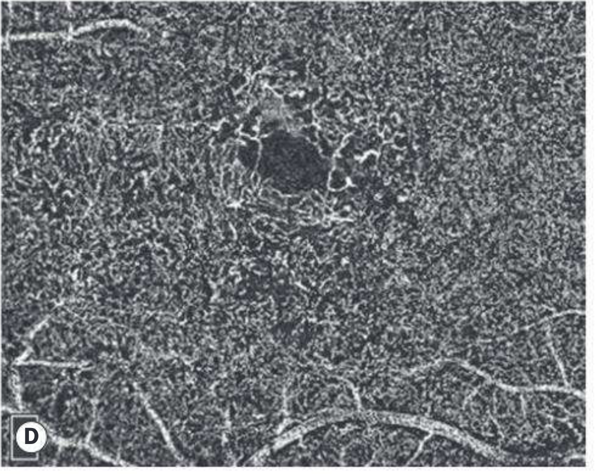
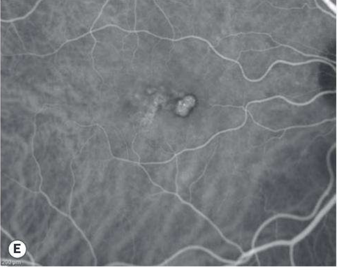
Fig. E - OCT-A of choroidal layer showing polypoidal choroidal vasculopathy (the dark "polyp" area lacks blood flow signal due to turbulence within):
Clinical Applications
| Condition | What OCT-A Shows |
|---|---|
| Macular neovascular membrane (AMD) | Abnormal flow in outer retina and choroid; detects non-exudative (dry) neovascular membranes |
| Diabetic retinopathy | Preretinal neovascularization; differentiates IRMA from new vessels; detects microvascular changes before clinical retinopathy appears |
| Macular oedema | Assesses deep retinal capillary plexus and microcirculation |
| Macular ischaemia | Maps perfusion deficits in perifoveal network |
| Polypoidal choroidal vasculopathy (PCV) | Identifies choroidal polyps and abnormal choroidal vessels |
| Chronic central serous retinopathy | Choroidal flow abnormalities |
| Macular telangiectasia | Vascular changes |
| Proliferative retinopathy | Objective monitoring after treatment |
Advantages
- No dye injection - safer, faster, no risk of allergic reaction
- Provides depth-resolved layer-by-layer vascular maps (superficial vs. deep capillary plexus, outer retina, choriocapillaris)
- Can detect subclinical vascular changes (e.g. diabetic microvascular changes before clinical signs)
- Rapid acquisition
Limitations
- No leakage, staining, or pooling visible (dye-based signs are absent)
- Small field of view (though newer widefield OCT-A is expanding this)
- Requires good patient cooperation and fixation
- Flow that is too slow or too fast may not register
- Susceptible to motion artifacts
OCT vs. OCT-A at a Glance
| Feature | Standard OCT | OCT-A |
|---|---|---|
| What it shows | Tissue structure/layers | Blood flow/vasculature |
| Dye needed? | No | No |
| Leakage visible? | No | No |
| Layer separation | Yes | Yes |
| Best for | Macular edema, layer anatomy | Neovascularization, ischemia |
Recent Literature (2023-2025)
- Hormel & Jia (2023) reviewed OCT-A retinal biomarkers - PMID 37791289
- Crincoli et al. (2024) published an update focused on diabetic retinopathy - PMID 38376579
- Jia et al. (2025) covered widefield OCT-A advances - PMID 40517946
Sources: Kanski's Clinical Ophthalmology 10th Ed., pp. 594-596; The Wills Eye Manual, p. 1105
Would you like me to go deeper on any specific disease application (e.g., diabetic retinopathy, AMD, glaucoma) or on the technical aspects of OCT-A?
This is a shared conversation. Sign in to Orris to start your own chat.