
Enumerate the causes of dysphagia. How will you clinically differentiate between them? Describe barium swallow appearance of oesophagus in different causes of dysphagia.
Your credits are exhausted. You can purchase a top-up or subscribe to a plan to continue using Orris. If you're already subscribed, your daily credits will reset tomorrow.
Enumerate the causes of dysphagia. How will you clinically differentiate between them? Describe barium swallow appearance of oesophagus in different causes of dysphagia.
Reading File
Finding Sources
Finding Sources
Searching PubMed
"Deglutition Disorders"[MeSH Terms] AND "diagnosis"
Reading File
Reading File
Searching Images
barium swallow achalasia bird beak narrowing dilated oesophagus
Searching Images
barium swallow carcinoma oesophagus irregular shouldered stricture apple core
Reading File
Reading File
Dysphagia: causes, clinical differentiation and barium swallow findings
1. Causes of dysphagia
A. Oropharyngeal (transfer) dysphagia
- Neuromuscular: stroke, Parkinson disease, motor neurone disease, myasthenia gravis, multiple sclerosis, bulbar palsy, inclusion body myositis, myotonic dystrophy.
- Muscular or cricopharyngeal: cricopharyngeal achalasia or spasm, polymyositis, dermatomyositis.
- Structural: pharyngeal pouch (Zenker diverticulum), post-cricoid web (Plummer-Vinson or Paterson-Brown-Kelly syndrome), tumours of the tongue, tonsil, pharynx or larynx, retropharyngeal abscess, post-radiotherapy or post-surgical change, cervical osteophytes.
- Mouth and pharynx lesions: tonsillitis, quinsy, painful ulcers, xerostomia.
B. Oesophageal dysphagia
Intraluminal
- Foreign body, food bolus.
Intrinsic mural (mechanical)
- Malignant: carcinoma of the oesophagus (squamous or adenocarcinoma), carcinoma of the cardia, leiomyoma and other rare tumours.
- Benign stricture: peptic (GORD), corrosive, post-radiation, post-intubation, post-sclerotherapy, drug-induced, Barrett stricture.
- Rings and webs: Schatzki lower oesophageal ring.
- Inflammatory: eosinophilic oesophagitis, candidal, herpetic or CMV oesophagitis.
- Diverticula: mid-oesophageal traction or epiphrenic.
Extrinsic compression
- Enlarged left atrium (mitral stenosis), aortic aneurysm, dysphagia lusoria (aberrant right subclavian artery), mediastinal lymph nodes (malignant or tuberculous), bronchial carcinoma, goitre, paraoesophageal hernia, cervical spine disease (osteophytes, Pott disease).
Motility disorders
- Primary: achalasia, diffuse oesophageal spasm, jackhammer oesophagus, ineffective oesophageal motility.
- Secondary: scleroderma (systemic sclerosis), Chagas disease (secondary achalasia), diabetes, hypothyroidism, amyloidosis, pseudo-achalasia from a cardia carcinoma.
C. Functional or psychogenic
- Globus, anxiety. This is a diagnosis of exclusion.
2. Clinical differentiation
Step 1: Is it true dysphagia? Separate it from globus (a lump in the throat that is present between swallows and eases with eating), odynophagia, and dysphagia from a dry mouth.
Step 2: Localise the level.
- Difficulty initiating the swallow, with coughing, choking, nasal regurgitation or hoarseness, points to oropharyngeal dysphagia.
- Food sticking behind the sternum after the swallow has started points to oesophageal dysphagia.
Step 3: Use the history.
| Feature | Pointer |
|---|---|
| Solids only, progressive over weeks to months, then semisolids and liquids | Mechanical obstruction, especially carcinoma or stricture |
| Solids and liquids from the start, intermittent | Motility disorder (achalasia, spasm) |
| Short history (under 3 months), age over 50, marked weight loss, anorexia | Carcinoma |
| Long history of heartburn or reflux, then dysphagia that is slowly progressive or non-progressive | Peptic stricture (or Barrett oesophagus with adenocarcinoma) |
| Past ingestion of acid or alkali | Corrosive stricture |
| Young patient, long history, regurgitation of undigested food, nocturnal cough or aspiration, relief with drinking water or standing, weight loss that is mild relative to the duration | Achalasia |
| Chest pain with dysphagia, triggered by hot or cold fluids, episodic | Diffuse spasm or jackhammer oesophagus |
| Intermittent dysphagia for solids, with episodes of sudden bolus impaction (meat), in an atopic patient | Eosinophilic oesophagitis or Schatzki ring |
| Gurgling in the neck, regurgitation of food eaten hours earlier, halitosis, a swelling in the neck that appears during meals | Pharyngeal pouch |
| Middle-aged woman, iron deficiency anaemia, dysphagia high in the throat | Post-cricoid web (Plummer-Vinson syndrome) |
| Raynaud phenomenon, skin tightness, severe reflux | Scleroderma |
| Dysphagia with painful swallowing, oral thrush, HIV or immunosuppression, steroid inhaler use | Infective oesophagitis |
| Dysphagia with neurological signs, dysarthria, ptosis, fatigable weakness | Neurological or bulbar cause |
| Dysphagia for both solids and liquids with a cardiac murmur | Left atrial enlargement |
Step 4: Examine the patient. The following points are from S Das, A Manual on Clinical Surgery, 13th Edition, in the dysphagia section on examination.
- Mouth and pharynx: tonsils and fauces for lesions, palatal mobility, and the posterior pharyngeal wall for a retropharyngeal abscess.
- Neck: goitre, nodes, and a swelling that appears during meals above the left clavicle (late pharyngeal pouch). "Tracheal tugging" suggests an aortic arch aneurysm. Palpate the left supraclavicular fossa (Virchow node), which may be the only sign of oesophageal carcinoma.
- Chest: pleural effusion in late carcinoma. Aspiration pneumonitis, lung abscess or bronchiectasis in achalasia. Dyspnoea from a hugely dilated oesophagus. Look for a hiatal or paraoesophageal hernia.
- Abdomen: epigastric mass or hepatomegaly from cardia carcinoma and metastases.
- Spine: cervical tenderness or deformity if Pott disease is suspected.
- General: cachexia, anaemia (koilonychia and glossitis in Plummer-Vinson), skin changes of scleroderma, and cranial nerve or limb signs for neurological disease.
Step 5: Investigations that complete the differentiation.
- Endoscopy with biopsy is the first investigation for most oesophageal dysphagia. It confirms or excludes carcinoma and stricture, and diagnoses oesophagitis and eosinophilic oesophagitis.
- Barium swallow is described below.
- High-resolution manometry is the gold standard for motility disorders.
- CT or endoscopic ultrasound is used for staging and extrinsic compression.
- Videofluoroscopy and fibreoptic endoscopic evaluation of swallowing (FEES) are used for oropharyngeal dysphagia.
- A normal endoscopy does not exclude achalasia, because 30 to 40% of endoscopies are reported normal before the final diagnosis (Bailey & Love, 28th Edition, achalasia section). Endoscopy is still needed to exclude pseudo-achalasia from a cardia cancer.
3. Barium swallow appearances
Technique points from Grainger & Allison's Diagnostic Radiology:
- If the history suggests obstruction but the routine images look normal, a barium-coated biscuit or a barium tablet may reveal an occult stricture.
- Use thin barium for a pharyngeal pouch. Thick barium is not washed out of the pouch (S Das, Manual on Clinical Surgery).
Achalasia
- Early: normal-calibre oesophagus with defective distal peristalsis, and barium retained for more than 5 minutes (Grainger & Allison).
- Established: a dilated oesophagus with a smooth, tapering narrowing at the lower end. This is described as a "bird's beak" or "rat's tail" (Bailey & Love, 28th Edition).
- Retention of contrast, an absent gastric air bubble, and abnormal contractions in the body. Progressive dilatation gives a sigmoid-shaped oesophagus.
- A chest X-ray may show an air-fluid level in a widened mediastinum.
- The narrowing is smooth and symmetrical. The mucosa is intact, and the segment relaxes after hot water or amyl nitrite.
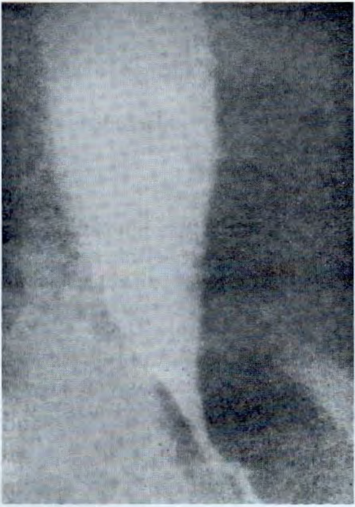
Carcinoma of the oesophagus
- A short, irregular, rigid stricture with an "apple core" appearance.
- Abrupt "shouldering" at the upper and lower margins, with destroyed or ulcerated mucosa.
- Proximal dilatation is modest. There may be an eccentric mass, a filling defect, or a fistula to the trachea.
- Features that favour cancer over achalasia are a short history, an irregular narrowing, shouldering, and a non-relaxing segment.
- Pseudo-achalasia (a cardia carcinoma mimicking achalasia) can look like a bird's beak. Endoscopy and CT are needed.
Benign (peptic or corrosive) stricture
- A long, smooth or tapered narrowing with mild proximal dilatation, and no shouldering (S Das, Fig. 31.3).
- A peptic stricture is in the lower third and often accompanies a hiatus hernia, and may be associated with reflux. A corrosive stricture is long and may be multiple.
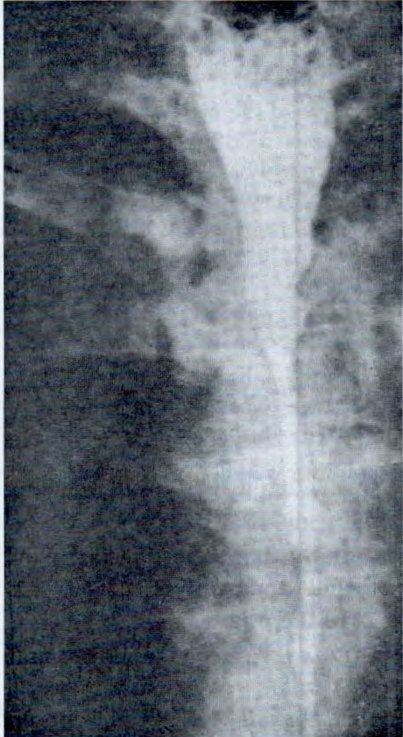
Diffuse oesophageal spasm and jackhammer oesophagus
- A "corkscrew" or "rosary bead" oesophagus from non-peristaltic, simultaneous contractions (Grainger & Allison). Transit is delayed.
- The study may be normal if the spasm does not occur during the examination.
Scleroderma
- Reduced or absent peristalsis, a patulous gastro-oesophageal junction, and free reflux with its complications (Grainger & Allison).
- The oesophagus is dilated and atonic, and a stricture may be present.
Eosinophilic oesophagitis
- A small-calibre ("narrow-calibre") oesophagus. Fixed rings (the "ringed" or "feline" oesophagus) and strictures are common (Grainger & Allison).
Pharyngeal pouch (Zenker diverticulum)
- A posterior outpouching at the pharyngo-oesophageal junction, above the cricopharyngeus.
- The pouch fills first, then spills over. It may be large enough to displace and compress the oesophagus.
Post-cricoid web
- A thin, anterior, shelf-like filling defect at the C5 to C6 level, best seen on lateral views during rapid swallowing.
Schatzki ring
- A thin, smooth, symmetrical ring at the squamocolumnar junction, just above a hiatus hernia. It is best seen with the oesophagus distended with barium.
Extrinsic compression
- A smooth, extramucosal indentation that is usually pulsatile in vascular lesions.
- An enlarged left atrium causes a posterior indentation at the level of the heart.
- Dysphagia lusoria shows an oblique posterior indentation at the level of the aortic arch.
Oesophageal diverticula (traction or epiphrenic)
- A traction diverticulum is a small, tented outpouching in the mid-oesophagus, often from old tuberculous nodes.
- An epiphrenic diverticulum is a larger pouch above the diaphragm, often with an underlying motility disorder.
Candida or other infective oesophagitis
- A shaggy, "cobblestone" or plaque-like irregular mucosa, with a linear or granular filling-defect pattern, particularly in the upper and mid oesophagus.
Oropharyngeal dysphagia
- Videofluoroscopy shows delayed or absent initiation of swallow, nasal regurgitation, vallecular and pyriform pooling, aspiration into the larynx and trachea, and a prominent cricopharyngeal bar.
- The sequence of findings is often more informative than the static image.
4. Summary
| Cause | Typical barium finding |
|---|---|
| Achalasia | Dilated oesophagus, smooth bird's beak, retention |
| Carcinoma | Irregular shouldered stricture, apple core, mucosal destruction |
| Benign stricture | Smooth, long, tapered narrowing, no shouldering |
| Spasm or jackhammer | Corkscrew or rosary-bead oesophagus |
| Scleroderma | Atonic, patulous oesophagus with reflux |
| Eosinophilic oesophagitis | Narrow-calibre, ringed oesophagus |
| Pharyngeal pouch | Posterior pouch above the cricopharyngeus |
| Web or ring | Thin anterior shelf (web) or circumferential ring (Schatzki) |
| Extrinsic compression | Smooth extramucosal indentation |
I did not find a recent systematic review that changes this approach. A quick PubMed check of the last two years returned only tangentially related reviews. The image library search found no matching figures, so the two images above are from the S Das textbook.
Sources: S Das, A Manual on Clinical Surgery, 13th Edition (dysphagia: examination, special investigations and differential diagnosis); Grainger & Allison's Diagnostic Radiology (oesophageal fluoroscopy, achalasia, jackhammer oesophagus, scleroderma, eosinophilic oesophagitis); Bailey & Love's Short Practice of Surgery, 28th Edition (achalasia). I added the broader list of causes and the descriptions of web, ring, Zenker and carcinoma appearances from standard teaching, and did not check them against the library text.
This is a shared conversation. Sign in to Orris to start your own chat.