
Simple diagram of minimal change disease, acute proliferative glomerulonephritis, focal segmental glomerulonephritis, membranous glomerulonehpropathy
glomerulonephritis AND nephrotic syndrome
NIDDK glomerular diseases minimal change disease FSGS membranous nephropathy acute poststreptococcal glomerulonephritis
A simple educational four-panel diagram comparing glomerular diseases: minimal change disease, acute proliferative glomerulonephritis, focal segmental glomerulosclerosis, and membranous nephropathy. Show a clear glomerulus cross-section in each panel with labels for the characteristic light microscopy pattern and a short clinical pattern: minimal change - normal-looking glomerulus and diffuse podocyte foot-process effacement, nephrotic; acute proliferative glomerulonephritis - enlarged hypercellular glomerulus from inflammatory cells, nephritic; focal segmental glomerulosclerosis - scarred segments in some glomeruli, nephrotic; membranous nephropathy - diffuse thickened capillary walls with subepithelial immune deposits, nephrotic. Include a concise legend: nephrotic = heavy proteinuria and edema; nephritic = hematuria and reduced GFR. Correct spelling, clean medical textbook diagram.
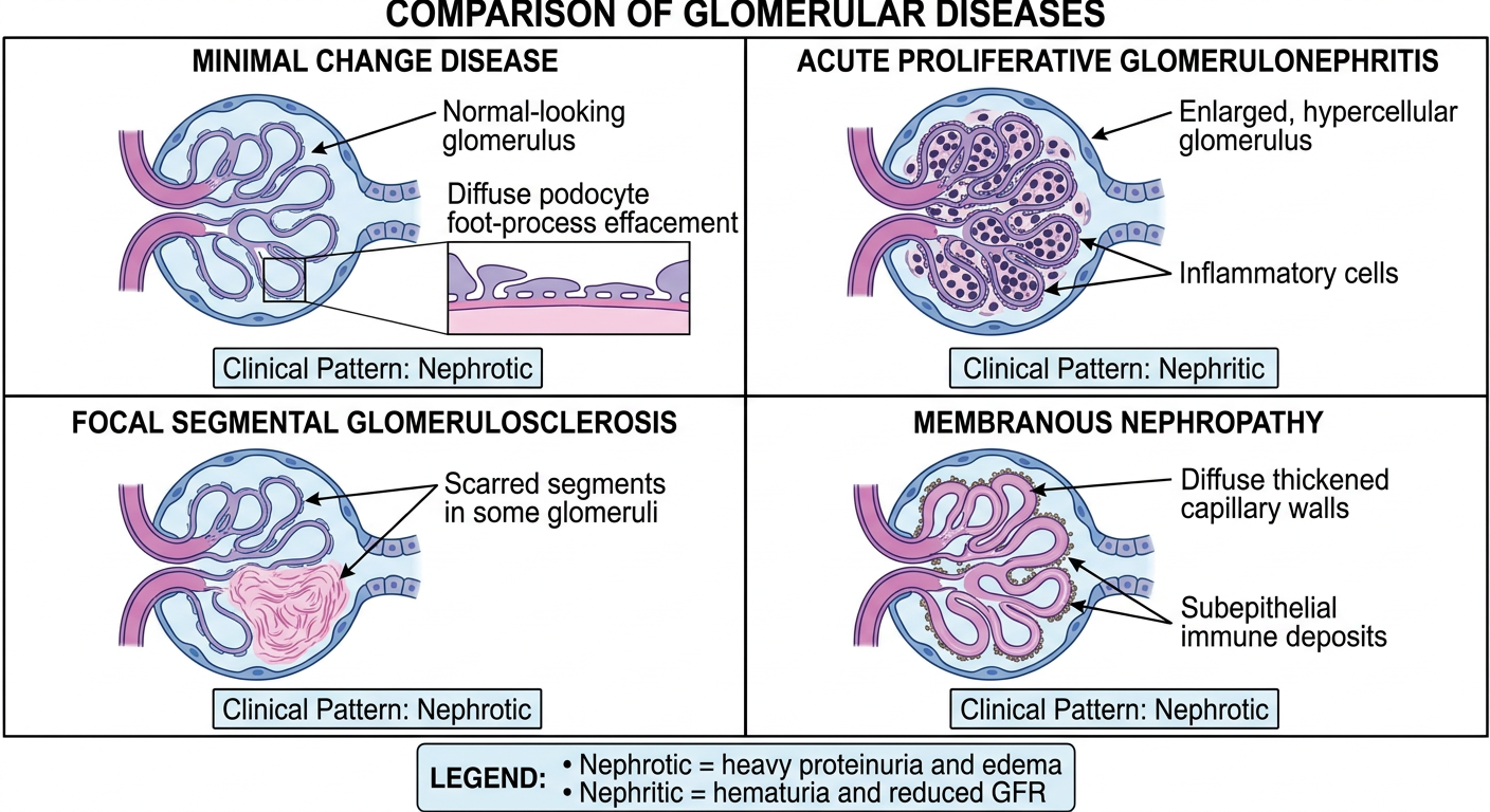
- Minimal change disease (MCD): glomeruli look essentially normal on light microscopy; electron microscopy shows diffuse podocyte foot-process effacement. Nephrotic syndrome.
- Acute proliferative glomerulonephritis: diffuse glomerular hypercellularity and inflammation. Nephritic syndrome, typically hematuria, hypertension, and reduced GFR.
- Focal segmental glomerulosclerosis (FSGS): sclerosis affects some glomeruli (focal) and part of each affected glomerulus (segmental). Nephrotic syndrome.
- Membranous nephropathy: diffuse capillary-wall/GBM thickening due to subepithelial immune deposits. Nephrotic syndrome.
Simple diagram of small cell, adeno, squamous cell carcinomas of lung
simple histology comparison diagram small cell carcinoma adenocarcinoma squamous cell carcinoma lung
lung neoplasms AND histology
NCI non-small cell lung cancer adenocarcinoma squamous cell carcinoma small cell lung cancer histology
A simple educational three-panel diagram comparing the main lung cancer types: small cell carcinoma, adenocarcinoma, and squamous cell carcinoma. Include a small lung silhouette showing typical location, simple microscopic-style drawings, and concise labels. Small cell carcinoma: central/hilar, sheets of small blue cells, nuclear molding, neuroendocrine, strongly associated with smoking, early metastasis. Adenocarcinoma: peripheral, gland formation and mucin, most common type, may occur in nonsmokers. Squamous cell carcinoma: central, keratin pearls and intercellular bridges, strongly associated with smoking, may cavitate. Include a footer: small cell = small-cell lung cancer; adeno and squamous = non-small-cell lung cancer. Accurate spelling, clean easy-to-memorize medical exam diagram.
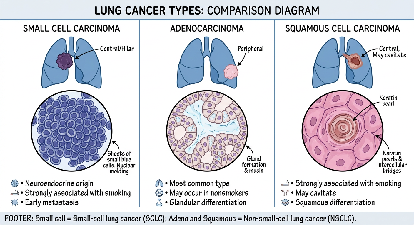
| Feature | Small cell carcinoma | Adenocarcinoma | Squamous cell carcinoma |
|---|---|---|---|
| Group | SCLC | NSCLC | NSCLC |
| Usual site | Central/hilar | Peripheral | Central bronchus |
| Hallmark microscopy | Small cells, scant cytoplasm, nuclear molding | Glands and/or mucin | Keratin pearls, intercellular bridges |
| Smoking link | Strong | May occur in never-smokers | Strong |
| Behavior | Rapid growth, early metastasis | Commonest subtype | May cavitate |
Simple diagram of papillary, clear cell, chromophobe renal cell carcinoma And wilms tumor
histology diagram clear cell papillary chromophobe renal cell carcinoma Wilms tumor comparison
renal cell carcinoma AND Wilms tumor
NCI renal cell cancer clear cell papillary chromophobe Wilms tumor histology
A simple educational four-panel medical pathology diagram comparing clear cell renal cell carcinoma, papillary renal cell carcinoma, chromophobe renal cell carcinoma, and Wilms tumor. Each panel should include a small kidney silhouette with typical gross location, a clear simplified histology-style sketch, and concise accurate labels. Clear cell RCC: golden yellow cortical tumor, nests/alveoli of cells with clear cytoplasm and delicate branching vessels, VHL chromosome 3p association, most common adult renal cancer. Papillary RCC: papillae with fibrovascular cores, foamy macrophages, often multifocal/bilateral, trisomy 7 and 17. Chromophobe RCC: pale cells with distinct cell borders and perinuclear halos, from collecting duct intercalated cells, generally favorable prognosis. Wilms tumor: pediatric embryonal renal tumor, triphasic histology showing blastemal small blue cells, epithelial tubules, and stromal tissue, WT1 or WT2 association. Footer: first three are adult renal cell carcinoma types; Wilms tumor is primarily a childhood tumor. Correct spelling, uncluttered, easy-to-memorize exam diagram.
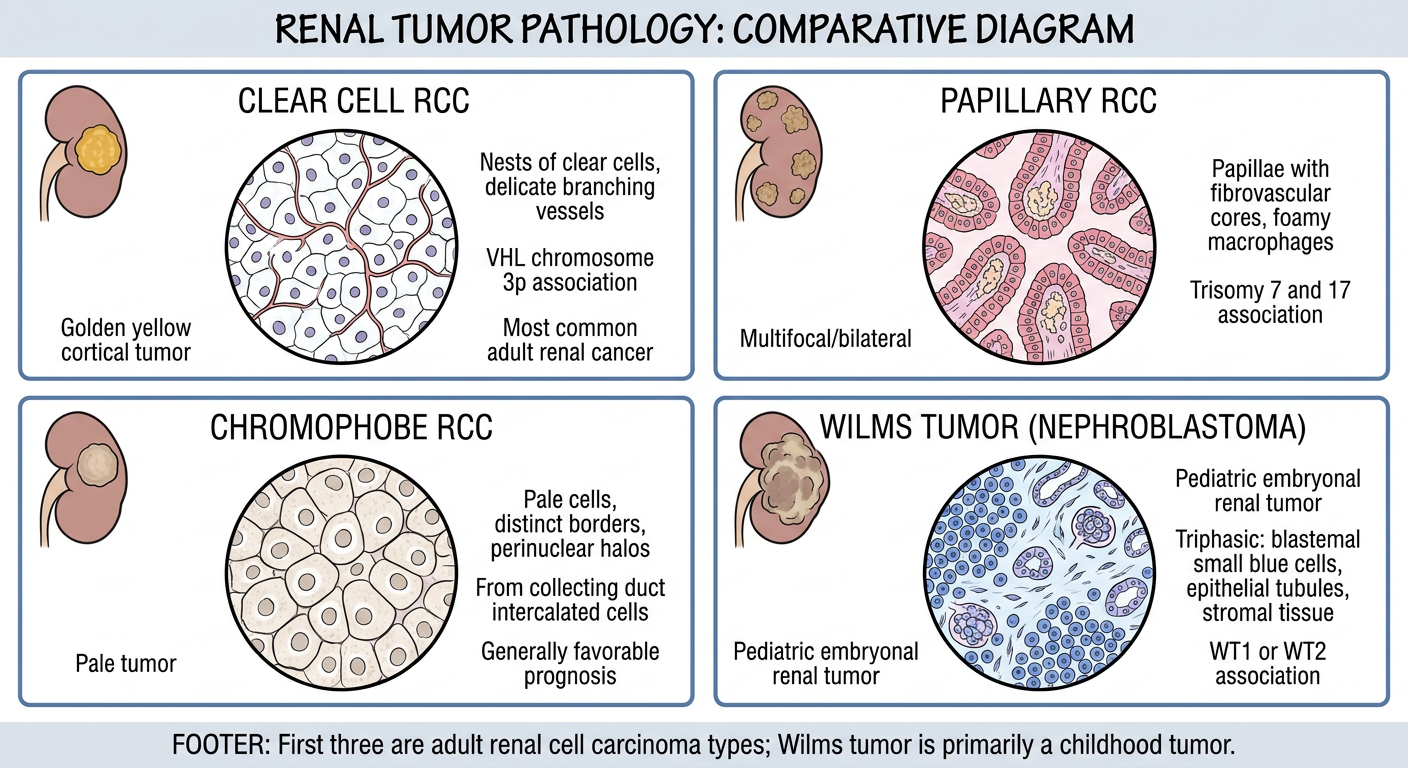
| Tumor | Key histology | Typical setting / association |
|---|---|---|
| Clear cell RCC | Nests of cells with clear cytoplasm and delicate vasculature | Most common adult RCC; VHL alteration, chromosome 3p |
| Papillary RCC | Papillae with fibrovascular cores, foamy macrophages | Often multifocal/bilateral; trisomy 7 and 17, MET in some cases |
| Chromophobe RCC | Pale cells, sharply outlined borders, perinuclear halos | Collecting-duct intercalated-cell origin; generally better prognosis |
| Wilms tumor | Triphasic: blastemal cells + epithelial tubules + stroma | Childhood embryonal renal tumor; WT1/WT2-related alterations |
- Clear cell = clear cytoplasm
- Papillary = papillae
- Chromophobe = cell border + halo
- Wilms = 3 components in a child
Simple diagram of gastric adenocarcinoma(interstitial and diffuse type) and crohns disease and ulcerative collitis
educational pathology diagram gastric adenocarcinoma intestinal diffuse type Crohn disease ulcerative colitis comparison
inflammatory bowel disease AND pathology
NCI stomach cancer intestinal diffuse gastric adenocarcinoma Crohn disease ulcerative colitis pathology
A single clean, simple medical educational comparison diagram in four panels: gastric adenocarcinoma intestinal type, gastric adenocarcinoma diffuse type, Crohn disease, and ulcerative colitis. Panel 1 intestinal-type gastric adenocarcinoma: show an irregular gland-forming tumor in the distal stomach/antrum, with a sketch of malignant glands and label 'glands, often H. pylori / intestinal metaplasia related'. Panel 2 diffuse-type gastric adenocarcinoma: show diffusely thick rigid linitis plastica stomach wall, scattered signet-ring cells, label 'loss of cohesion, CDH1 association'. Panel 3 Crohn disease: show mouth-to-anus involvement with terminal ileum commonly affected, skip lesions, deep linear ulcers/cobblestone surface, transmural inflammation, fistula and noncaseating granuloma icon. Panel 4 ulcerative colitis: show continuous inflammation beginning at rectum and extending proximally through colon, mucosal/submucosal disease, broad superficial ulcers, pseudopolyps, crypt abscess icon. Footer mnemonic: Crohn = skip + transmural; UC = continuous + mucosal. Correct spelling: intestinal type, ulcerative colitis. Keep text concise, large and legible, easy to memorize for pathology students.
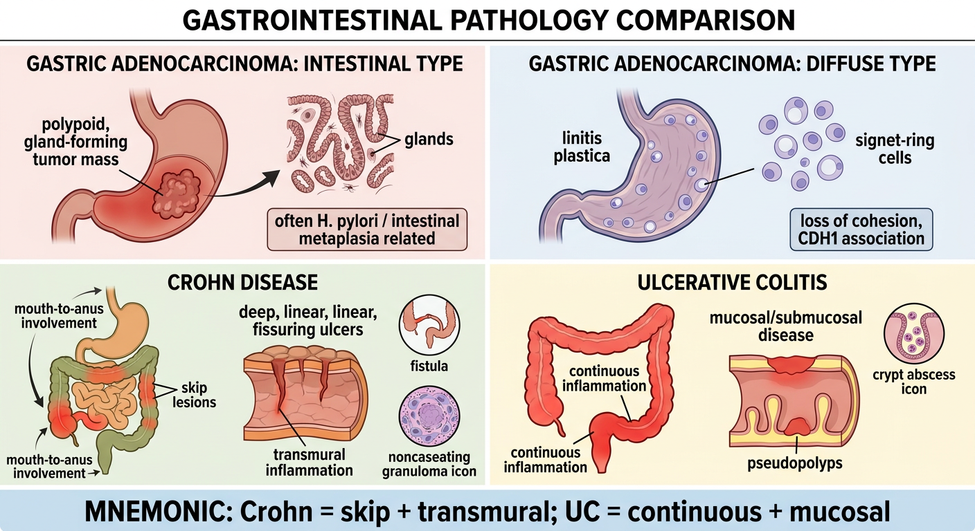
| Condition | High-yield visual clue |
|---|---|
| Gastric adenocarcinoma, intestinal type | Gland-forming malignant tumor, usually linked to chronic gastritis, intestinal metaplasia, and often H. pylori |
| Gastric adenocarcinoma, diffuse type | Poorly cohesive signet-ring cells infiltrate the wall, producing rigid linitis plastica; associated with loss of E-cadherin function, including CDH1 alterations |
| Crohn disease | Skip lesions, any site from mouth to anus, transmural inflammation, deep fissures, cobblestoning, fistulas; noncaseating granulomas may occur |
| Ulcerative colitis | Continuous inflammation starting in the rectum, limited to colon; mucosal and submucosal disease, superficial ulcers, pseudopolyps, crypt abscesses |
Crohn = Cobblestones, Cracks, full-thickness, skip lesions
UC = Uniform, Unbroken, limited to Colon
Simple diagram of ductal carcinoma insitu and lobar carcinoma insitu
simple histology diagram comparing ductal carcinoma in situ DCIS and lobular carcinoma in situ LCIS breast
ductal carcinoma in situ AND lobular carcinoma in situ
NCI ductal carcinoma in situ DCIS lobular carcinoma in situ LCIS breast overview
A clean, simple two-panel educational pathology diagram comparing ductal carcinoma in situ (DCIS) and lobular carcinoma in situ (LCIS) of the breast. Panel 1 DCIS: show a breast duct cross-section expanded and filled by cohesive atypical epithelial cells, an intact basement membrane and intact myoepithelial layer, plus a small mammogram icon showing branching microcalcifications. Label: 'Ductal carcinoma in situ (DCIS)', 'ducts filled by cohesive malignant cells', 'may show comedo necrosis and calcifications', 'confined by basement membrane', 'direct precursor to invasive ductal carcinoma'. Panel 2 LCIS: show multiple lobules/acini distended by small uniform discohesive cells, intact basement membrane, label loss of E-cadherin. Label: 'Lobular carcinoma in situ (LCIS)', 'lobules filled by discohesive cells', 'usually incidental, often multifocal and bilateral', 'confined by basement membrane', 'risk marker for invasive carcinoma in either breast'. Include shared footer: 'In situ = basement membrane intact: no stromal invasion.' Correct all spelling, uncluttered, highly legible, easy-to-memorize exam diagram.
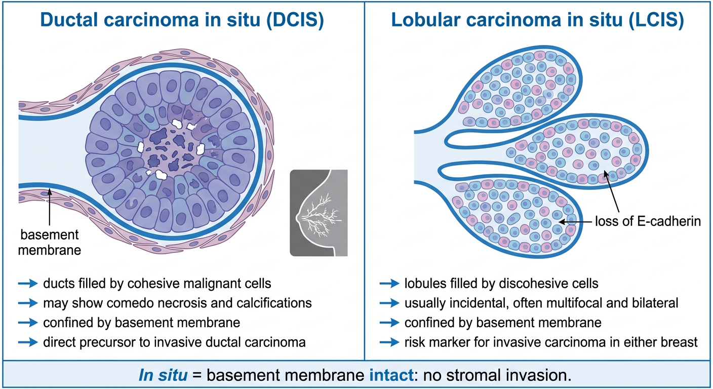
| Feature | Ductal carcinoma in situ (DCIS) | Lobular carcinoma in situ (LCIS) |
|---|---|---|
| Site | Milk ducts | Lobules/acini |
| Cell pattern | Cohesive atypical cells fill ducts | Small, uniform discohesive cells distend lobules |
| Basement membrane | Intact, so no stromal invasion | Intact, so no stromal invasion |
| Typical clue | Comedo necrosis and microcalcifications may be present | Loss of E-cadherin; often incidental, multifocal, and bilateral |
| Clinical meaning | A noninvasive lesion and direct precursor of invasive breast carcinoma | Primarily a marker of increased risk of future invasive carcinoma in either breast |
DCIS = Duct Distended, Calcification
LCIS = Lobules, Loss of E-cadherin
Simple diagram of solid, comedo, papillary, cribriform pattern in ductal carcinoma insitu
ductal carcinoma in situ solid comedo papillary cribriform patterns histology schematic
ductal carcinoma in situ AND histopathology
NCI DCIS comedo cribriform papillary solid architectural patterns pathology
A simple, accurate, clean four-panel pathology teaching diagram showing architectural patterns of ductal carcinoma in situ (DCIS) in breast ducts. All panels must show an intact basement membrane and myoepithelial layer, with no stromal invasion. Panel 1 SOLID DCIS: a round duct completely filled by a solid sheet of cohesive atypical epithelial cells, no lumen. Panel 2 COMEDO DCIS: a distended duct filled by high-grade atypical cells with a central plug of necrotic debris, label 'central comedo necrosis' and tiny calcifications. Panel 3 PAPILLARY DCIS: malignant epithelial cells growing on delicate fibrovascular cores projecting into the duct lumen, label 'fibrovascular cores'. Panel 4 CRIBRIFORM DCIS: a duct filled by uniform cohesive cells containing many evenly spaced round punched-out lumina, label 'sieve-like / Swiss-cheese pattern'. Include a concise footer: 'DCIS: malignant cells confined within ducts. Basement membrane intact.' Correct medical spelling, large readable labels, uncluttered, easy-to-memorize histology diagram for medical students.
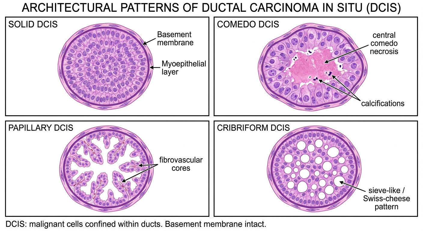
| Pattern | Histologic appearance | Memory clue |
|---|---|---|
| Solid | Duct lumen completely filled by cohesive neoplastic cells | Solid = no spaces |
| Comedo | High-grade cells with central luminal necrosis, often with calcification | Comedo = central dead core |
| Papillary | Tumor cells project into lumen around fibrovascular cores | Papillary = fronds with cores |
| Cribriform | Uniform cells form many rounded, punched-out lumina | Cribriform = Swiss cheese |