
Sdh in detail
Asking for Preferences
Which 'SDH' do you want covered in detail?
Reading File
Finding Sources
Searching PubMed
"subdural hematoma"[MeSH Terms] AND management
Searching the Web
subdural hematoma acute chronic management classification
Reading File
Reading File
Subdural Hematoma (SDH)
(Note: "SDH" can also stand for Succinate Dehydrogenase or Social Determinants of Health — this answer covers Subdural Hematoma, the most common clinical usage. Let me know if you meant one of the others.)
Definition and Pathophysiology
A subdural hematoma is a collection of blood that accumulates in the subdural space, between the arachnoid mater and the dura mater. The dura has two layers - an outer collagenous layer and an inner cellular layer containing fibroblasts. Bridging veins travel from the cerebral cortex, through the subarachnoid space and dura, to drain into the dural venous sinuses. Because the brain floats in CSF while the venous sinuses are fixed to the skull, sudden acceleration-deceleration of the head (trauma) can shear these bridging veins at their point of penetration through the dura. Extravasated blood then dissects between the two dural layers (Robbins & Kumar Pathologic Basis of Disease, p. 3945-3951).
Older adults and infants are especially susceptible:
- Elderly: cerebral atrophy stretches and elongates the bridging veins, increasing their vulnerability even to minor trauma.
- Infants: bridging veins are thin-walled and fragile (a key finding in abusive head trauma / shaken baby syndrome).
Less commonly, SDH can arise from arterial bleeding rather than venous, and unlike epidural hemorrhage (which is typically arterial and progresses rapidly), rising intracranial pressure (ICP) usually self-tamponades venous bleeding, which is why some subdural bleeds are self-limited (Adams and Victor's Principles of Neurology, p. ~2401).
Classification
SDH is classified by time since injury/onset, which correlates with CT density and drives management:
| Type | Timing | CT appearance |
|---|---|---|
| Acute | 0-3 days after injury | Hyperdense (bright) |
| Subacute | 3 days - 3 weeks | Isodense (may be missed - visible mainly by mass effect/midline shift) |
| Chronic | >3 weeks | Hypodense (dark) |
| Acute-on-chronic | Fresh bleed into a pre-existing chronic SDH | Mixed - hyperdense areas admixed with hypodense/isodense old collection |
On MRI, the acute clot is T2-hypointense (deoxyhemoglobin), becomes hyperintense over subsequent weeks as methemoglobin forms, then the chronic clot becomes hypointense again on T1 (Adams and Victor's Principles of Neurology, p. 2403).
Gross/microscopic evolution of a subdural clot (Robbins Pathologic Basis of Disease, p. 3954-3960):
- Clot lysis - about 1 week
- Fibroblast ingrowth from the dural surface into the hematoma - about 2 weeks
- Development of a granulation tissue membrane, which can rebleed and lead to chronic enlargement of the collection
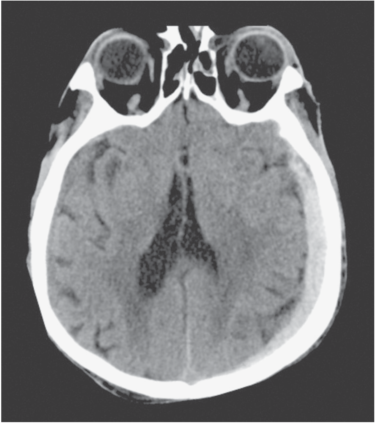
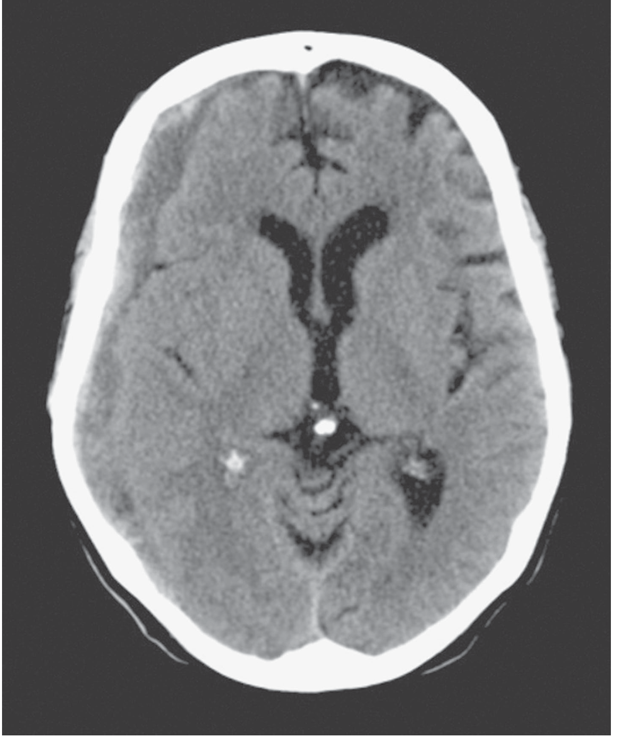
Note the classic radiologic distinction from epidural hematoma: SDH is crescent (concave) shaped and can cross suture lines (since it's not limited by dural attachments), while an epidural hematoma is biconvex (lentiform) and is limited by suture lines.
Clinical Features
Acute SDH:
- May be unilateral or bilateral
- Presentation ranges from a brief lucid interval followed by deterioration, to coma from the moment of injury, to minimal symptoms if the collection is small
- Often coexists with cerebral contusion, laceration, or epidural hemorrhage, making it hard to distinguish clinically from other traumatic lesions before imaging
- A large unilateral clot causes midline shift and lateral ventricle compression; bilateral clots may cause symmetric compression without shift
- Rarely, SDH occurs in the posterior fossa, causing headache, vomiting, pupillary inequality, dysphagia, and cranial nerve findings
Chronic SDH:
- Classic population: elderly patients, often with minimal or forgotten trauma, alcoholism, anticoagulant/antiplatelet use, coagulopathy, epilepsy, or arachnoid cysts as risk factors
- Presents insidiously over weeks with headache, cognitive decline/confusion, gait disturbance, focal weakness, or seizures - frequently mimics dementia or stroke in older patients
Management
Acute symptomatic SDH:
- Immediate neurosurgical consultation; transfer to a neurocritical care setting if not locally available
- Medical management: neuroprotective measures, ICP management if elevated ICP is present, correction of coagulopathy/reversal of anticoagulation
- Surgical evacuation (craniotomy) for hematomas causing significant mass effect, midline shift, or neurological decline
- Small, asymptomatic acute SDH may be managed conservatively with serial imaging
Chronic SDH:
- Minimally symptomatic/small collections may be managed conservatively (with consideration of atorvastatin, tranexamic acid, or corticosteroids in select cases per recent literature - though evidence is still evolving)
- Symptomatic or enlarging chronic SDH is typically treated with burr-hole drainage or craniotomy; recurrence risk depends on hematoma architecture on CT (homogeneous vs. laminar/mixed density collections carry higher recurrence risk, roughly 10-15%)
Recent Evidence (last 2 years - systematic reviews/meta-analyses)
A few points from current literature that supplement (and in some cases refine) the classic textbook approach:
- A 2025 systematic review found ongoing uncertainty regarding the comparative effectiveness of different invasive interventions (burr-hole vs. craniotomy vs. subdural evacuating port) for chronic SDH - no single approach is definitively superior across all patients (PMID: 40103530).
- A 2024 umbrella review/meta-analysis of chronic SDH surgical outcomes offers pooled guidance for neurosurgeons on optimizing surgical technique and reducing recurrence (PMID: 39179695).
- A 2024 systematic review/meta-analysis found relatively high failure rates of conservative management (including adjuncts like atorvastatin, tranexamic acid, corticosteroids, mannitol) in minimally symptomatic chronic SDH, suggesting caution before assuming medical therapy alone is sufficient even in "minimally symptomatic" patients (PMID: 39245136).
If your intended management plan for a specific patient relies heavily on "conservative treatment of minimally symptomatic chronic SDH," it's worth weighing this newer failure-rate data against older textbook assumptions of benign conservative management.
Let me know if you'd like me to go deeper into a specific angle - e.g., surgical technique (burr hole vs. craniotomy vs. middle meningeal artery embolization), pediatric/abusive head trauma SDH, or differentiating SDH from epidural hematoma in detail.
Management
Finding Sources
Finding Sources
Searching PubMed
"subdural hematoma" AND surgery
Searching the Web
subdural hematoma management guidelines surgical indications burr hole craniotomy middle meningeal artery embolization 2025
Reading File
Reading File
Management of Subdural Hematoma (SDH)
Management hinges on acuity (acute vs. chronic), size/thickness, midline shift, neurologic status (GCS, focal deficits), and trajectory of clinical/radiographic change. Below is the detailed approach.
1. Initial Resuscitation and Assessment (All Patients)
- Standard trauma/ABCs approach: airway protection if GCS <8, maintain normocapnia and oxygenation
- Avoid hypotension; track systolic and mean arterial pressure closely to maintain cerebral perfusion
- Non-contrast CT head immediately - assess for fracture, hemorrhage, midline shift, and effacement of basal/perimesencephalic cisterns; concurrently image the cervical spine to rule out associated fracture/dislocation
- Correct coagulopathy urgently - reverse anticoagulants/antiplatelets (e.g., vitamin K/PCC for warfarin, idarucizumab or PCC for DOACs, platelets if on antiplatelets and undergoing surgery) since many chronic SDH patients are older adults on these agents
- If a polytrauma patient has impaired GCS with a unilateral fixed, dilated pupil suggesting a mass lesion, an exploratory burr hole can rarely be performed concurrently with emergency laparotomy/thoracotomy, though CT-guided evacuation is preferred whenever feasible (Mulholland and Greenfield's Surgery, p. 803-804).
2. Acute SDH
Surgical evacuation (craniotomy) is indicated for any of:
- Hematoma thickness >1 cm
- Midline shift >5 mm
- GCS drop of 2 or more points from time of injury to hospitalization
- Neurologic decline in combination with a clot of at least 1 cm or any midline shift
(Schwartz's Principles of Surgery, p. 1518-1527; Mulholland and Greenfield's Surgery, p. 1440)
- Acute SDH and EDH are both treated with a formal craniotomy centered on the clot (not a simple burr hole)
- Depending on brain swelling, the surgeon may leave the bone flap off (decompressive craniectomy) rather than replacing it (craniotomy) to allow room for swelling
- A recent international trial reached equipoise between routine craniectomy vs. craniotomy for evacuated acute SDH - neither is clearly superior, and a 2024 meta-analysis of decompressive craniectomy vs. craniotomy for acute SDH similarly found no definitive advantage of one over the other (PMID: 38436794)
- ICP monitoring is often placed intraoperatively, especially if preoperative GCS <8
- Decompressive craniectomy for elevated ICP without a mass lesion is not well supported - the DECRA (2011) and RESCUEicp (2016) RCTs both showed ICP reduction with craniectomy but worse neurologic outcomes at 6 months, so this is reserved for refractory tier-3 ICP management at select centers
Nonoperative management of acute SDH is reasonable if none of the above thresholds are met:
- Requires frequent serial neurologic exams and serial CT scans until the clot stabilizes
- Many small acute SDHs stabilize and reabsorb, or evolve into chronic SDH over weeks
- Prognosis after acute SDH is worse than epidural hematoma, largely because SDH is typically associated with more severe underlying parenchymal brain injury (bridging vein tears occur with greater acceleration-deceleration forces)
3. Chronic SDH
Surgery is the recommended treatment when:
- Hematoma thickness >10 mm
- Midline shift >5 mm
- Pronounced neurologic symptoms are present
Surgery rapidly relieves intracranial hypertension and prevents herniation. Options include:
- Burr hole drainage - simplest, effective for most chronic SDH; a twist-drill craniotomy is a similar minimally invasive variant
- Craniotomy - more invasive; recent data suggest it may reduce recurrence compared to burr holes but at the cost of more short-term morbidity (Schwartz's Principles of Surgery, p. 1518-1527)
- A large meta-analysis (Almenawer et al., ~34,829 patients) found comparable outcomes across techniques with variable recurrence rates
Minimally symptomatic / small chronic SDH:
- Can often be managed conservatively with observation and serial imaging
- Adjunctive medical therapies studied: atorvastatin, tranexamic acid, corticosteroids (dexamethasone), mannitol - evidence for these remains mixed
- Caution: a 2024 meta-analysis found relatively high failure rates of conservative management even in minimally symptomatic chronic SDH (PMID: 39245136), so close follow-up is essential if this route is chosen
- A separate 2024 meta-analysis found surgery generally outperforms dexamethasone alone for chronic SDH, with steroids carrying their own complication risks (PMID: 38692115)
4. Middle Meningeal Artery Embolization (MMAE) - Newer Adjunct/Alternative
This is the most significant recent development in chronic SDH management:
- MMAE occludes the middle meningeal artery, which supplies the vascularized outer membrane that drives hematoma growth/recurrence
- A landmark 2024 NEJM RCT found that adjunctive MMAE after surgical evacuation significantly reduced the risk of recurrence or progression of chronic SDH compared to surgery alone (PMID: 39565988)
- Multiple 2025 trials (MAGIC-MT, EMBOLISE, STEM) are further defining its role - as a standalone treatment in select non-emergent patients, or combined with burr-hole/craniotomy
- A 2025 consensus guideline from the Society of Vascular and Interventional Neurology recommends surgery for CSDH with thickness >10 mm, marked symptoms, or midline shift >5 mm, and positions MMAE as either a standalone option in appropriately selected nonsurgical candidates or as an adjunct to reduce recurrence after surgical evacuation
- This is a rapidly evolving area - if you're managing a real patient, current institutional/interventional neuroradiology availability will determine whether MMAE is an option
5. Special Considerations
- Elderly patients on anticoagulation: reversal is a priority before any intervention; decisions about resuming anticoagulation post-SDH must balance thromboembolic risk against rebleeding risk, typically deferred several weeks
- Recurrence: chronic SDH recurrence risk is roughly 10-15% for homogeneous or laminar (mixed-density) collections on CT; this population is a good target for MMAE
- Posterior fossa SDH: rare, but can cause rapid deterioration (headache, vomiting, pupillary inequality, cranial nerve findings) and warrants urgent neurosurgical involvement given limited space for mass effect
Summary Table
| Scenario | First-line management |
|---|---|
| Acute SDH, thickness >1cm, shift >5mm, or GCS drop ≥2 | Emergent craniotomy ± craniectomy |
| Acute SDH, small, stable, no exam decline | Serial CT + neuro exams |
| Chronic SDH, thickness >10mm, shift >5mm, symptomatic | Burr hole drainage or craniotomy; consider adjunctive MMAE |
| Chronic SDH, minimally symptomatic/small | Observation ± medical therapy (limited evidence, higher failure rate than expected) |
| Recurrent/high-recurrence-risk chronic SDH | Consider MMAE as adjunct |
Evidence flag: The MMAE data (PMID: 39565988) and the 2025 SVIN consensus statement represent a genuine shift from purely surgical/observational paradigms in the older textbooks - worth incorporating if you're forming a current management plan rather than relying solely on textbook-era protocols.
Medical management
Finding Sources
Searching PubMed
"subdural hematoma" AND (dexamethasone OR "tranexamic acid" OR atorvastatin)
Searching the Web
medical management chronic subdural hematoma dexamethasone tranexamic acid atorvastatin conservative treatment anticoagulation reversal
Medical (Non-Surgical) Management of Subdural Hematoma
"Medical management" here refers to the non-operative, pharmacologic, and supportive measures used either as primary treatment (for small, asymptomatic, or high-surgical-risk cases) or as an adjunct to surgery (to reduce recurrence). No medical therapy reliably resolves an SDH on its own, and surgery remains the definitive treatment for symptomatic/large hematomas - but several agents have a real, evidence-based role.
1. Correction of Coagulopathy (Highest Priority)
This applies to both acute and chronic SDH and takes precedence over any other medical measure:
- Warfarin: vitamin K plus 4-factor prothrombin complex concentrate (PCC) for rapid reversal
- DOACs: idarucizumab (dabigatran) or andexanet alfa/PCC (factor Xa inhibitors)
- Antiplatelet agents: platelet transfusion considered if urgent surgery is planned, though evidence for benefit is mixed
- Correcting coagulopathy and thrombopathy is considered crucial to prevent hematoma expansion regardless of what other therapy is chosen (Rosen's Emergency Medicine, ROSEN's Q&A p.163-173)
2. Corticosteroids (Dexamethasone)
- Mechanism: chronic SDH growth is driven by an inflammatory, angiogenic outer membrane; corticosteroids are thought to reduce this inflammatory/neovascular process and membrane permeability
- The landmark Dex-CSDH trial (Miah et al., NEJM 2023, PMID 37314705) directly compared dexamethasone vs. surgery for chronic SDH and found dexamethasone resulted in worse functional outcomes and more adverse events than surgery, largely because it delayed definitive treatment and carried steroid-related complications (hyperglycemia, infection, psychiatric effects) without adequately shrinking hematomas in many patients
- A related 2023 prospective study (PMID 36029208) found the hematoma-size response to dexamethasone varied by CSDH subtype - some architectural subtypes responded better than others
- Current standing: dexamethasone is not recommended as a substitute for surgery in symptomatic chronic SDH. It may still be considered in select patients unfit for surgery, but should not be assumed to be a safe "lesser" alternative.
3. Tranexamic Acid (TXA)
- Mechanism: antifibrinolytic that may stabilize the fragile neomembrane vasculature and reduce microbleeding within the hematoma capsule
- Several 2024-2025 systematic reviews/meta-analyses have evaluated TXA:
- A 2025 systematic review (PMID 40577507) and a 2024 meta-analysis (PMID 38711565) both suggest TXA may reduce chronic SDH recurrence, particularly when used as an adjunct after evacuation
- A 2024 JNS meta-analysis (PMID 38579356) similarly supports a possible benefit but notes the evidence is still based on relatively small, heterogeneous studies
- Caution: most trials excluded patients on concurrent anticoagulation, so TXA's safety profile in that population is unclear, and thromboembolic risk (DVT/PE) is a theoretical concern with any antifibrinolytic
4. Statins (Atorvastatin)
- Mechanism: HMG-CoA reductase inhibitor with anti-inflammatory and pro-angiogenic-stabilizing effects thought to reduce capillary leakiness within the hematoma membrane
- Typical studied regimen: atorvastatin 20 mg once daily for approximately 8 weeks
- A 2024 updated systematic review/meta-analysis (PMID 38759787) found atorvastatin reduces hematoma volume and may lower recurrence, particularly when combined with surgery or dexamethasone
- An earlier trial (Xu et al. 2016) found conservative treatment plus atorvastatin significantly reduced hematoma volume in non-surgical patients, though it did not show significant added benefit in the surgical arm
- Ongoing RCTs (REACH, ATOCH2) are expected to better define its role
- A 2021 expert consensus statement identifies atorvastatin and dexamethasone as the two most-studied pharmacologic agents for chronic SDH, while cautioning on contraindications for each
5. Other Agents (Weaker Evidence)
- ACE inhibitors: proposed anti-angiogenic effect on the CSDH membrane; evidence is limited to small studies
- Mannitol: generally not useful for routine chronic SDH management; reserved for patients with impending herniation as a temporizing measure, not as primary therapy. Note also that rapid osmotic diuresis (mannitol) can itself rarely cause a subdural hematoma in older adults via rupture of fragile bridging veins, and rebound cerebral edema can follow its use (Morgan and Mikhail's Clinical Anesthesiology, p. 3993-3994) - so it is a double-edged tool.
- Platelet-activating factor receptor antagonists: mentioned in the literature but only studied in small trials
6. Supportive / Adjunctive Measures
- Seizure prophylaxis: antiepileptic drugs (AEDs) considered in patients at high risk for seizures, not routine for all SDH patients
- Serial imaging: essential whenever conservative management is chosen - CT is convenient and inexpensive; MRI may be more sensitive for monitoring evolution
- Mobilization: early mobilization may modestly increase recurrence risk but helps prevent immobilization-related complications (DVT, pneumonia, deconditioning) - a trade-off requiring individualized judgment
- Blood pressure and ICP management: maintain adequate cerebral perfusion, avoid hypotension, treat elevated ICP if present using standard neurocritical care measures
Who Is a Candidate for Medical Management Alone
Per the literature, conservative/medical therapy alone is generally reserved for:
- Asymptomatic or minimally symptomatic small hematomas
- Patients who decline surgery
- Patients at high operative risk
Even so, a 2024 meta-analysis found relatively high failure rates of conservative management in this "minimally symptomatic" population, so close follow-up with a low threshold to convert to surgical drainage if there's any clinical or radiographic progression is important.
Bottom Line
- No drug reliably makes an SDH disappear on its own; think of medical therapy as bridging, adjunctive, or alternative-for-poor-surgical-candidates, not as first-line for symptomatic hematomas.
- Dexamethasone monotherapy is now relatively discredited as a surgery substitute after the 2023 Dex-CSDH NEJM trial.
- Atorvastatin and TXA have the most encouraging (though still modest-quality) recent evidence, especially as adjuncts to reduce recurrence after evacuation or MMA embolization.
- Coagulopathy reversal is the one "medical management" step that is essentially universal and non-negotiable regardless of whether surgery follows.
How long manitol can be given
Finding Sources
Searching the Web
mannitol duration of therapy raised intracranial pressure how many days maximum dose monitoring
Duration of Mannitol Therapy
There is no fixed universal "day limit" for mannitol - duration is guided by clinical response, serum osmolality/osmolar gap, and renal function, not a set calendar endpoint. Here's the detailed picture:
Typical Dosing Regimen
- Initial (bolus) dose: 0.25-1 g/kg IV (up to 1.5-2 g/kg in some protocols), given over 20-30 minutes
- Maintenance dosing: 0.25-0.5 g/kg IV every 4-6 hours, repeated as needed based on ICP response
- Onset of ICP reduction: 15-30 minutes; peak effect: 20-60 minutes; duration of effect: 3-8 hours - this is why dosing is typically spaced every 4-6 hours (Katzung's Basic and Clinical Pharmacology, p. 2715)
What Actually Limits the Duration
1. Serum osmolality ceiling
- Mannitol is generally continued only while serum osmolality stays below 320 mOsm/kg. Once it exceeds this threshold, mannitol is typically withheld to protect renal function (Bradley and Daroff's Neurology in Clinical Practice, p. 215-218)
- A desirable target range during therapy is roughly 300-315 mOsm/L (Morgan and Mikhail's Clinical Anesthesiology, p. 3993-3994)
- Some protocols instead track the osmolar gap (measured minus calculated osmolality) rather than absolute osmolality, holding the next dose if the gap exceeds ~55 mOsm/L, since mannitol itself elevates measured osmolality independent of the physiologic effect you're trying to achieve
2. Renal function
- In patients with significant renal impairment, mannitol cannot be excreted normally and accumulates in the blood, drawing water out of cells and causing hyponatremia without truly lowering the physiologic osmotic gradient - a sign therapy needs to stop (Katzung's Basic and Clinical Pharmacology, p. 2733)
- Monitor renal function throughout; discontinue if renal, cardiac, or pulmonary status worsens, or if CNS toxicity develops
3. Clinical/ICP response
- If ICP monitoring is available, treatment is ICP-directed - mannitol is continued only as long as it's producing a measurable ICP reduction
- If ICP is not being directly monitored, clinicians rely on scheduled dosing plus serial neuro exams and imaging, tapering once the underlying process (edema, mass effect) stabilizes or once definitive treatment (e.g., surgical evacuation of a hematoma) has occurred
4. Practical duration in practice
- Most acute courses run for a few days (typically 24-72 hours) during the acute herniation-risk window, tapering as the patient stabilizes or after surgery addresses the underlying lesion
- Repeated/prolonged dosing beyond this increases risk of the complications below, so most protocols avoid open-ended, multi-day continuous use without re-evaluating the osmolality/renal picture at least before every dose
Monitoring While On Therapy
- Serum osmolality (or osmolar gap) and serum sodium checked before each repeat dose
- Renal function (creatinine/BUN)
- Fluid balance / hemodynamics (mannitol has mild vasodilating and diuretic effects and can transiently drop blood pressure)
- Watch for rebound cerebral edema and rebound ICP elevation, which can follow discontinuation or even prolonged use itself - a recognized risk cited across neuro-anesthesia references
Important Caveat Specific to Your SDH Context
As noted earlier, mannitol is not considered a mainstay medical therapy for chronic subdural hematoma itself - it's reserved as a temporizing measure only for impending herniation, not for shrinking the SDH long-term. Ironically, rapid osmotic diuresis with mannitol has itself been reported to cause subdural hematoma in older adults via rupture of stretched, fragile bridging veins - so it's a short-term rescue tool, not something used for extended courses in this population.
Bottom line: mannitol is dosed every 4-6 hours based on response, and continued only as long as serum osmolality stays under ~320 mOsm/kg, renal function is preserved, and it is still producing meaningful ICP/clinical benefit - typically a few days in the acute setting, reassessed before every single dose rather than following a fixed multi-day protocol.
This is a shared conversation. Sign in to Orris to start your own chat.