
Amyloidosis , types , symptoms , diagnosis and diagnostic test
amyloidosis Congo red staining apple green birefringence histology
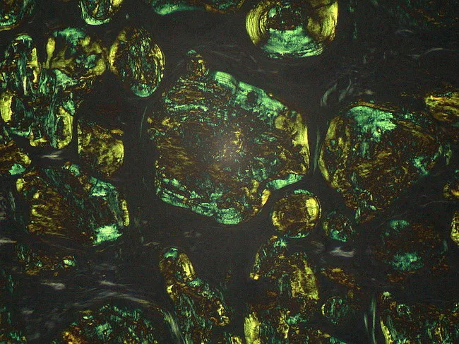
Light microscopy image of a lymph node biopsy stained with Congo Red and examined under polarized light. The tissue reveals extracellular, amorphous eosinophilic deposits distributed throughout nodal architecture consistent with amyloid. The deposits assume a dense, granular to plaque-like appearance on routine brightfield, and demonstrate distinctive apple-green birefringence when illuminated with polarized light, confirming the beta-pleated sheet rich configuration of amyloid fibrils. Congo Red positivity with birefringence distinguishes amyloid from other PAS-positive glycoprotein material and helps differentiate from non-amyloid nodal deposits. The beta-pleated sheet conformation accounts for the characteristic staining behavior and optical properties used in diagnosis. Clinically, identification of nodal amyloid supports systemic or localized amyloidosis and prompts workup for light-chain (AL) or serum amyloid A (AA) subtypes, including serum free light chains, immunofixation, and mass spectrometry-based typing. This image is relevant for educational illustrations of amyloid histology, pathophysiology, and diagnostic pathology workflows, including differential diagnosis with glycoprotein-rich PAS-positive deposits that are Congo Red negative. Potential clinical scenarios include workup of lymphadenopathy in patients with chronic inflammatory states, plasma cell dyscrasias, and lymphoproliferative disorders. High quality images like this support interdepartmental communication, teaching, and reproducible reporting of amyloid histology for junior pathologists and medical trainees in pathology education programs.
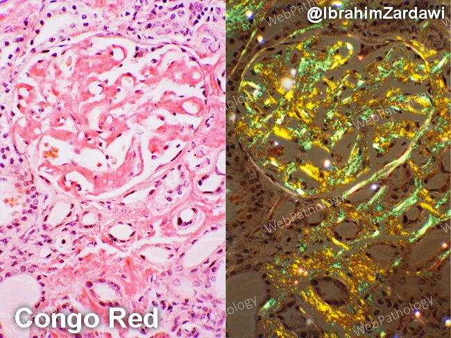
This renal cortical histology specimen demonstrates AL-type amyloidosis with kidney involvement in a patient with multiple myeloma. The left panel shows hematoxylin and eosin–stained tissue with amorphous, eosinophilic extracellular deposits within the glomerular mesangium and extending into the tubulointerstitium, causing mesangial expansion and variable basement membrane thickening. The right panel uses Congo Red staining to highlight the amyloid deposits, which appear salmon-pink on bright-field examination. Under polarized light, the same deposits display the diagnostic apple-green birefringence, a hallmark of Congo Red–positive amyloid. The deposits are diffuse, involving multiple glomeruli and extending into the tubulointerstitium, sometimes outlining capillary walls. In AL amyloidosis associated with plasma cell dyscrasia, the deposited light-chain–derived proteins disrupt normal renal architecture and contribute to proteinuria and progressive renal impairment. Differential considerations include AA (secondary) amyloidosis and other renal amyloidoses, but the combination of a known plasma cell disorder, Congo Red positivity, and apple-green birefringence strongly supports AL-type amyloidosis. This image serves as a valuable teaching reference for renal pathology, nephrology, and hematology-oncology, illustrating classic Congo Red histology, amyloid distribution patterns within glomeruli and interstitium, and the polarizing light signature. Correlates with proteinuria and renal dysfunction in many myeloma patients, guiding targeted therapy and monitoring of clonal plasma cell burden.
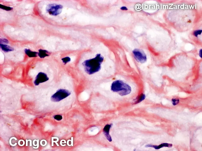
This histology image depicts human cardiac tissue (myocardium) stained with Congo Red and examined under bright-field microscopy. The image highlights extracellular, eosinophilic amyloid deposits that encircle and encroach upon individual cardiomyocytes (perimyocytic/interstitial deposition). The deposits appear as amorphous, orange to salmon-pink material on Congo Red, consistent with amyloid. When the same tissue is examined under polarized light, Congo Red–positive material exhibits characteristic apple-green birefringence, a pathognomonic feature of amyloidosis. The prevailing pattern is perimyocytic, with amyloid accumulating in the interstitium around myocytes and near small vessels, potentially contributing to myocardial stiffness and diastolic impairment. Histologic architecture shows preserved cellular morphology of myocytes with basophilic nuclei contrasted against the pink-red amyloid matrix. This image is diagnostic for cardiac amyloidosis on biopsy and would typically prompt typing (AL vs ATTR) by immunohistochemistry or mass spectrometry, as well as correlation with serum free light chains or transthyretin testing. Clinically, such infiltration predisposes to restrictive cardiomyopathy, diastolic dysfunction, arrhythmias, and heart failure symptoms. This slide underscores the utility of Congo Red staining in identifying myocardial amyloid deposits and the diagnostic value of polarized light birefringence for definitive confirmation. Useful for teaching, differential diagnosis, and research into infiltrative cardiomyopathies and treatment monitoring.
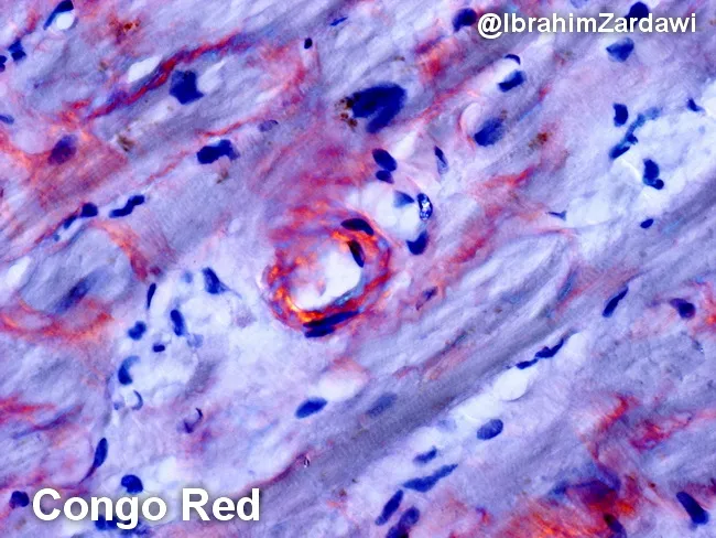
Imaging modality and technique: histopathology using light microscopy with Congo red staining of cardiac tissue, demonstrating characteristic amyloid deposition in the perimyocytic (pericellular) region of the myocardium. The deposited material appears orange to salmon pink on conventional hematoxylin-eosin–like chromogen, and exhibits classic apple-green birefringence under polarized light, confirming amyloid protein with a beta-pleated sheet configuration. The pattern is typically diffuse to pericellular around individual myocytes, producing a subtle, eosinophilic, amorphous coating that can obscure underlying cardiomyocyte detail. In this specimen, the amyloid is evident as an acellular, amorphous, hyaline-like material hugging muscle fibers, with irregular but cohesive contours and occasional nodular clusters. Congo red positivity combined with birefringence is diagnostic of systemic or cardiac amyloidosis when correlated with clinical data. Typing by immunohistochemistry, immunofluorescence, or mass spectrometry is often required to distinguish AL (light-chain) from ATTR (transthyretin) amyloidosis, which has implications for prognosis and treatment. Clinically, these deposits stiffen the myocardium, contributing to restrictive physiology and heart failure with preserved ejection fraction. The image illustrates foundational histopathologic criteria used in diagnosing cardiac amyloidosis and serves as educational material for pathology residents, cardiology fellows, and medical students studying protein misfolding diseases and Congo red histology. This image supports diagnostic confidence globally.
amyloidosis diagnosis treatment
amyloidosis macroglossia clinical signs systemic amyloid
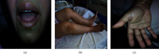
A three-part clinical photograph series demonstrating physical manifestations of systemic amyloidosis. Panel (a) presents an oral view showing macroglossia (enlarged tongue) with characteristic lateral scalloping, where tooth indentations are visible along the tongue's margins. Panel (b) shows the lower extremities of a patient in a supine position, highlighting a flexion contracture of the knee joint and associated muscle stiffness. Panel (c) displays a palmar view of the hand, illustrating sclerodactyly with thickened, stiffened skin on the palm and tapering of the fingers. These multi-system findings—encompassing macroglossia, joint contractures, and skin changes resembling scleroderma—are classic clinical signs of AL amyloidosis (light chain amyloidosis) and related plasma cell dyscrasias. The imagery serves as an educational resource for identifying the diverse musculoskeletal and soft tissue presentations of systemic amyloid deposition.
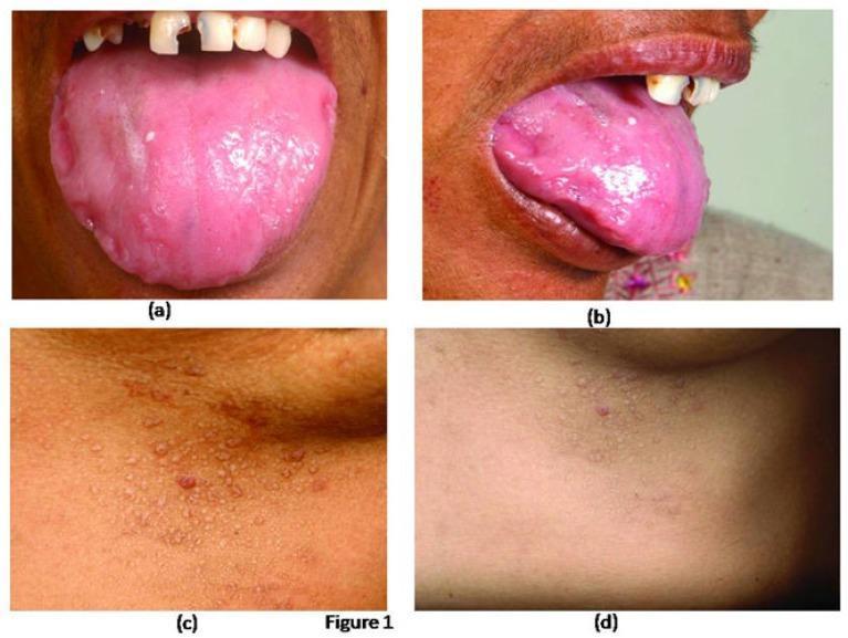
A composite of four clinical photographs demonstrating cutaneous and mucosal manifestations of systemic amyloidosis. Panels (a) and (b) show an intraoral and lateral view of the tongue, exhibiting significant macroglossia (enlargement). The tongue surface is irregular and bumpy, with prominent scalloping (crenation) along the lateral margins caused by indentations from the adjacent teeth. Panels (c) and (d) provide close-up views of the lower chest wall, displaying a dense distribution of small, discrete, waxy, papular lesions. These papules are flesh-colored to slightly erythematous and appear well-circumscribed. These findings represent classic diagnostic signs: macroglossia with dental indentations and characteristic papular amyloid skin deposits. The image is highly relevant for medical education concerning restrictive physiology disorders, plasma cell dyscrasias, and the physical examination of multisystemic protein deposition diseases.
Amyloidosis: Types, Symptoms, Diagnosis and Diagnostic Tests
What is Amyloidosis?
- Binds Congo red dye, producing apple-green birefringence under polarized light (pathognomonic)
- Stains with thioflavin T (produces fluorescence)
- Consists of randomly oriented, 8-12 nm nonbranching fibrils on electron microscopy
- Contains the glycoprotein serum amyloid P component (SAP) in all deposits
- Deposits cause organ dysfunction mainly by disrupting tissue architecture without triggering inflammation
Classification / Types of Amyloidosis
1. AL Amyloidosis (Primary Amyloidosis)
- Most common systemic type (~2,000-3,000 new US cases/year)
- Amyloid protein: immunoglobulin light chains (usually λ > κ) from a clonal plasma cell proliferation
- Occurs in 5-15% of patients with multiple myeloma; also occurs without overt myeloma (previously called "primary" amyloidosis) - but monoclonal immunoglobulins are detectable in serum or urine in virtually all cases
- Organs commonly involved: heart, GI tract, kidneys, peripheral nerves, liver, spleen, tongue, skin
2. AA Amyloidosis (Reactive/Secondary Amyloidosis)
- Amyloid protein: serum amyloid A (SAA), an acute-phase reactant
- Triggered by chronic inflammation - cytokines (IL-6, IL-1) stimulate hepatic SAA synthesis; long-standing inflammation sustains elevated SAA, eventually leading to fibril deposition
- Common causes:
- Rheumatoid arthritis (most common; ~3% of RA patients develop amyloidosis)
- Ankylosing spondylitis, IBD (Crohn's, ulcerative colitis)
- Familial Mediterranean Fever (FMF)
- Chronic infections (TB, bronchiectasis, osteomyelitis)
- Subcutaneous heroin injection (chronic skin infections)
- Tumors: renal cell carcinoma, Hodgkin lymphoma
- Organs commonly involved: kidneys (most prominent), liver, spleen, lymph nodes, adrenals, thyroid
3. ATTR Amyloidosis (Transthyretin Amyloidosis)
- Amyloid protein: transthyretin (TTR), a transport protein for thyroid hormone and retinol
- Two subtypes:
- ATTRwt (wild-type): Caused by age-related misfolding of normal TTR; predominantly in men >70 years old; affects the heart and connective tissues (e.g., carpal tunnel). The second most common systemic amyloidosis and increasing with aging populations
- ATTRv (variant/hereditary): Autosomal dominant gene mutation in TTR; specific mutations target different organs - some cause familial amyloid polyneuropathy (peripheral/autonomic nerves), others cause familial cardiomyopathy. A V122I variant in TTR is carried by ~4% of African-Americans and causes restrictive cardiomyopathy
4. Hereditary/Familial Amyloidoses (other proteins)
- Fibrinogen Aα-chain (Afib) - predominantly renal
- Apolipoprotein A-I or A-II (Apo A-I, A-II)
- Lysozyme (ALys)
- Gelsolin (Agel)
- LECT2 (ALECT2) - more prevalent in Latinos (southwest US), Punjabis, Native Americans, Egyptians
5. Localized Amyloidoses
- Deposits restricted to a single organ (lung, larynx, skin, bladder, tongue, eye region)
- Often consist of AL protein from local plasma cell infiltrates
6. Endocrine Amyloidosis
- Deposits in endocrine tumors: medullary thyroid carcinoma (amyloid is diagnostically helpful), pancreatic islet tumors, pheochromocytoma
- Also in type 2 diabetes mellitus - islet amyloid polypeptide (IAPP) deposits in pancreatic islets
7. Aβ-Amyloid (Alzheimer's Disease)
- β-amyloid (Aβ) protein, derived from amyloid precursor protein (APP), forms cerebral plaques and deposits in cerebral blood vessel walls
8. Aβ2m Amyloidosis (Dialysis-Related)
- β2-microglobulin (a component of MHC class I) deposits in joints and soft tissues of patients on long-term hemodialysis
| Type | Protein | Clinical Setting |
|---|---|---|
| AL | Ig light chain | Multiple myeloma, plasma cell dyscrasia |
| AA | Serum amyloid A | Chronic inflammation (RA, FMF, IBD) |
| ATTRwt | Wild-type transthyretin | Elderly men, cardiac |
| ATTRv | Mutant transthyretin | Familial neuropathy, cardiomyopathy |
| Aβ | β-amyloid | Alzheimer disease |
| Aβ2m | β2-microglobulin | Long-term hemodialysis |
Clinical Features / Symptoms
Kidney (Most Common and Serious)
- Proteinuria - often the first sign; progresses to nephrotic syndrome (massive proteinuria, edema, hypoalbuminemia)
- Progressive renal insufficiency and end-stage renal disease (ESRD)
- Kidneys may be normal or enlarged early, then shrunken from vascular ischemia
Heart
- Restrictive cardiomyopathy - myocardial stiffness from perimyocytic amyloid deposition
- Diastolic heart failure (HFpEF) - exertional dyspnea, peripheral edema
- Arrhythmias and conduction disturbances
- "Sparkling" or granular appearance on echocardiography
Peripheral Nervous System
- Peripheral neuropathy - progressive sensorimotor neuropathy, especially in ATTR amyloidosis
- Autonomic neuropathy - orthostatic hypotension, impotence, GI dysmotility
Gastrointestinal Tract
- Macroglossia (enlarged, firm tongue) - almost pathognomonic of AL amyloidosis
- GI dysmotility, malabsorption, diarrhea
- GI bleeding (mucosal deposits)
- Hepatomegaly (liver deposits can be massive)
Skin (particularly AL amyloidosis)
- Waxy papular lesions on the skin of the eyelids, face, neck, and trunk
- Easy bruising ("raccoon eyes" - periorbital purpura after minor trauma or Valsalva)
- Nail dystrophy
Musculoskeletal
- Carpal tunnel syndrome (common in ATTRwt)
- Joint stiffness, arthralgias (shoulder-pad sign - amyloid deposits around the shoulder)
- Muscle weakness (amyloid myopathy)
Other Organs
- Splenomegaly
- Adrenal insufficiency (adrenal involvement)
- Thyroid enlargement
- Recurrent fevers and serositis (FMF-associated AA amyloidosis)
Clinical Signs (Classic Physical Findings)
Diagnosis
Step 1 - Suspect Amyloidosis (Clinical)
- Unexplained nephrotic syndrome + cardiomyopathy + neuropathy
- Plasma cell dyscrasia / myeloma
- Chronic inflammatory disease with proteinuria
- Macroglossia + periorbital purpura
Step 2 - Screen for Monoclonal Protein (AL workup)
- Serum protein electrophoresis (SPEP) - detects M-spike
- Serum immunofixation electrophoresis (IFE) - more sensitive; identifies monoclonal immunoglobulin
- 24-hour urine IFE - detects Bence Jones protein (free light chains)
- Serum free light chain (FLC) assay - measures κ and λ light chains and κ/λ ratio; highly sensitive for AL
Step 3 - Tissue Biopsy (Definitive Diagnosis)
Staining Methods
| Test | Finding |
|---|---|
| Congo red stain | Pink/red color under ordinary light → apple-green birefringence under polarized light (pathognomonic) |
| Thioflavin T/S | Green fluorescence under UV light |
| H&E stain | Amorphous, eosinophilic extracellular deposits |
| Crystal violet / methyl violet | Metachromatic staining (violet→red) |

Biopsy Sites (in order of preference)
- Abdominal fat pad aspiration (subcutaneous fat) - safest, least invasive; sensitivity ~70-80%
- Rectal biopsy - high yield (~75%), relatively safe
- Bone marrow biopsy - often done alongside plasma cell workup
- Organ biopsy (kidney, liver, heart) - highest sensitivity but more invasive; needed if fat/rectal biopsy negative and clinical suspicion high
- Labial salivary gland biopsy
Step 4 - Typing the Amyloid (Critical - Determines Treatment)
Methods for Typing
| Test | Purpose |
|---|---|
| Immunohistochemistry (IHC) on biopsy | Stains for AL (κ/λ light chains), AA (SAA protein), TTR; limited sensitivity (~65-85%), false positives occur in up to 35% |
| Laser microdissection + mass spectrometry (LMD-MS) | Gold standard for typing; can identify all amyloid types including rare hereditary forms; available in specialized centers |
| Immunofluorescence (IF) | Used in renal biopsies; detects light chain restriction |
| Genetic testing | Identifies TTR mutations (ATTRv) and other hereditary mutations; recommended in all ATTR cases |
| Serum free light chain assay + SPEP/IFE | Supports AL diagnosis |
Step 5 - Additional Diagnostic Tests
| Test | What it Detects |
|---|---|
| Bone marrow biopsy + aspirate | Plasma cell percentage; clonality (AL) |
| Echocardiography | "Sparkling" granular myocardium, concentric hypertrophy, diastolic dysfunction - cardiac amyloid |
| Cardiac MRI | Diffuse subendocardial late gadolinium enhancement (LGE) - very characteristic pattern |
| Serum NT-proBNP / BNP | Cardiac biomarker; elevated in cardiac involvement; used for staging (Mayo clinic staging) |
| Serum cardiac troponin (cTnT/cTnI) | Also part of AL amyloidosis staging |
| 24-hour urine protein | Quantifies proteinuria |
| Serum creatinine / eGFR | Renal function assessment |
| SAA level | Elevated in AA amyloidosis; useful for monitoring treatment response |
| Radiolabeled SAP scintigraphy (123I-SAP) | Non-invasive whole-body scan to map amyloid burden and distribution; SAP component binds all amyloid deposits - available mainly in UK |
| 99mTc-DPD/PYP/HMDP scintigraphy (bone scan) | Highly specific for ATTR cardiac amyloidosis (grade 2-3 uptake in heart); non-invasive; can diagnose ATTRwt without biopsy when combined with absence of monoclonal protein |
| Serum transthyretin (TTR) level | Low in hereditary ATTR |
| Nerve conduction studies / EMG | Documents peripheral neuropathy pattern |
Diagnosis Summary Algorithm
Clinical suspicion
        ↓
Screen: SPEP, IFE, serum FLC, 24h urine IFE
        ↓
Tissue biopsy (fat pad → rectal → organ)
        ↓
Congo red stain → apple-green birefringence?
        ↓ YES
Typing: IHC or LMD-mass spectrometry
        ↓
AL?          AA?           ATTR?
→ FLC, BM   → SAA, CRP   → Genetic test
   biopsy     inflam.        echocardiogram
              workup         DPD scan
Prognosis (Brief)
- AL amyloidosis: Median survival ~2 years without treatment; poorer if myeloma-associated. Newer plasma cell-targeted therapies (bortezomib, daratumumab-based regimens) have significantly improved outcomes
- AA amyloidosis: Depends on control of the underlying inflammatory disease; renal function can stabilize or improve with anti-inflammatory therapy
- ATTRv amyloidosis: Liver transplant (removes source of mutant TTR); tafamidis (TTR stabilizer) and RNA-interference agents (patisiran, inotersen) have shown efficacy in randomized trials
- ATTRwt amyloidosis: Tafamidis approved and significantly reduces mortality in cardiac ATTR
Recent Evidence: A 2025 systematic review [PMID: 39819351] on ATTR amyloidosis epidemiology confirms the increasing prevalence of ATTRwt with population aging, and highlights that the disease remains under-diagnosed. No recent meta-analyses contradict the established diagnostic approach above.