Radius bone easily explained with attachments notes
Reading File
Finding Sources
Searching Images
radius bone anatomy attachments labeled diagram
Finding Sources
Finding Sources
Reading File
Reading File
Reading File
Finding Sources
Loading Image
Loading Image
Excellent — two superb textbook diagrams retrieved. Now I have everything needed for comprehensive notes. Let me compile the full answer.
The Radius — Easy Explained Notes with Attachments
Source: Imaging Anatomy Text and Atlas Vol. 3 — Bones, Joints, Vessels and Nerves
Overview
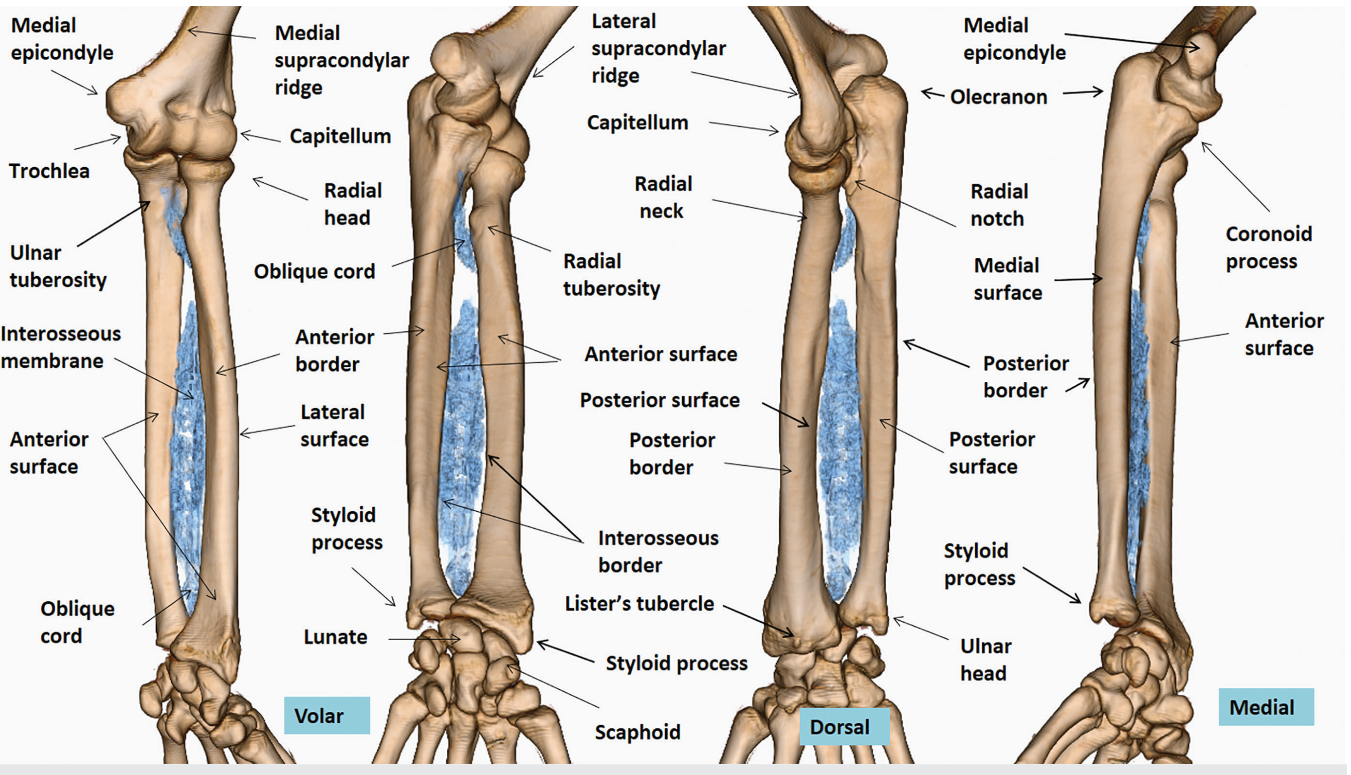
The radius is a long, paired, asymmetrical bone forming the lateral framework of the forearm. It is wider distally (opposite to the ulna, which is wider proximally). It rotates around the ulna during pronation/supination.
Bony Landmarks (Proximal → Distal)
| Landmark | Description |
|---|---|
| Radial head | Broad, rounded cylindrical structure at the top; has a central concavity — the fovea articularis radii — which articulates with the capitulum of humerus |
| Radial neck | Narrow constriction just below the radial head |
| Radial tuberosity | Ovoid bony protuberance on the anteromedial surface just below the neck; posterior rough surface = tendon insertion site |
| Radial shaft | Triangular in cross-section — 3 surfaces + 3 borders |
| Styloid process | Palpable lateral bony projection at the distal end |
| Lister's tubercle | Palpable dorsal bony projection at the distal end |
| Sigmoid notch | Narrow concave surface on the medial side of distal radius — articulates with the ulnar head |
Surfaces of the Radial Shaft
| Surface | Key Feature |
|---|---|
| Anterior (volar) | Smooth and concave proximally; broadens distally |
| Posterior (dorsal) | Flat; slight proximal and distal convexities |
| Lateral | Convex; upper segment = rounded and convex |
Borders of the Radial Shaft
| Border | Description |
|---|---|
| Anterior border | From inferior margin of radial tuberosity → anterior aspect of radial styloid |
| Posterior border | Obtuse/indistinct; from posterior radial neck → posterior base of styloid |
| Interosseous border | Sharp in mid-portion; gives attachment to the interosseous membrane |
Muscle Attachments (The Most Important Part!)
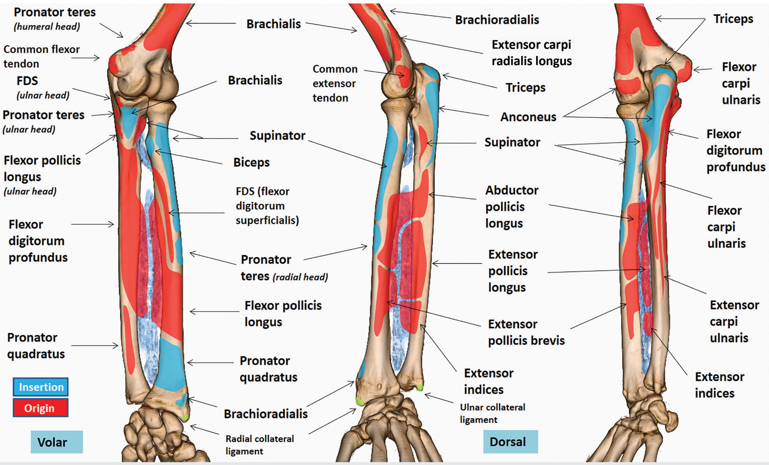
INSERTIONS onto the Radius (muscles that pull on/into the radius)
| Muscle | Where on Radius | Action |
|---|---|---|
| Biceps brachii | Posterior/ulnar margin of radial tuberosity | Flexion + supination |
| Supinator | Upper lateral surface + proximal posterior surface | Supination |
| Pronator teres | Mid-lateral surface (radial head = insertion) | Pronation |
| Pronator quadratus | Distal anterior surface | Pronation |
| Brachioradialis | Base of styloid process (distal radius) | Flexion of elbow |
ORIGINS from the Radius (muscles that arise from the radius)
| Muscle | Where on Radius | Runs to |
|---|---|---|
| Flexor digitorum superficialis (FDS) | Superior half of anterior border and anterior surface | Middle phalanges of fingers 2–5 |
| Flexor pollicis longus (FPL) | Superior half of anterior surface | Distal phalanx of thumb |
| Abductor pollicis longus (APL) | Middle third of posterior surface | Base of 1st metacarpal |
| Extensor pollicis brevis (EPB) | Middle third of posterior surface (just distal to APL) | Proximal phalanx of thumb |
| Extensor indices | Distal posterior surface | Extensor expansion of index finger |
Ligament Attachments at the Distal Radius
| Ligament | Attachment |
|---|---|
| Brachioradialis tendon | Base of radial styloid |
| Radial collateral ligament | Apex of radial styloid |
| Palmar radiocarpal ligament | Volar surface of distal radius |
| Dorsal radiocarpal ligament | Dorsal surface of distal radius |
Articulations of the Radius
| Joint | Partner Bone | Type |
|---|---|---|
| Humeroradial joint (proximal) | Capitulum of humerus | Ball & socket (pivot) |
| Proximal radioulnar joint | Radial notch of ulna | Pivot |
| Distal radioulnar joint | Sigmoid notch ↔ ulnar head | Pivot |
| Radiocarpal joint | Scaphoid + Lunate | Condyloid/ellipsoid |
Interosseous Membrane (Quick Note)
- Arises from the interosseous border of the radial shaft (distal ¾)
- Separates the anterior (flexor) and posterior (extensor) compartments of the forearm
- Proximal fibers form the oblique cord — runs from the radial tubercle to the proximal ulna
- Perforated by the anterior and posterior interosseous vessels
Memory Tricks
| Mnemonic | What to Remember |
|---|---|
| "Biceps Twists the Bottle" | Biceps inserts at radial tuberosity → supination + flexion |
| "Supinator wraps the top" | Supinator covers the upper radius like a wrap |
| "Pronator Teres crosses the mid" | PT inserts at the mid-lateral radius |
| "PQ at the bottom" | Pronator quadratus = distal anterior radius |
| Radius = wider DISTALLY | Opposite to ulna (ulna = wider proximally) |
Clinical Pearl
The radial head is the most commonly fractured part of the radius in adults after a fall on an outstretched hand (FOOSH). It is identified on X-ray by the fracture line or displacement of the posterior fat pad sign.
Notes compiled from: Imaging Anatomy Text and Atlas Vol. 3 — Bones, Joints, Vessels and Nerves; General Anatomy and Musculoskeletal System (THIEME Atlas)
Ulna detailed notes esaily explained with all attachments joints ligaments nerves
Finding Sources
Finding Sources
Reading File
Finding Sources
Reading File
Reading File
Reading File
Finding Sources
Finding Sources
Loading Image
Loading Image
Finding Sources
Reading File
Finding Sources
Reading File
Reading File
I now have comprehensive information from multiple authoritative textbooks. Let me compile the full detailed notes.
The Ulna — Detailed Easy Notes with All Attachments, Joints, Ligaments & Nerves
Sources: Imaging Anatomy Atlas Vol. 3 | Gray's Anatomy for Students | Miller's Review of Orthopaedics | Bradley & Daroff's Neurology
Quick Overview
The ulna is a long bone on the medial (little finger) side of the forearm. It is wider proximally and narrows distally — the opposite of the radius. The ulna is the primary load-bearing bone of the forearm and acts as the fixed axis around which the radius rotates during pronation/supination.
Bony Landmarks — Proximal to Distal
| Landmark | What it is | Key Point |
|---|---|---|
| Olecranon | Large posterior hook at the proximal end | The "tip of the elbow" you can feel; insertion of triceps brachii |
| Coronoid process | Triangular anterior projection | Brachialis inserts here; forms the lower lip of the trochlear notch |
| Trochlear (semilunar) notch | Large C-shaped concavity between olecranon + coronoid | Articulates with the trochlea of humerus |
| Radial notch | Narrow oval hollow on the lateral side of the coronoid process | Articulates with the head of the radius |
| Tuberosity of ulna | Roughened apex of the coronoid process | Main insertion of brachialis |
| Sublime tubercle | Anteromedial facet of coronoid | Insertion of the anterior bundle of the medial (ulnar) collateral ligament |
| Supinator crest | Posterior margin below the radial notch | Gives origin to the supinator muscle |
| Ulnar head | Distal lateral rounded knob | Articulates with sigmoid notch of radius + articular disc (TFCC) |
| Ulnar styloid process | Posteromedial bony projection at distal end | Attachment of ulnar collateral ligament |
Shaft — Surfaces & Borders
The ulnar shaft is triangular in cross-section proximally and mid-portion; cylindrical distally.
Three Surfaces
| Surface | Location | Muscle Attachments |
|---|---|---|
| Anterior surface | Between anterior & interosseous borders | Upper ¾ → Flexor digitorum profundus (FDP) origin; lower ¼ → Pronator quadratus origin |
| Posterior surface | Between interosseous & posterior borders | Proximal portion → Anconeus insertion + Supinator origin; lateral subdivision → Extensor pollicis longus, Abductor pollicis longus origins |
| Medial surface | Between anterior & posterior borders | Upper ¾ → one head of Flexor digitorum profundus |
Three Borders
| Border | Description | Attachment |
|---|---|---|
| Anterior border | Medial coronoid process → base of styloid | Upper ¾ → FDP origin; lower ¼ → Pronator quadratus origin |
| Posterior border | From confluence of olecranon borders → base of styloid | Aponeurosis shared by FCU, ECU, FDP on upper ¾ |
| Interosseous border | Lateral crest from radial notch | Origin of supinator (proximally); interosseous membrane attaches along its length |
Muscle Attachments — Complete Table
INSERTIONS onto the Ulna (muscles that pull into the ulna)
| Muscle | Where on Ulna | Nerve | Action |
|---|---|---|---|
| Triceps brachii | Rough superior surface of olecranon | Radial nerve (C7, C8) | Elbow extension |
| Anconeus | Lateral surface of olecranon | Radial nerve | Assists elbow extension |
| Brachialis | Tuberosity of ulna + anterior-inferior coronoid | Musculocutaneous (+ small radial branch) | Elbow flexion |
| Pronator teres (ulnar head) | Medial surface of coronoid process | Median nerve | Pronation |
| Flexor carpi ulnaris (FCU) | Medial surface of olecranon | Ulnar nerve | Wrist flexion + adduction |
ORIGINS from the Ulna (muscles arising from ulna)
| Muscle | Where on Ulna | Nerve | Action |
|---|---|---|---|
| Flexor digitorum profundus (FDP) | Anterior surface + medial surface (upper ¾) + anterior border | Median (radial 2 digits) + Ulnar (ulnar 2 digits) | Finger flexion (DIP) |
| Flexor digitorum superficialis (FDS) — ulnar head | Medial surface of coronoid | Median nerve | Finger flexion (PIP) |
| Flexor carpi ulnaris (FCU) | Medial olecranon + posterior border (aponeurosis) | Ulnar nerve | Wrist flexion |
| Extensor carpi ulnaris (ECU) | Posterior border aponeurosis (upper ¾) | Radial nerve (posterior interosseous) | Wrist extension + adduction |
| Supinator | Supinator crest + interosseous border (proximal) | Radial nerve (posterior interosseous, C6) | Supination |
| Anconeus | Originates from lateral epicondyle → inserts on lateral olecranon | Radial nerve | Elbow extension |
| Abductor pollicis longus (APL) | Posterior surface (lateral subdivision, mid-shaft) | Radial nerve (posterior interosseous) | Thumb abduction |
| Extensor pollicis longus (EPL) | Posterior surface (lateral subdivision, mid-shaft) | Radial nerve (posterior interosseous) | Thumb extension |
| Pronator quadratus (PQ) | Distal anterior border + lower ¼ anterior surface | Median nerve (anterior interosseous) | Pronation |
Memory tip: "The ulna feeds BOTH the deep flexors AND the deep extensors." FDP on the front, APL/EPL on the back.
Joints of the Ulna
| Joint | Ulnar Surface | Partner | Type | Movement |
|---|---|---|---|---|
| Humeroulnar joint | Trochlear (semilunar) notch | Trochlea of humerus | Hinge (ginglymoid) | Flexion / Extension |
| Proximal radioulnar joint | Radial notch | Head of radius | Pivot (trochoid) | Pronation / Supination |
| Distal radioulnar joint | Sigmoid notch of radius ← note the ulnar HEAD goes INTO this | Sigmoid notch of radius | Pivot | Pronation / Supination |
| Wrist (ulnocarpal) | Via triangular fibrocartilage complex (TFCC) | Separated from carpus by articular disc | Not direct — via TFCC | Load transmission |
Key fact: The ulna does not directly articulate with the carpal bones. The TFCC (articular disc) separates the ulnar head from the carpus. Normally only 18% of wrist load passes through the ulnocarpal joint.
Ligaments of the Ulna
Medial (Ulnar) Collateral Ligament Complex — 3 bundles
| Bundle | Origin | Insertion on Ulna | Function |
|---|---|---|---|
| Anterior bundle (most important!) | Anteroinferior medial epicondyle | Sublime tubercle (18 mm distal to coronoid tip) | Primary restraint to valgus stress; most important static stabilizer |
| Posterior bundle (Bardinet's ligament) | Posterior medial epicondyle | Medial surface of olecranon | Secondary stability; forms floor of cubital tunnel |
| Transverse bundle (Cooper's ligament) | Proximal medial olecranon | Just distal to coronoid | Ulna-to-ulna; least important clinically |
Lateral (Radial) Collateral Ligament Complex
| Ligament | Attachment to Ulna | Function |
|---|---|---|
| Lateral ulnar collateral ligament (LUCL) | Supinator crest of ulna | Primary restraint against posterolateral rotatory instability; reconstructed in "Tommy John" surgery |
| Annular ligament | Anterior + posterior margins of radial notch | Encircles radial head; stabilizes proximal radioulnar joint |
Other Ligaments
| Ligament | Where | Function |
|---|---|---|
| Interosseous membrane | Attaches along interosseous border of ulna | Binds radius and ulna; transmits force; separates flexor/extensor compartments |
| Oblique cord | From radial tuberosity to proximal ulna | Reinforces interosseous membrane proximally |
| Ulnar collateral ligament of wrist | Styloid process | Medial support for wrist joint |
| TFCC (articular disc) | Distal ulna — styloid base | Separates ulna from carpus; cushions |
Nerves Related to the Ulna
Ulnar Nerve — The Most Important Nerve
| Feature | Detail |
|---|---|
| Origin | Medial cord of brachial plexus — C8, T1 roots |
| Course at elbow | Passes through the ulnar groove between the medial epicondyle and the olecranon |
| Cubital tunnel | Enters the forearm under the humeral-ulnar aponeurosis (two heads of FCU) — this is where it gets compressed in cubital tunnel syndrome |
| Forearm branches | Motor to FCU + medial two digits of FDP |
| Dorsal cutaneous branch | Exits 5–8 cm proximal to wrist → dorsal medial hand + little & ring fingers |
| Palmar cutaneous branch | At ulnar styloid level → proximal medial palm |
| Wrist | Passes through Guyon's canal (between pisiform + hook of hamate) |
| Terminal branches | Deep palmar (motor): hypothenar muscles, interossei, 3rd & 4th lumbricals, adductor pollicis; Superficial (sensory): little finger + medial ring finger |
Other Nerves at Key Ulnar Landmarks
| Nerve | Relationship to Ulna | Clinical Note |
|---|---|---|
| Posterior interosseous nerve (deep radial) | Passes around radial neck → winds through supinator (near supinator crest of ulna) | Can be compressed in radial tunnel syndrome |
| Anterior interosseous nerve (branch of median) | Runs on anterior interosseous membrane near ulna | Damage → loss of FDP to index + FPL ("OK sign" weakness) |
Ulnar Variance — Clinical Concept
| Term | Meaning | Clinical Link |
|---|---|---|
| Ulnar neutral | Ulnar head cortex = level with lunate fossa | Normal; 82% load through radiocarpal, 18% ulnocarpal |
| Negative ulnar variance (>2.5 mm shorter) | Ulnar head is cone-shaped and shorter | Associated with avascular necrosis of lunate (Kienböck's) |
| Positive ulnar variance (>2.5 mm longer) | More stress on ulnocarpal joint (+42% load) | Predisposes to early degeneration + ulnar impaction syndrome |
Summary Memory Table
| Structure | Key Muscle(s) | Memory Cue |
|---|---|---|
| Olecranon (superior surface) | Triceps insertion | "Tri = 3 heads, all go to the olecranon" |
| Tuberosity of ulna / Coronoid | Brachialis insertion | "Brachialis ALWAYS inserts on ulna" |
| Medial olecranon + posterior border | FCU origin | "FCU follows the ulna's medial/posterior edge" |
| Anterior surface (upper ¾) | FDP origin | "Deep flexors = deep anterior ulna" |
| Posterior surface (lateral) | APL, EPL, Supinator | "Back of ulna = thumb movers" |
| Sublime tubercle | Anterior bundle MCL | "Sublime = sublime stabilizer against valgus" |
| Ulnar groove | Ulnar nerve | "Between medial epicondyle and olecranon = the nerve's groove" |
Notes compiled from: Imaging Anatomy Text and Atlas Vol. 3 | Gray's Anatomy for Students | Miller's Review of Orthopaedics 9th Ed. | Bradley & Daroff's Neurology in Clinical Practice
Upper end shaft lower end separately
Finding Sources
Finding Sources
Reading File
Finding Sources
Reading File
Loading Image
Loading Image
Loading Image
All the material needed is in context. Here are the structured notes divided into the three regions as requested:
ULNA — Upper End | Shaft | Lower End
Sources: Gray's Anatomy for Students | Imaging Anatomy Atlas Vol. 3 | Miller's Review of Orthopaedics
━━━━━━━━━━━━━━━━━━━━━━━━━━━━━━
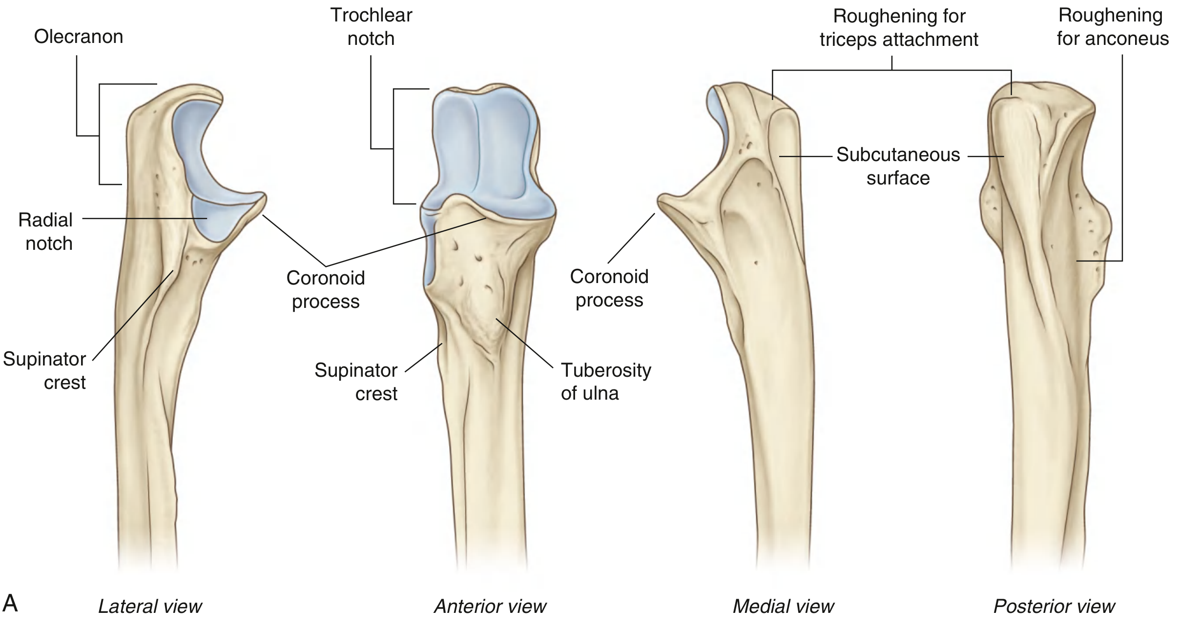
1. UPPER END (Proximal End)
━━━━━━━━━━━━━━━━━━━━━━━━━━━━━━
The proximal end is the largest, widest part of the ulna. It forms the elbow joint.
Parts of the Upper End
| Part | Description |
|---|---|
| Olecranon | Large posterior hook; "tip of the elbow" you palpate; projects proximally from the ulna |
| Coronoid process | Triangular beak projecting anteriorly; forms the lower lip of the trochlear notch |
| Trochlear (semilunar) notch | Large C-shaped cavity between olecranon + coronoid; articulates with trochlea of humerus |
| Radial notch | Narrow oval depression on the lateral surface of the coronoid; articulates with the head of radius |
| Tuberosity of ulna | Rough apex of coronoid's anterior surface; biggest roughening |
| Sublime tubercle | Anteromedial facet of coronoid; small but crucial |
| Supinator crest | Bony ridge posterior to radial notch; origin of supinator |
Surfaces of the Olecranon
| Surface | Features / Attachments |
|---|---|
| Superior surface (rough, quadrilateral) | Triceps brachii — INSERTION ✅ |
| Anterolateral surface | Articular — forms upper half of trochlear notch |
| Posterior surface | Smooth, triangular — covered by olecranon bursa; subcutaneous |
| Medial surface | Flexor carpi ulnaris (FCU) — ORIGIN ✅; attachment of posterior + oblique bands of ulnar collateral ligament |
| Lateral surface | Anconeus — INSERTION ✅ |
Surfaces of the Coronoid Process
| Surface | Features / Attachments |
|---|---|
| Superolateral surface | Articular — forms lower half of trochlear notch |
| Lateral surface | Radial notch (articulates with radial head) + supinator crest just below |
| Anteromedial facet | Sublime tubercle → insertion of anterior bundle of MCL ✅ |
| Anterior surface (triangular) | Brachialis inserts at tuberosity of ulna (apex) ✅; also FDS (anterior margin), FDP, pronator teres origins |
| Medial surface | FDS, FDP, Pronator teres — ORIGINS ✅ |
Joint at Upper End
| Joint | Articular Surface | Partner | Type |
|---|---|---|---|
| Humeroulnar joint | Trochlear notch | Trochlea of humerus | Hinge (primary) |
| Proximal radioulnar joint | Radial notch | Head of radius | Pivot |
Ligaments at Upper End
| Ligament | Attachment | Function |
|---|---|---|
| Medial (Ulnar) Collateral Ligament — anterior bundle | Medial epicondyle → Sublime tubercle | Primary restraint to valgus stress |
| Medial (Ulnar) Collateral Ligament — posterior bundle | Posterior medial epicondyle → medial olecranon | Secondary valgus restraint; forms floor of cubital tunnel |
| Medial (Ulnar) Collateral Ligament — transverse bundle | Proximal olecranon → just distal to coronoid | Least important; ulna-to-ulna only |
| Annular ligament | Anterior + posterior margins of radial notch | Encircles radial head; stabilizes proximal radioulnar joint |
| Lateral ulnar collateral ligament (LUCL) | Lateral epicondyle → supinator crest | Prevents posterolateral rotatory instability |
Nerve at Upper End — KEY ⚠️
The ulnar nerve runs in the ulnar groove — the channel between the medial epicondyle and the olecranon. This is where it is most vulnerable to compression → cubital tunnel syndrome.
━━━━━━━━━━━━━━━━━━━━━━━━━━━━━━
2. SHAFT (Body)
━━━━━━━━━━━━━━━━━━━━━━━━━━━━━━
The shaft is triangular in cross-section proximally and mid-portion, becoming cylindrical distally. It is broad above and narrows toward the distal end.
Three Borders of the Shaft
| Border | Character | Attachment |
|---|---|---|
| Anterior border | Smooth, rounded | Upper ¾ → FDP origin; lower ¼ → Pronator quadratus origin |
| Posterior border | Sharp and palpable along its entire length | Upper ¾ → aponeurosis shared by FCU, ECU, FDP |
| Interosseous border | Sharp (especially mid-shaft) | Interosseous membrane attaches here; proximally also gives supinator origin |
Three Surfaces of the Shaft
| Surface | Location | Attachments |
|---|---|---|
| Anterior surface | Between anterior + interosseous borders; concave, narrows distally | Upper ¾ → FDP ORIGIN ✅; lower ¼ → oblique ridge for Pronator quadratus ORIGIN ✅; nutrient canal proximal to midpoint |
| Posterior surface | Between interosseous + posterior borders; divided by a vertical ridge | Proximal to ridge → Anconeus INSERTION + Supinator ORIGIN; Lateral subdivision below ridge → Supinator, APL, EPL ORIGINS |
| Medial surface | Between anterior + posterior borders; convex and smooth | Upper ¾ → one origin of FDP |
Muscle Attachments of the Shaft — Summary Table
| Muscle | Location on Shaft | Origin / Insertion | Nerve |
|---|---|---|---|
| Flexor digitorum profundus (FDP) | Anterior surface + medial surface + anterior border (upper ¾) | ORIGIN | Median (lateral 2) + Ulnar (medial 2) |
| Flexor carpi ulnaris (FCU) | Posterior border (aponeurosis, upper ¾) | ORIGIN | Ulnar nerve |
| Extensor carpi ulnaris (ECU) | Posterior border (aponeurosis, upper ¾) | ORIGIN | Posterior interosseous (radial) |
| Supinator | Interosseous border (proximal) + posterior surface | ORIGIN | Posterior interosseous (C6) |
| Anconeus | Posterior surface proximal to ridge | INSERTION | Radial nerve |
| Abductor pollicis longus (APL) | Posterior surface — lateral subdivision | ORIGIN | Posterior interosseous |
| Extensor pollicis longus (EPL) | Posterior surface — lateral subdivision | ORIGIN | Posterior interosseous |
| Pronator quadratus | Lower ¼ of anterior surface + anterior border | ORIGIN | Anterior interosseous (median) |
Interosseous Membrane (at the shaft)
- Attaches along the interosseous border of the ulnar shaft
- Separates anterior (flexor) and posterior (extensor) compartments
- Central band = thickest and strongest
- Perforated by anterior and posterior interosseous vessels
- Prevents proximal migration of the radius
━━━━━━━━━━━━━━━━━━━━━━━━━━━━━━
3. LOWER END (Distal End)
━━━━━━━━━━━━━━━━━━━━━━━━━━━━━━
The lower end is the smallest, narrowest part. It expands slightly to form the ulnar head and the styloid process.
Parts of the Lower End
| Part | Description |
|---|---|
| Ulnar head | Rounded distal end; lateral surface is convex |
| Styloid process | Rounded bony projection from posteromedial aspect; projects distally |
Articular Surfaces of the Lower End
| Surface | Articulates with | Joint |
|---|---|---|
| Lateral convex surface of ulnar head | Sigmoid notch of radius | Distal radioulnar joint (pivot) |
| Inferior surface of ulnar head | Articular disc (TFCC) | Not with carpus directly — TFCC separates them |
⚠️ The ulna does NOT directly touch the carpal bones. The TFCC (triangular fibrocartilage complex) sits between the ulnar head and the carpus.
Muscle Attachment at Lower End
| Muscle | Attachment | Type |
|---|---|---|
| Pronator quadratus | Distal anterior surface of ulnar shaft (this is its origin on the ulna) | ORIGIN |
| Extensor carpi ulnaris tendon | Passes in a groove on the posterior ulnar head in its own synovial sheath | Passes over (not attached) |
Ligaments at Lower End
| Ligament | Attachment | Function |
|---|---|---|
| Triangular fibrocartilage complex (TFCC) / Articular disc | Apex attached to depression at base of styloid process (between styloid and articular surface) | Separates distal radioulnar joint from wrist; transmits load |
| Ulnar collateral ligament of wrist | Styloid process apex | Medial support of wrist |
| Dorsal radioulnar ligament | Via TFCC — connects ulna to radius | Stabilizes distal radioulnar joint |
| Palmar radioulnar ligament | Via TFCC — connects ulna to radius | Stabilizes distal radioulnar joint |
Ulnar Variance (Important Concept)
| Variance | Meaning | Association |
|---|---|---|
| Neutral | Ulnar head = same level as lunate fossa | Normal |
| Negative (>2.5 mm shorter) | Ulnar head small and cone-shaped | Kienböck's disease (lunate AVN) |
| Positive (>2.5 mm longer) | Ulnocarpal joint receives +42% load | Ulnar impaction syndrome; TFCC tears |
QUICK SUMMARY CARD
| Region | Key Landmark | Key Muscle | Key Joint | Key Ligament |
|---|---|---|---|---|
| Upper end | Olecranon + Coronoid | Triceps (insertion) + Brachialis (insertion) | Humeroulnar (hinge) | Medial collateral lig. (anterior bundle) |
| Shaft | Interosseous border | FDP + ECU + APL (origins) | — | Interosseous membrane |
| Lower end | Ulnar head + Styloid | Pronator quadratus (origin) | Distal radioulnar (pivot) | TFCC / Articular disc |
Notes from: Gray's Anatomy for Students | Imaging Anatomy Text and Atlas Vol. 3 | Miller's Review of Orthopaedics 9th Ed.
Features of each shaft upper lower end first then attachments exam oriented
Your credits are exhausted. Your daily credits reset tomorrow, or you can purchase a top-up for more.
This is a shared conversation. Sign in to Orris to start your own chat.