Give me detail idea on what dermatomes are …how they are related to cns system …what are never roots …levels of spinal cord where different neurons meet …what are the different tracts and all the relevant info tht i should know must know as a final year mbbs student to localise the stroke and the artery …refer to Hutchinson and other standard medicine textbooks …

Reading File
Finding Sources
Finding Sources
Finding Sources
Finding Sources
Reading File
Reading File
Reading File
Reading File
Reading File
Reading File
Reading File
Reading File
Reading File
Reading File
Finding Sources
Finding Sources
Searching Images

dermatome map of the human body anterior and posterior view

Searching Images

cerebral arterial territories MCA ACA PCA diagram brain
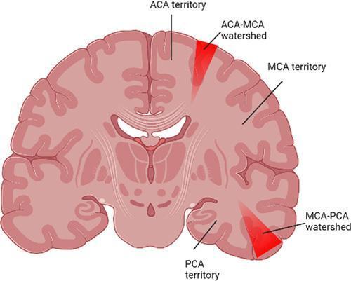
Anatomical diagram of a coronal brain section illustrating cerebral vascular territories and watershed (border zone) regions. The diagram identifies three primary arterial territories: the Anterior Cerebral Artery (ACA) territory in the medial cortex, the Middle Cerebral Artery (MCA) territory in the lateral cortex, and the Posterior Cerebral Artery (PCA) territory in the inferior-medial cortex. Two critical watershed zones are highlighted in red: the ACA-MCA watershed, appearing as a superior wedge-shaped area at the border of the medial and lateral hemispheres, and the MCA-PCA watershed, located at the inferior-lateral border. The illustration demonstrates the clinical concept of hypoperfusion injury, where these distal arterial junctions are most susceptible to ischemia during acute hypotension. Labels and anatomical landmarks including the cerebral cortex, ventricles, and subcortical structures provide orientation for neuroanatomical and radiological correlation in the study of watershed infarcts.
Anatomical diagram of a coronal brain section illustrating cerebral vascular territories and watershed (border zone) regions. The diagram identifies three primary arterial territories: the Anterior Cerebral Artery (ACA) territory in the medial cortex, the Middle Cerebral Artery (MCA) territory in the lateral cortex, and the Posterior Cerebral Artery (PCA) territory in the inferior-medial cortex. Two critical watershed zones are highlighted in red: the ACA-MCA watershed, appearing as a superior wedge-shaped area at the border of the medial and lateral hemispheres, and the MCA-PCA watershed, located at the inferior-lateral border. The illustration demonstrates the clinical concept of hypoperfusion injury, where these distal arterial junctions are most susceptible to ischemia during acute hypotension. Labels and anatomical landmarks including the cerebral cortex, ventricles, and subcortical structures provide orientation for neuroanatomical and radiological correlation in the study of watershed infarcts.

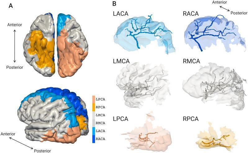
Educational diagram illustrating cerebral vascular territories and their respective image-based arterial structures. Panel A presents superimposed views (superior and lateral) of the human brain hemispheres, with color-coded regions identifying vascular territories: Anterior Cerebral Artery (ACA) in blue (LACA light blue, RACA dark blue), Middle Cerebral Artery (MCA) in gray/white (LMCA darker gray, RMCA lighter), and Posterior Cerebral Artery (PCA) in peach/orange (LPCA light peach, RPCA dark orange). These regions map the cortical distribution of blood supply across the cerebral surface. Panel B displays the reconstructed macroscale vascular geometries for each artery (LACA, RACA, LMCA, RMCA, LPCA, RPCA) isolated within their specific territories. The branching patterns and vessel densities reflect anatomical differences: ACA vessels predominantly track the superior and medial surfaces; MCA displays dense, complex networks along the lateral cortical surface; and PCA vessels occupy the posterior and inferior aspects. This model combines image-based geometries with mathematical algorithms to demonstrate the spatial relationship between major cerebral vessels and the gyri and sulci they supply.
Educational diagram illustrating cerebral vascular territories and their respective image-based arterial structures. Panel A presents superimposed views (superior and lateral) of the human brain hemispheres, with color-coded regions identifying vascular territories: Anterior Cerebral Artery (ACA) in blue (LACA light blue, RACA dark blue), Middle Cerebral Artery (MCA) in gray/white (LMCA darker gray, RMCA lighter), and Posterior Cerebral Artery (PCA) in peach/orange (LPCA light peach, RPCA dark orange). These regions map the cortical distribution of blood supply across the cerebral surface. Panel B displays the reconstructed macroscale vascular geometries for each artery (LACA, RACA, LMCA, RMCA, LPCA, RPCA) isolated within their specific territories. The branching patterns and vessel densities reflect anatomical differences: ACA vessels predominantly track the superior and medial surfaces; MCA displays dense, complex networks along the lateral cortical surface; and PCA vessels occupy the posterior and inferior aspects. This model combines image-based geometries with mathematical algorithms to demonstrate the spatial relationship between major cerebral vessels and the gyri and sulci they supply.

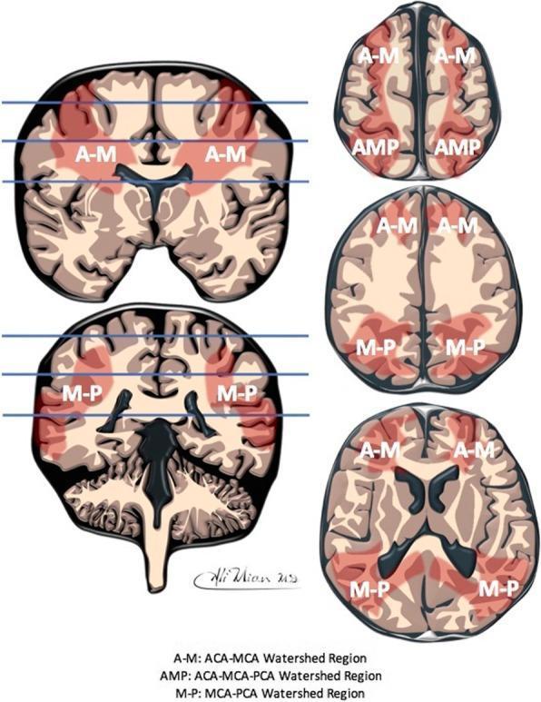
This medical illustration presents a schematic representation of the inter-arterial watershed regions of the human brain. The content is organized into two primary columns: the left column features two coronal cross-sections with horizontal blue lines indicating the anatomical levels of the corresponding three axial cross-sections shown in the right column. A red semi-transparent overlay highlights the boundary zones between major cerebral arterial territories. Letter annotations identify specific confluence zones: 'A-M' denotes the watershed between the Anterior Cerebral Artery (ACA) and Middle Cerebral Artery (MCA), primarily seen in the superior and lateral frontal regions; 'M-P' indicates the boundary between the MCA and Posterior Cerebral Artery (PCA) in the parieto-occipital and temporal regions; and 'AMP' signifies the triple-confluence of ACA, MCA, and PCA located at the vertex/midline. This diagram is designed for educational indexing in neurology and radiology, illustrating the anatomical distribution of regions most susceptible to hypoperfusion and watershed infarction in both adult and neonatal populations.

This medical illustration presents a schematic representation of the inter-arterial watershed regions of the human brain. The content is organized into two primary columns: the left column features two coronal cross-sections with horizontal blue lines indicating the anatomical levels of the corresponding three axial cross-sections shown in the right column. A red semi-transparent overlay highlights the boundary zones between major cerebral arterial territories. Letter annotations identify specific confluence zones: 'A-M' denotes the watershed between the Anterior Cerebral Artery (ACA) and Middle Cerebral Artery (MCA), primarily seen in the superior and lateral frontal regions; 'M-P' indicates the boundary between the MCA and Posterior Cerebral Artery (PCA) in the parieto-occipital and temporal regions; and 'AMP' signifies the triple-confluence of ACA, MCA, and PCA located at the vertex/midline. This diagram is designed for educational indexing in neurology and radiology, illustrating the anatomical distribution of regions most susceptible to hypoperfusion and watershed infarction in both adult and neonatal populations.

Your credits are exhausted. You can purchase a top-up or subscribe to a plan to continue using Orris. If you're already subscribed, your daily credits will reset tomorrow.
This is a shared conversation. Sign in to Orris to start your own chat.