
ADEM etiology, pathology , mechanisms, clinical and mri feature from standerd text book and article
Reading File
Now let me search the library and PubMed in parallel for comprehensive ADEM coverage.
Finding Sources
Searching PubMed
"acute disseminated encephalomyelitis" etiology pathology MRI
Excellent results. Now let me read the most comprehensive textbook sections in parallel - Adams & Victor, Bradley & Daroff (neurology block 11), Harrison's, and Grainger & Allison's (radiology block 11 which has the most paragraphs).
Reading File
Reading File
Reading File
Reading File
Excellent content. Now let me read the continuation of Harrison's (clinical manifestations + MRI), the Grainger & Allison radiology section (for detailed MRI features), and also fetch the ADEM MRI images.
Reading File
Reading File
Loading Image
Now let me fetch the ADEM vs MS comparison MRI image and also read the continuation of Grainger & Allison for spinal cord MRI details and the Adams & Victor continuation.
Loading Image
Reading File
Reading File
Let me now fetch the two remaining key MRI images (ADEM in child and spinal cord ADEM) and also get the PubMed abstract on the COVID-ADEM systematic review for current evidence.
Loading Image
Loading Image
Reading Article
PMID: 34452974
I now have comprehensive data from multiple authoritative textbooks and recent literature. Let me compile the complete answer.
Acute Disseminated Encephalomyelitis (ADEM)
Sources: Adams and Victor's Principles of Neurology (12th ed.), Bradley and Daroff's Neurology in Clinical Practice, Harrison's Principles of Internal Medicine (22nd ed., 2025), Grainger & Allison's Diagnostic Radiology, Plum and Posner's Stupor and Coma
1. ETIOLOGY
ADEM is a monophasic immune-mediated inflammatory-demyelinating disorder of the CNS. It is classified by trigger:
Postinfectious (Most Common)
The neurologic syndrome typically begins as the acute illness is resolving - often as the exanthem fades - after a latency of days to 2 weeks.
| Antecedent Infection | Approximate Risk |
|---|---|
| Measles (pre-vaccination era) | 1 in 800-2,000 cases |
| Varicella (chickenpox) | 1 in 4,000-10,000 cases |
| Rubella, mumps, influenza | Lower |
| EBV, CMV, HHV-6, HIV | Documented |
| Mycoplasma pneumoniae | Common non-specific URTI trigger |
| Dengue, Zika, SARS-CoV-2 | Also described |
- In developed countries today, varicella is now the most frequent antecedent.
- Non-specific upper respiratory tract infections account for 50-75% of cases.
- Occasional cases have no identifiable trigger.
Postvaccinal
- Historically associated with rabies and smallpox vaccines prepared with neural tissue (these are no longer used).
- A large Vaccine Safety DataLink study of >9 million individuals across 24 vaccines found no meaningful excess risk, with the possible exception of Tdap (estimated <1 case/million doses).
- Modern vaccines do not meaningfully cause ADEM.
Other (Rare)
- Drug reactions, paraneoplastic disorders, rheumatic diseases.
Important modern caveat (Harrison's 22nd ed., 2025): Many cases previously diagnosed as ADEM are now recognized as MOG antibody-associated disease (MOGAD). ADEM cases with anti-MOG serum/CSF antibodies are now reclassified under MOGAD.
2. PATHOLOGY
The hallmark pathologic lesion is innumerable foci of perivenular inflammation and demyelination scattered throughout the brain and spinal cord.
Microscopic features (Adams and Victor; Harrison's):
- Lesions are diffuse, poorly marginated, and of uniform age (in contrast to MS where lesions are multiple, distinct, and of mixed age)
- Lesions vary from miniscule to several millimeters; they invariably surround small and medium-sized veins (perivenular)
- Perivenular inflammatory infiltrate: primarily lymphocytes and mononuclear cells
- Adjacent white matter invaded by monocytes and microglia - corresponding zones of demyelination
- Axons and neurons are relatively preserved (demyelination without axonal destruction)
- Multifocal meningeal (pial) infiltration - a feature of ADEM not seen in MS
- Both white matter and gray matter structures can be involved (contrast with MS which is predominantly white matter)
- In the most explosive form - Acute Hemorrhagic Leukoencephalitis (AHLE / Hurst disease) - lesions are vasculitic and hemorrhagic
Distinction from MS pathology:
- MS lesions: multiple, distinct, well-circumscribed, irregularly distributed, mixed age
- ADEM lesions: diffuse, less distinct margins, uniform age, perivenular, meningeal involvement
3. PATHOGENESIS / MECHANISMS
The key mechanism is cross-reactive autoimmunity (molecular mimicry), not direct CNS viral invasion.
Evidence against direct viral CNS infection:
- An interval separates the exanthem from neurologic onset
- Pathologic changes differ completely from viral encephalitis
- Virus is rarely isolated from CSF or brain tissue
- Postmeasles ADEM patients show no intrathecal measles antibody synthesis
Molecular mimicry mechanism (Bradley and Daroff; Adams and Victor):
Microbial antigens structurally resemble myelin proteins, activating myelin-reactive T-cell clones that cross-react against CNS myelin:
| Pathogen | Mimicked Myelin Antigen |
|---|---|
| HHV-6, coronavirus, influenza hemagglutinin, EBV | MBP (Myelin Basic Protein) |
| Haemophilus influenzae | PLP (Proteolipid Protein) |
| Semliki Forest Virus peptides | MOG (Myelin Oligodendrocyte Glycoprotein) |
Animal model - Experimental Autoimmune Encephalomyelitis (EAE):
- Produced by inoculating animals with sterile brain tissue + adjuvants
- Appears 8-15 days after sensitization
- Produces identical perivenular demyelinating lesions
- T cells from ADEM patients are 10x more likely to react to MBP than controls
Humoral vs. cellular debate:
- Evidence of antibody binding, complement activation, and eosinophilic infiltration suggests a humoral component (unlike MS which is predominantly T-cell mediated)
- Anti-MBP antibodies detected in CSF
- Anti-MOG antibodies (~30% of ADEM patients) - these patients are now classified as MOGAD
- The MOG antibody titer drops rapidly after the acute attack, so its exact pathogenic role is uncertain
Why monophasic? Unlike MS, the immune response in ADEM appears to occur acutely without further CNS amplification - further inflammation is suppressed after the initial attack. This differentiates it from MS.
4. CLINICAL FEATURES
Epidemiology:
- More common in children than adults (median age 5-8 years)
- No sex preponderance (unlike MS, which favors females)
- Incidence: 0.3-0.6 per 100,000/year
Prodromal phase: 1-3 weeks before neurologic onset, patients have fever, malaise, headache, nausea/vomiting related to preceding infection.
Neurologic onset:
- Abrupt onset over hours to 1-2 days, rapid progression
- In postinfectious ADEM: fever reappears as the exanthem fades
- Maximum deficits within 2-5 days
Core clinical features (Harrison's; Adams and Victor; Bradley and Daroff):
| Feature | Description |
|---|---|
| Encephalopathy | Hallmark - required for diagnosis; ranges from lethargy to stupor/coma; behavioral change |
| Headache & meningismus | Common at onset |
| Seizures | Common (more so than in MS) |
| Hemiparesis or quadriparesis | Reflects disseminated disease |
| Sensory loss | Present |
| Brainstem involvement | Cranial nerve palsies, ataxia |
| Cerebellar ataxia | Especially prominent in varicella-associated ADEM |
| Optic neuritis | Generally bilateral (unlike MS where it is usually unilateral) |
| Transverse myelopathy | Complete (unlike MS where it is usually partial) |
| Extensor plantar responses | Hyperreflexia or hyporeflexia |
CSF findings:
- Normal opening pressure
- Lymphocytic pleocytosis in 80% of patients, generally ≥200 cells/μL; mixed PMN-lymphocytic pattern in early days
- Protein: modestly elevated - 0.5-1.5 g/L (50-150 mg/dL)
- Glucose: normal
- Oligoclonal bands (OCBs): transient, minority of cases; if present, follow for MS
- Red blood cells: if present, suggest hemorrhagic leukoencephalitis (AHLE)
Outcome:
- Most children have favorable outcome with full recovery despite severity
- Mortality 10-20% in severe cases historically; significant disability in survivors
- Adults often make better recoveries than children paradoxically (except severe cases)
- Follow-up: 25-30% of adults initially diagnosed with ADEM later develop MS or another chronic inflammatory disorder (vasculitis, sarcoidosis, lymphoma, MOGAD, NMOSD)
5. MRI FEATURES
MRI is the cornerstone of diagnosis. ADEM lesions are characteristically different from MS lesions.
Brain MRI (Grainger & Allison; Harrison's; Bradley and Daroff):
| Feature | ADEM | MS (for comparison) |
|---|---|---|
| Lesion size | Large, >1-2 cm | Smaller, variable |
| Margins | Poorly defined, patchy | Well-defined |
| Location | Subcortical and central WM, cortical grey-white junction | Periventricular, corpus callosum (Dawson fingers) |
| Periventricular | Less common | Characteristic |
| Corpus callosum | Less common | Common |
| Ovoid lesions (Dawson fingers) | Much less frequent | Characteristic |
| Grey matter involvement | Common - thalamus, basal ganglia (especially in children), cortex (30%) | Rare |
| Symmetry | Relatively symmetric in severe cases | Asymmetric |
| Signal | T2/FLAIR hyperintense; T1 hypointense lesions should NOT be present | T1 hypointense "black holes" present |
| Mass effect | Mild or absent even in large lesions | Usually absent |
| Age of lesions | All same age (uniform enhancement) | Mixed age |
Gadolinium enhancement:
- Present in 14-30% of cases (less common than in MS)
- When present: all lesions enhance simultaneously (same age - key differentiating feature from MS)
- Enhancement patterns: complete ring, incomplete ring, nodular, gyral, or spotty
Sequence-specific findings:
- T2/FLAIR: multifocal white matter hyperintensities - primary sequence for detection
- T1 post-contrast: enhancement in active lesions (14-30%)
- DWI: may show restricted diffusion in acute lesions
- T1 hypointense "black holes" should NOT be seen in ADEM (their presence suggests MS)
Distribution:
- Cerebrum (subcortical and central white matter)
- Cerebellum
- Brainstem
- Thalamus and basal ganglia (particularly in children; may be symmetric)
- Cortical grey matter (30% of cases)
- Spinal cord (1/3 of patients)
Spinal cord MRI:
- Thoracic region predominantly affected
- Long extensive transverse myelitis (LETM) - extending over multiple segments (similar to NMOSD)
- Cord swelling
- Variable enhancement
- Lesions may be confined to grey matter, white matter, or both
Follow-up MRI - critical diagnostic criterion (IPMSSG 2012 criteria):
- Most lesions appear early and support diagnosis
- In ~50% of cases, new lesions and enlargement can occur within the first 3 months (still consistent with ADEM)
- No new T2 lesions after 3 months from onset - if new lesions appear after 3 months, reclassify
- Complete MRI resolution within 6 months: strongly supports ADEM diagnosis
- Persistence or incomplete resolution: associated with subsequent MS diagnosis
- A reference MRI at 3 months post-onset is recommended
IPMSSG 2012 Diagnostic MRI Criteria:
- Diffuse, poorly demarcated, large (>1-2 cm) lesions
- Predominantly cerebral white matter
- No T1 hypointense lesions
- Deep grey matter lesions (thalamus/basal ganglia) can be present, especially in children
MRI Images (from Grainger & Allison's Diagnostic Radiology)
Fig. 58.22 - ADEM vs MS brain MRI comparison (T2-FLAIR and contrast T1):
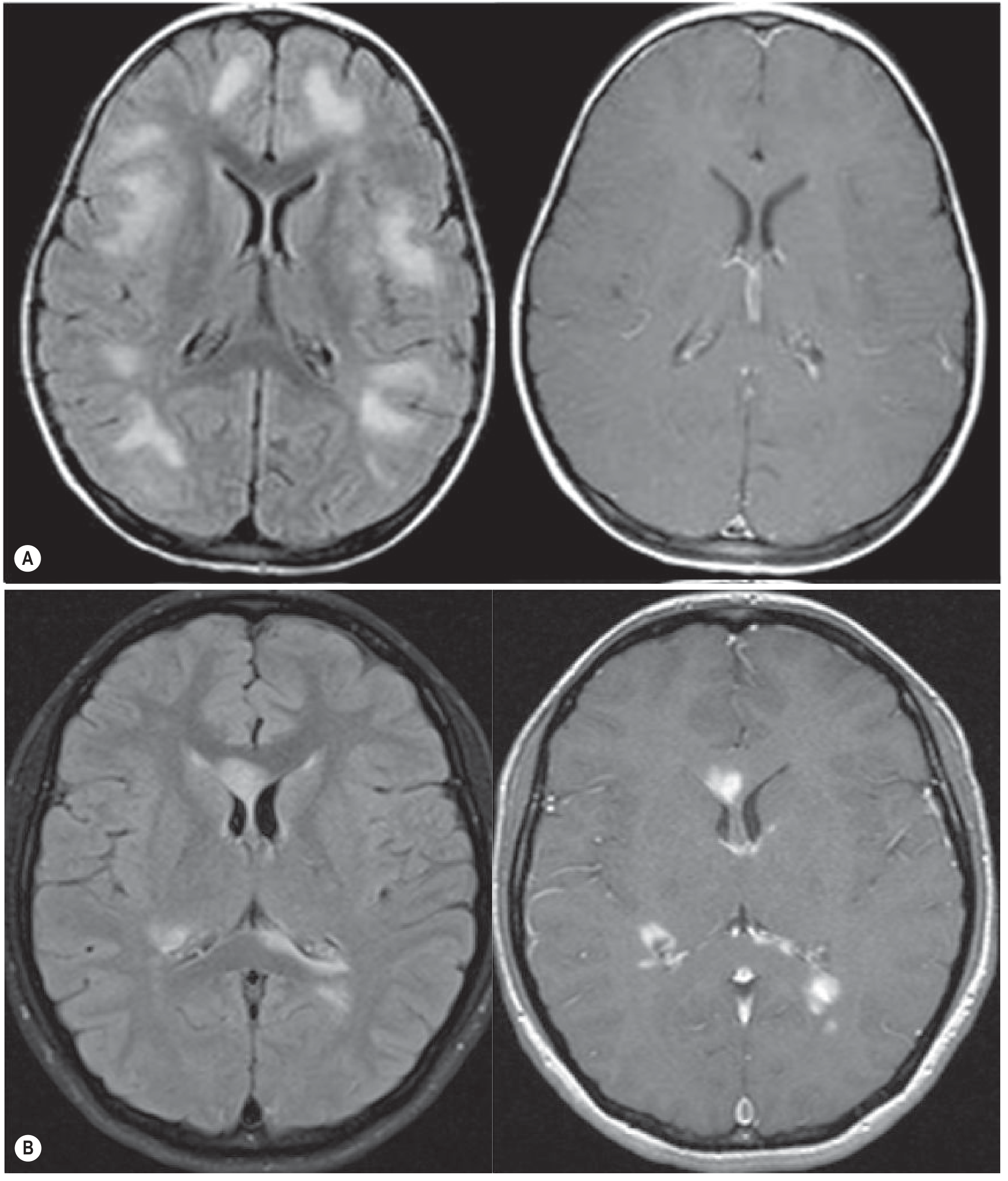
ADEM (A): larger, poorly marginated lesions with predominant subcortical location, no contrast enhancement. MS (B): smaller, well-defined periventricular lesions with contrast enhancement.
Fig. 58.23 - ADEM in a 12-year-old boy (axial T2):
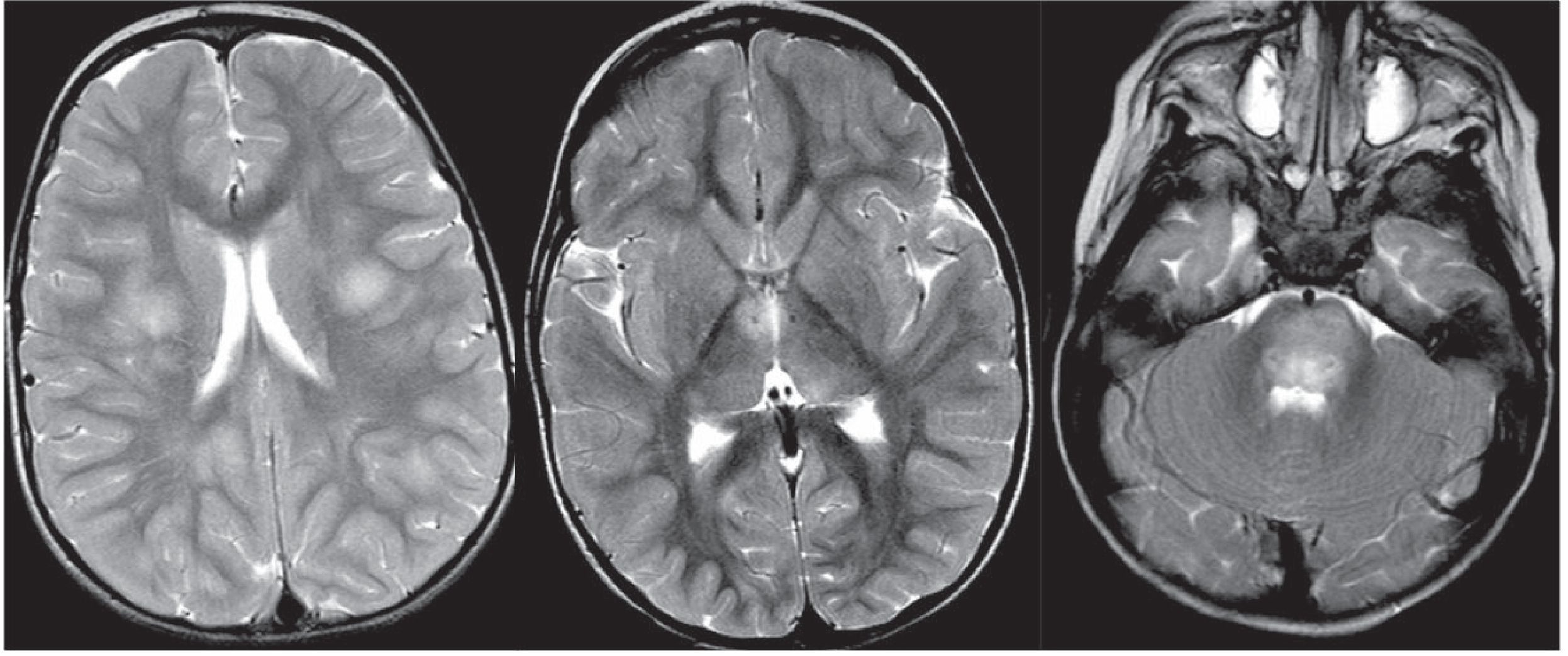
Asymmetrical involvement of subcortical hemispheric white matter, both thalami, and diffuse pontine involvement - characteristic pediatric ADEM pattern.
Fig. 58.24 - Spinal cord ADEM in a 25-year-old woman (sagittal T2):
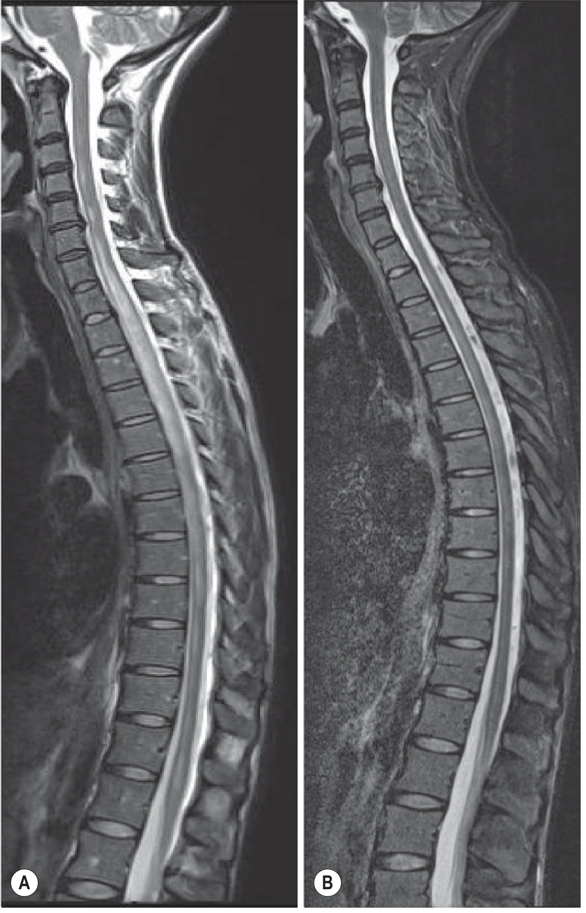
Extensive spinal cord hyperintensity involving the thoracic-cervical segment at presentation (A), with complete resolution weeks later (B) - hallmark of the monophasic ADEM course.
6. DIAGNOSIS (Summary Criteria - IPMSSG 2012)
All of the following must be present:
- First event of encephalopathy + polyfocal neurological deficits
- Presumed inflammatory demyelinating cause
- Encephalopathy: alteration in consciousness or behavior unexplained by fever, systemic illness, or postictal symptoms
- Brain MRI abnormalities consistent with demyelination during acute phase (within 3 months)
- No new clinical and MRI findings ≥3 months after initial event
- Typical MRI lesions (described above)
Key features favoring ADEM over MS:
- Simultaneous onset of disseminated symptoms (rare in MS)
- Encephalopathy (uncommon in MS)
- Bilateral optic neuritis (unilateral in MS)
- Complete transverse myelopathy (partial in MS)
- Extensive symmetric white matter changes + basal ganglia/cortical GM lesions
- Enhancement of ALL abnormal areas simultaneously
- No T1 black holes
- Resolution of lesions on follow-up
7. RECENT EVIDENCE - Post-COVID ADEM
A 2021 systematic review (Manzano et al., Neurology Neuroimmunol Neuroinflamm, PMID 34452974) analyzed 46 post-COVID-19 ADEM/AHLE cases:
- Median age 49.5 years (older than typical ADEM)
- Anti-MOG seropositive in only 1/15 tested
- Hemorrhage on MRI in 42% (unusually high)
- 32% mortality, 64% had final mRS ≥4 (poor outcome)
- Post-COVID ADEM differs from prepandemic ADEM: older, severe antecedent infection, high hemorrhage rate, poor outcomes
8. ACUTE HEMORRHAGIC LEUKOENCEPHALITIS (AHLE / Hurst Disease) - Severe Variant
- The most explosive form of ADEM
- Lesions are vasculitic and hemorrhagic
- Clinical course is devastating
- CSF: red blood cells present
- MRI: hemorrhagic foci (susceptibility-weighted imaging/GRE sensitive)
- High mortality
Sources
- Adams and Victor's Principles of Neurology, 12th Edition - Chapter on inflammatory demyelinating diseases
- Bradley and Daroff's Neurology in Clinical Practice - Chapter 79
- Harrison's Principles of Internal Medicine, 22nd Edition (2025) - Chapter on demyelinating diseases
- Grainger & Allison's Diagnostic Radiology - Chapter 58 (Demyelinating diseases MRI)
- Plum and Posner's Diagnosis and Treatment of Stupor and Coma
- Manzano GS et al. Neurol Neuroimmunol Neuroinflamm. 2021 Nov [PMID: 34452974] - Post-COVID ADEM systematic review
ADEM vS Ms VS NMO vs MOG progression , etiopathology, nature of illness , from standerd
Your credits are exhausted. You can purchase a top-up or subscribe to a plan to continue using Orris. If you're already subscribed, your daily credits will reset tomorrow.
This is a shared conversation. Sign in to Orris to start your own chat.