
Endoscopic decompression for terminal ileum obstruction
endoscopic decompression small bowel obstruction terminal ileum
endoscopic decompression ileus obstruction colonoscopy
endoscopic decompression colonoscopy small bowel obstruction terminal ileum
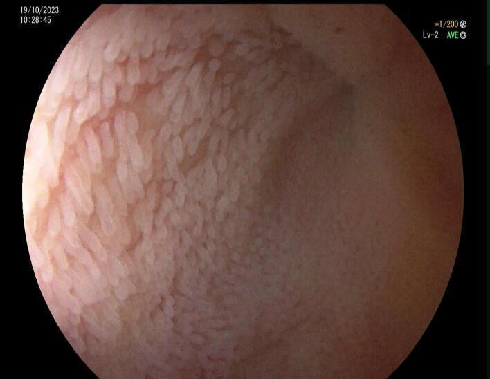
This diagnostic endoscopic image (colonoscopy) captures the terminal ileum mucosa. The visual shows a healthy, pink-colored mucosa with well-defined, elongated, pale intestinal villi characteristic of normal small bowel anatomy. There is no evidence of inflammation, ulceration, erythema, or visible lesions. Notably, the ileal lumen appears collapsed or significantly narrowed at the distal aspect shown, which in this clinical context signifies a complete luminal obstruction approximately 10 cm proximal to the ileocecal valve. The diagnostic focus is the contrast between the unremarkable, healthy appearance of the mucosal surface and the mechanical obstruction of the lumen. This image is relevant for gastroenterology and general surgery education, illustrating that mechanical small bowel obstructions (such as those caused by extrinsic adhesive bands or volvulus) may present with normal intraluminal mucosal findings despite severe clinical obstruction.
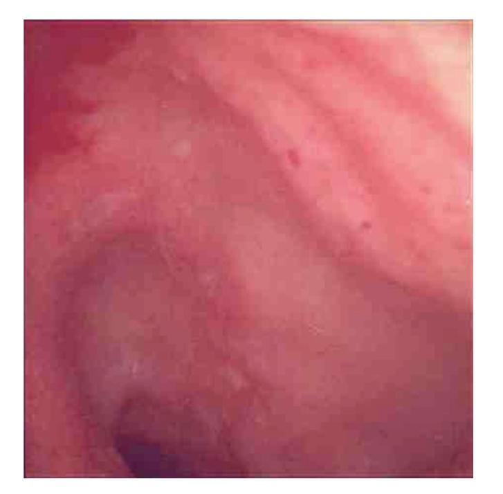
This diagnostic endoscopic image captures the terminal ileum during a colonoscopy, highlighting mucosal manifestations of inflammatory bowel disease, specifically Crohn's disease. The image reveals significant mucosal erythema and diffuse edema, giving the tissue a predominantly reddish-pink appearance. Distinct pathological features include multiple shallow, irregular erosions and superficial breaks in the mucosal lining. Small, petechial-like red spots and textural irregularities are visible across the surface, indicating active inflammation and patchy acute-on-chronic inflammatory changes. These visual markers are critical for the endoscopic assessment of ileitis and help differentiate inflammatory conditions from infectious etiologies such as tuberculosis. The visual evidence of mucosal disruption without obvious granulomata on initial inspection serves as an educational example of the diverse endoscopic presentations of ileal Crohn's disease.
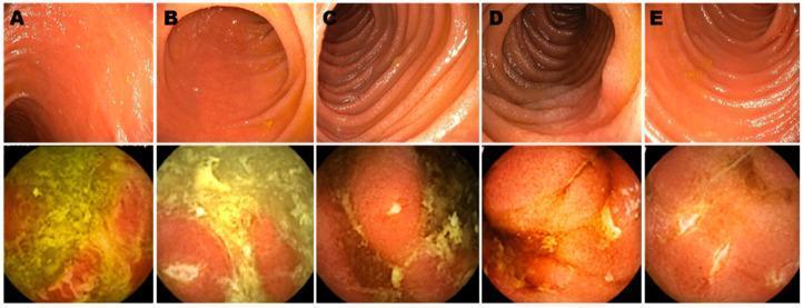
This Comparison Chart features a series of endoscopic images labeled A through E, contrasting traditional ileo-colonoscopy (upper panels) with wireless capsule endoscopy (WCE, lower panels) in post-operative Crohn's disease patients. The upper panels display the terminal ileum with a relatively smooth, pinkish mucosal surface and regular vascular patterns, consistent with endoscopic remission (Rutgeerts scores i0-i1). In contrast, the lower panels show WCE images of the proximal small bowel from the same patients, revealing significant pathological changes. These abnormalities include patchy erythema, friable mucosa, and prominent white or yellowish inflammatory exudates and fibrinous deposits. Linear ulcerations and cobble-stoning texture are visible in frames B and E, indicating active Crohn's recurrence. The comparison demonstrates the clinical utility of WCE in detecting proximal small bowel inflammation that may be missed by standard colonoscopy, emphasizing the importance of small bowel evaluation in disease staging and postoperative monitoring.
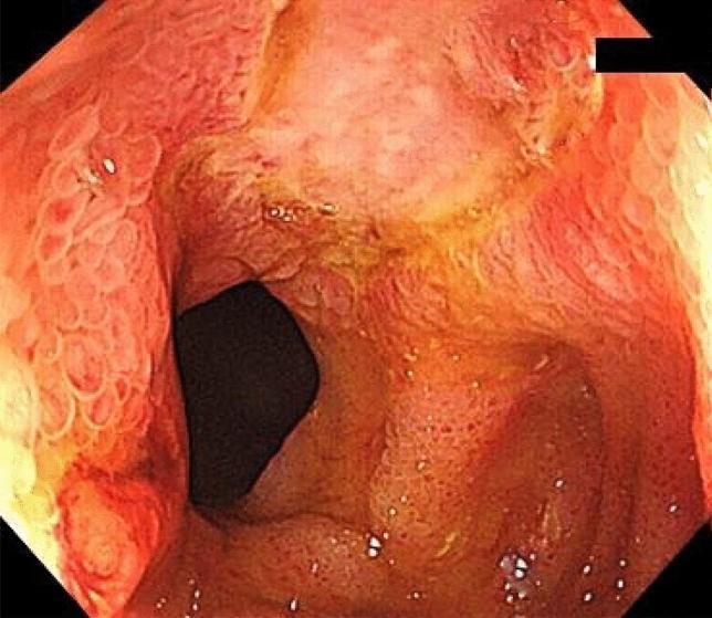
This diagnostic endoscopic image (colonoscopy) showcases the terminal ileum in a patient with inflammatory bowel disease-unclassified (IBDU). The mucosal surface exhibits significant pathological changes, including multiple small, round ulcerations and diffuse erythema. The mucosa appears predominantly pinkish-red with yellowish patches of exudate or discoloration. Evidence of chronic inflammation and repair is visible through irregular scar tissue and a distorted mucosal texture. Notably, the ileal lumen is visibly narrowed, indicating a pathological stricture that alters its normal circular shape. These features—ulceration, scarring, and luminal narrowing—are key endoscopic indicators of active and chronic intestinal inflammation, often necessitating interventions like endoscopic balloon dilation (EBD). The image provides a high-level educational example of ileal stricture morphology and mucosal remodeling in the context of chronic gastrointestinal disorders.
Endoscopic Decompression for Terminal Ileum Obstruction
1. Relevant Causes of Terminal Ileum Obstruction
| Cause | Endoscopic Role |
|---|---|
| Meconium ileus / DIOS (CF) | Retrograde contrast enema ± colonoscopy |
| Crohn's stricture | Endoscopic balloon dilation (EBD) |
| Adhesive SBO (extrinsic) | No endoscopic role |
| Ileocecal tuberculosis | EBD for short strictures |
| Gallstone ileus (at ileocecal valve) | Usually surgical; retrograde endoscopy rarely feasible |
| Cecal carcinoma obstructing ICV | Stenting via colonoscopy |
| Colonic pseudo-obstruction (Ogilvie) spilling into ileum | Colonoscopic decompression |
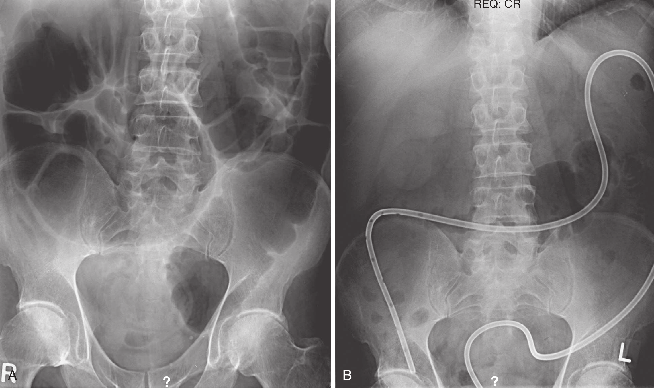
2. Colonoscopic Decompression - Technique and Indications
- Performed without prior bowel preparation (to reduce perforation risk)
- Minimal air insufflation - preferably CO2 to reduce distension
- Air and liquid stool aspirated through a large-bore suction channel
- Scope advanced to the proximal transverse colon under fluoroscopic guidance is sufficient; this achieves decompression in ~50% of cases by simple aspiration
- A guidewire is then passed through the accessory channel and the scope is withdrawn slowly under fluoroscopy; a decompression tube (14 Fr) is left over the wire with low intermittent suction + saline flushes every 4-6 hours
- Sabiston Textbook of Surgery, p. 2086
- Current Surgical Therapy 14e (Colonoscopic Decompression section)
3. Endoscopic Management of Meconium Ileus / DIOS at the Terminal Ileum
- Water-soluble contrast enema (e.g., Gastrografin - sodium and meglumine amidotrizoate) or dilute N-acetylcysteine enema
- Reflux of contrast into the terminal ileum dissolves/dislodges the inspissated meconium pellets
- Success rate ~60-70% in neonates; older patients (DIOS) treated similarly
- Sequential enemas may be needed
- Rare complications: intestinal perforation, mucosal injury, persistent obstruction
- Mulholland and Greenfield's Surgery 7e, p. 5512
4. Endoscopic Balloon Dilation (EBD) for Strictures at the Terminal Ileum
- Colonoscope is advanced through the ileocecal valve into the terminal ileum
- Guide wire placed across the stricture under fluoroscopic guidance
- Through-the-scope (TTS) balloon dilator deployed - applies purely radial force
- General rule: limit to 3 successive sizes per session (incremental approach)
- Steroid injection (triamcinolone/Kenalog) into the stricture margins reduces post-dilation fibrosis and re-stricture
- Fluoroscopic guidance recommended to confirm balloon positioning and assess full dilation (waist effacement)
- Short strictures (<4-5 cm), non-fistulizing, non-phlegmonous
- Benign anastomotic strictures (post-ileocecal resection)
- TB, radiation, ischemia-related short strictures at terminal ileum
- Provides short-term relief; repeat dilation required in a significant minority
- Not suitable for long strictures, active inflammation with abscess/fistula, or suspicion of malignancy
- Maingot's Abdominal Operations, p. 63 (Endoscopic Dilation)
- Goldman-Cecil Medicine: "Endoscopic dilation will usually alleviate symptoms, but repetitive dilation is required in a significant minority of patients"
5. Endoscopic Stenting at the Ileocecal Region
- Obstructing cecal/right colon carcinoma blocking the ICV (palliative bridge-to-surgery)
- Rare cases of extrinsic obstruction at the terminal ileum
6. What Endoscopy CANNOT Treat
- Mechanical adhesive SBO (the most common cause of small bowel obstruction) - requires surgery or laparoscopic lysis of adhesions
- Strangulated obstruction with ischemia (peritonitis, fever, leukocytosis, CT with devascularized loop) - emergency surgery
- Volvulus with compromised mesentery (e.g., ileosigmoid knotting): attempted colonoscopic decompression will fail because the mesentery is physically wrapped, and the attempt may delay surgery
- Perforation or peritonitis at presentation
Current Surgical Therapy 14e notes: "Ileosigmoid knotting may be confused for sigmoid volvulus on imaging prompting attempted endoscopic decompression. In these cases, endoscopic decompression will fail due to the wrapping of the mesentery, further raising..."
7. Management Algorithm
Terminal Ileum Obstruction
         |
         ├── Mechanical SBO (adhesions, hernia) → Surgery / laparoscopy
         |
         ├── DIOS / Meconium Ileus → Retrograde Gastrografin/NAC enema
         |         └── Fails → Operative enterotomy + irrigation
         |
         ├── Crohn's / anastomotic stricture → Ileoscopic balloon dilation
         |         └── + steroid injection; repeat PRN
         |
         ├── Ileocecal obstruction (neoplasm) → Colonoscopic SEMS (palliation)
         |
         └── Pseudo-obstruction extending to cecum/ileum
                   → Conservative → Neostigmine → Colonoscopic decompression
                   └── Tube left in situ; PEG after decompression to prevent recurrence
Key Points Summary
- Colonoscopic decompression is most evidence-based for Ogilvie's syndrome (ACPO) - success 61-95%, done with CO2, minimal insufflation, decompression tube left over guidewire.
- Retrograde contrast enema is the first-line endoscopic approach for terminal ileum DIOS/meconium ileus.
- Endoscopic balloon dilation via ileoscopy is the procedure of choice for short benign strictures (Crohn's, anastomotic, TB) at the terminal ileum.
- All endoscopic approaches are contraindicated when there is peritonitis, perforation, strangulation, or ischemia.
- After successful endoscopic decompression, polyethylene glycol (PEG) twice daily for 7 days reduces recurrence of colonic distension.
- Failure of colonoscopic decompression leads to percutaneous cecostomy (endoscopic or IR-guided) as a bridge before surgery.