
Facial fractures classification
facial fractures classification Le Fort
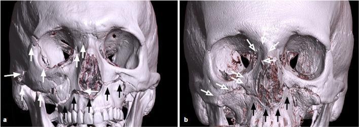
This diagnostic image displays 3D-CT reconstructions of two different patients (labeled a and b) highlighting various patterns of Le Fort facial fractures.
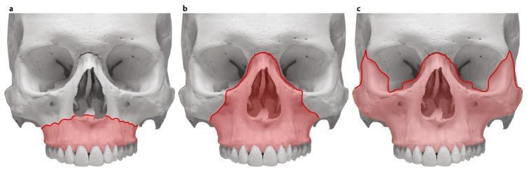
This Comparison Chart consists of three anatomical diagrams (a, b, and c) of the human skull, viewed from an anterior perspective. It illustrates the René Le Fort classification of midfacial fractures through shaded red regions. Diagram (a) represents a Le Fort I fracture, showing a transverse fracture line through the maxilla, isolating the alveolar process and palate. Diagram (b) represents a Le Fort II fracture (pyramidal fracture), with the shaded area extending superiorly through the nasal bones and infraorbital rims, involving the maxillary body. Diagram (c) represents a Le Fort III fracture (craniofacial dysjunction), where the shaded region encompasses the entire midface, including the zygomatic bones, orbital floors, and nasal bones, effectively separating the facial skeleton from the skull base. This visual educational resource is designed for maxillofacial surgery and emergency medicine training to differentiate the varying degrees of midfacial skeletal instability and anatomical involvement across these three fracture types.
mandible fracture classification zones
nasoorbital ethmoid NOE fracture orbital blowout
Facial Fractures - Classification
Anatomical Framework
I. UPPER FACIAL FRACTURES
Frontal Bone / Frontal Sinus Fractures
- Anterior table fractures - involve only the anterior wall of the frontal sinus. Isolated, nondisplaced fractures can be observed. Displaced fractures causing nasofrontal outflow tract obstruction require ORIF.
- Posterior table fractures - involve the wall adjacent to the anterior cranial fossa. These are complex, often associated with dural injury and CSF rhinorrhea. May require cranialization of the sinus or obliteration.
- Combined anterior + posterior table fractures - both walls involved; significant risk of intracranial complications.
II. MIDFACE FRACTURES
1. Nasal Fractures
2. Naso-Orbito-Ethmoid (NOE) Fractures
- Type I - single large central fragment bearing the medial canthal tendon; treated with ORIF
- Type II - central fragment comminuted but medial canthal tendon attached to one fragment
- Type III - severe comminution of the central fragment; medial canthal tendon insertion disrupted (transnasal canthopexy required)
3. Orbital Fractures (Blowout Fractures)
- Pure blowout: only the internal orbital wall, not the rim
- Impure blowout: orbital wall + rim
- Orbital floor fracture - entrapment of the inferior rectus causes diplopia and limited upgaze
- Medial wall fracture - easily missed; orbital emphysema common
- Orbital roof fracture - rare in adults (3-9.3% of facial fractures), more common in children (18-35% of pediatric facial fractures); divided into "pure" (roof only) and "impure" (roof + rim)
- Complications: enophthalmos, hypophthalmos, retro-orbital hematoma (compartment syndrome), superior orbital fissure syndrome (CN III, IV, V1, VI), orbital apex syndrome (adds optic nerve compression - surgical emergency)
4. Zygomatic (Tripod / Trimalar) Fractures
5. Le Fort Fractures

| Level | Name | Fracture Line | Mobility on Exam |
|---|---|---|---|
| Le Fort I | Transverse / Guérin's fracture | Horizontal fracture through the maxilla above the tooth roots; extends from pterygoid plates through lateral wall of maxillary sinus and piriform aperture of nose. Separates the alveolar process and palate from the rest of the midface. | Mobile alveolar ridge only |
| Le Fort II | Pyramidal fracture | Extends from pterygoid plates superiorly across the nasal bones, lacrimal bones, orbital floor, and inferior orbital rim, creating a pyramid-shaped fragment (maxilla + nasal complex). | Nasal complex + maxilla moves as a unit |
| Le Fort III | Craniofacial dysjunction | Complete separation of the entire midface from the skull base. Fracture extends from nasal bridge posteriorly along medial orbital wall (ethmoids), along orbital floor, through lateral orbital wall, and through the zygomatic arch. Associated with CSF leak. | Entire face (including zygomas) moves |
III. LOWER FACIAL FRACTURES
Mandible Fractures
| Region | Description |
|---|---|
| Symphysis | At the midline between the two mental foramina (some define symphysis as the exact midline and parasymphysis as off-midline to the foramen) |
| Parasymphysis | Between the midline and the mental foramen |
| Body | Between the mental foramen and the angle of the mandible |
| Angle | At/posterior to the region of the third molar, behind the tooth-bearing segment (dentoalveolar buttress unavailable) |
| Ramus | Posterior to the angle, in the vertical portion of the mandible |
| Subcondylar | Traverses the sigmoid notch and exits behind the angle; most common site due to the thin bone here |
| Condyle | Fracture of the condylar head itself |
| Coronoid | Fracture of the coronoid process (rare; extends anteriorly from sigmoid notch) |
- Fist/blunt trauma or fall: symphysis, parasymphysis, body
- A symphysis fracture often produces a contralateral condylar fracture (indirect force)
- Angle fractures have reduced dentition support and often require ORIF
- Subcondylar fractures may be managed conservatively (closed) or operatively depending on displacement and occlusion
IV. PANFACIAL FRACTURES
Summary Table
| Region | Fracture Type | Key Classification |
|---|---|---|
| Upper face | Frontal sinus | Anterior table / Posterior table / Combined |
| Midface (central) | Nasal | Displaced / Non-displaced |
| Midface (central) | NOE | Markowitz Types I / II / III |
| Midface (orbital) | Blowout | Pure / Impure; Floor / Medial / Lateral / Roof |
| Midface (lateral) | Zygomatic | Tripod / Isolated arch |
| Midface (maxilla) | Le Fort | I (transverse), II (pyramidal), III (craniofacial dysjunction) |
| Lower face | Mandible | Symphysis / Parasymphysis / Body / Angle / Ramus / Subcondylar / Condyle / Coronoid |
| All three thirds | Panfacial | No sub-classification; described by component fractures |
- Current Surgical Therapy, 14e - "Evaluation and Management of the Patient with Craniomaxillofacial Trauma"
- Bailey and Love's Short Practice of Surgery, 28e - Chapter 31: Maxillary, Zygomatic, and Orbital Fractures
- Rosen's Emergency Medicine, 9e - Chapter 34: Facial Trauma
- K.J. Lee's Essential Otolaryngology - Facial Fracture Classification
- Cummings Otolaryngology Head and Neck Surgery - Midface and Mandible Fractures