
Syringomyelia
syringomyelia
syringomyelia symptoms diagnosis treatment NINDS
MRI syringomyelia cervical spinal cord syrinx sagittal T2
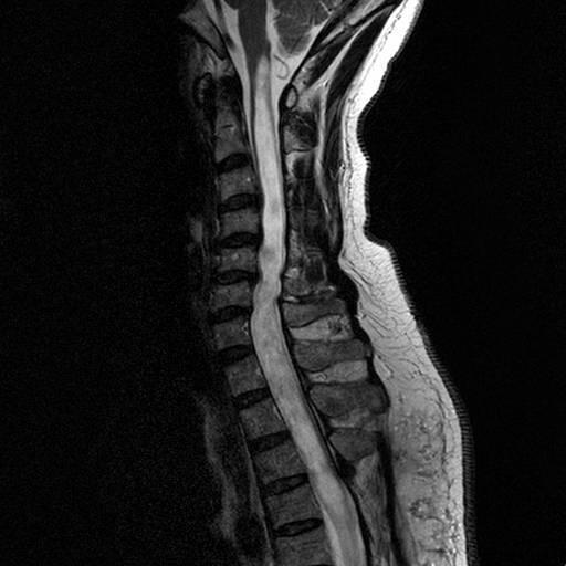
This diagnostic image is a T2-weighted sagittal MRI of the cervical spine, demonstrating a significant longitudinal syrinx (syringomyelia). The spinal cord is centrally located within the spinal canal and shows a large, well-defined, tubular hyperintense signal (indicating cerebrospinal fluid content) extending from the cervicomedullary junction through the cervical levels. The syrinx causes focal expansion of the spinal cord diameter at several levels. Anatomical landmarks include the rectangular vertebral bodies anterior to the cord and the visible posterior elements. Notably, the high-intensity fluid signal within the cord is distinct from the surrounding gray and white matter parenchyma. The superior margin of the syrinx is visualized near the obex of the medulla, but there is no direct communication with the fourth ventricle. The image serves as a clinical illustration of cervical syringomyelia and its impact on spinal cord morphology, essential for neurosurgical and neurological education.
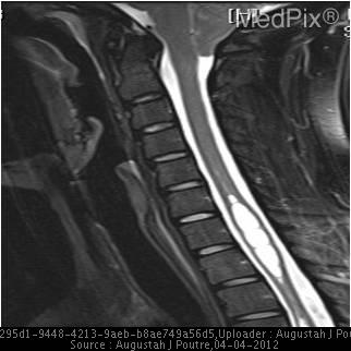
**Imaging Modality:** Sagittal T2-weighted Magnetic Resonance Imaging (MRI) of the cervical spine.
https://www.ninds.nih.gov/health-information/disorders/syrin…
Syringomyelia
Causes
- Chiari I malformation: the common association, due to impaired cerebrospinal fluid flow at the foramen magnum.
- Previous spinal cord trauma.
- Intramedullary spinal cord tumor.
- Arachnoiditis, meningitis, or myelitis.
- Congenital spinal abnormalities such as tethered cord.
- Sometimes no cause is found, termed idiopathic syringomyelia.
Typical features
- Dissociated sensory loss: loss of pain and temperature with preserved light touch, vibration, and joint-position sense.
- A bilateral, segmental “cape-like” sensory loss across the shoulders, arms, and hands is classic for a cervical syrinx.
- Burning neck, shoulder, or arm pain.
- Hand weakness and wasting due to anterior horn cell involvement.
- Reduced reflexes at the affected spinal level.
- Later, spastic weakness and increased reflexes in the legs from corticospinal tract involvement.
- Scoliosis, especially in children or adolescents.
- Horner syndrome can occur with a lower cervical or upper thoracic lesion.
- Repeated painless burns or injuries to the hands because pain sensation is reduced.
Diagnosis
- MRI of the entire spinal cord and brain is the key test. It demonstrates the syrinx and identifies causes such as Chiari malformation, tumor, or post-traumatic changes.
- Cine or phase-contrast MRI may assess cerebrospinal fluid flow when Chiari malformation is suspected.
- Neurological examination assesses sensory loss, weakness, reflex changes, gait, and autonomic dysfunction.
Management
- Observation: reasonable for an incidental, stable, asymptomatic syrinx, with clinical review and interval MRI.
- Treat the underlying obstruction or lesion:
- Chiari I with symptomatic/progressive syringomyelia: posterior fossa decompression, often with duraplasty, to restore CSF flow.
- Tumor: treatment directed at the tumor.
- Post-traumatic or arachnoid-related obstruction: individualized neurosurgical treatment.
- Syrinx shunting may be considered in selected persistent or recurrent cases, but it is not usually the first treatment when a correctable cause is present.
- Rehabilitation, pain management, and occupational therapy can help residual symptoms.
Seek urgent assessment if there is
- New or rapidly worsening limb weakness
- Difficulty walking or frequent falls
- New bladder or bowel dysfunction
- New severe neck pain with neurological symptoms
- Symptoms after a spinal cord injury