
Role of imaging in invasive fungal rhino sinusitis in detail according to ent standard textbooks
Reading File
Now let me search the medical library for invasive fungal rhinosinusitis imaging content in parallel.
Finding Sources
Finding Sources
Finding Sources
Excellent. Now let me read the most relevant sections from Cummings, Scott-Brown's, and KJ Lee in parallel.
Reading File
Reading File
Reading File
Let me now read the core imaging sections from Cummings and Scott-Brown's, plus the KJ Lee AIFS/CIFS imaging content:
Reading File
Reading File
Reading File
Now let me read the Scott-Brown's AIFS imaging section and the Cummings block 10 (Invasive Fungal Rhinosinusitis section) plus granulomatous section from KJ Lee:
Reading File
Reading File
Reading File
Now let me read the Cummings block 9 (AIFS imaging section) and look for the Scott-Brown's block 2 AIFS imaging section:
Reading File
Reading File
Finding Sources
Now let me fetch the key images and read more sections for granulomatous IFS imaging:
Loading Image
Loading Image
Reading File
Let me now fetch the black turbinate sign image from Scott-Brown's and search for more specific imaging content:
Reading File
Finding Sources
Now I have comprehensive information from three major ENT textbooks. Let me fetch the key black turbinate sign image from Scott-Brown's to include in the response:
Loading Image
I now have comprehensive information from all three major ENT textbooks (Cummings, Scott-Brown's, and KJ Lee's Essential Otolaryngology). Let me compile the detailed answer.
Role of Imaging in Invasive Fungal Rhinosinusitis (IFRS) - A Textbook Review
Sources: Cummings Otolaryngology Head and Neck Surgery, Scott-Brown's Otorhinolaryngology Head & Neck Surgery (Vol. 1), KJ Lee's Essential Otolaryngology
Overview and Classification
Invasive fungal rhinosinusitis (IFRS) is classified into three distinct forms, each with characteristic imaging patterns:
- Acute Invasive Fungal Sinusitis (AIFS) - fulminant, in severely immunocompromised patients
- Chronic Invasive Fungal Sinusitis (CIFS) - indolent >4 weeks, mildly immunocompromised or immunocompetent
- Chronic Granulomatous Invasive Fungal Sinusitis (CGIFS) - rarest form, immunocompetent individuals, geographically distinct
The hallmark pathology is angioinvasion and perineural invasion by fungi - this drives the characteristic imaging findings, particularly the loss of contrast enhancement in devitalized tissue.
General Principles of Imaging in IFRS
Imaging plays a key adjunctive role and should never substitute for nasal endoscopy and tissue biopsy. As Cummings states: "Imaging studies should be considered as an adjunct to assess for extent of disease, but should not substitute for a thorough endoscopic examination."
Two modalities are used in a complementary fashion:
| Modality | Primary Role |
|---|---|
| CT (High-Resolution) | Study of choice for bony erosion, early mucosal changes, extrasinus extension |
| MRI (with Gadolinium) | Superior for soft tissue involvement, orbital and intracranial extent, detecting devitalized tissue |
A case-controlled study cited in Cummings found MRI had higher sensitivity than CT (85% vs. 63%) with similar specificity (83%) for detecting IFRS.
1. ACUTE INVASIVE FUNGAL SINUSITIS (AIFS)
CT Findings
CT is typically the initial study but findings may be subtle and nonspecific early in disease.
Early CT findings (most common):
- Severe unilateral nasal mucosal inflammatory changes - marked mucosal thickening or complete nasal cavity opacification - these are the most frequent early findings on CT (Scott-Brown's, citing retrospective review)
- Unilateral soft tissue mucosal thickening along turbinates, septum, and nasal walls
- Sinus opacification (nonspecific)
Worrisome/advanced CT findings:
- Bony erosion - a late-phase finding, suggesting established disease
- Soft tissue emphysema
- Extrasinus fat stranding or infiltration - must be evaluated carefully for spread:
- Periantral soft tissue infiltration (obliteration of fat planes, nasal floor, palatal defect)
- Intraorbital extension from ethmoid disease (may occur even without lamina papyracea dehiscence)
- Masticator space involvement
- Pterygopalatine and infratemporal fossa extension
As Cummings notes: "CT scans may reveal nonspecific opacification, with more worrisome findings such as bony erosion typically occurring late in the disease process." The key early CT sign is therefore unilateral, marked mucosal thickening - not bone destruction.
MRI Findings
MRI with gadolinium contrast is the modality of choice for soft tissue extension assessment (KJ Lee's).
Signal characteristics:
- T1-weighted: isointense signal in affected regions (same as bacterial sinusitis - not useful alone for differentiation)
- T2-weighted: low to intermediate signal intensity in fungal infection (contrast with high T2 signal in bacterial sinusitis - this difference is diagnostically useful)
- Post-gadolinium T1: loss of contrast enhancement in areas of fungal angioinvasion (devitalized mucosa does not enhance)
The "Black Turbinate Sign" - Pathognomonic MRI Feature:
The most important and widely discussed MRI finding is the "black turbinate sign" - non-enhancement of the sinonasal mucosa (particularly the middle turbinate) on post-contrast T1-weighted MRI. This represents devascularized, devitalized tissue caused by fungal angioinvasion.
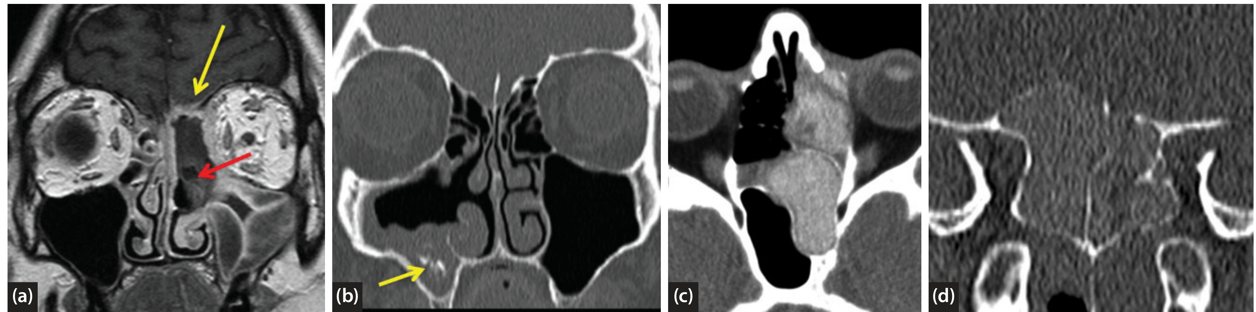
Scott-Brown's Fig. 117.9 - Composite fungal sinusitis imaging showing the black turbinate sign (a), fungal ball CT (b), and allergic fungal sinusitis CT (c,d)
As KJ Lee's describes: "'Black turbinate sign' or the lack of enhancement with Gd in the sinonasal mucosa on T1/T2 signal is highly suggestive of mucosal necrosis from fungal angioinvasion. On the other hand, extrasinus invasion would typically enhance with Gd."
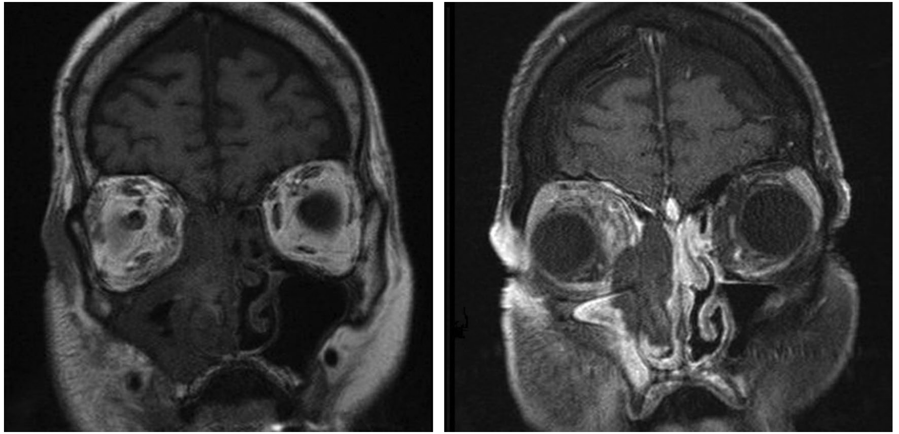
KJ Lee's Fig. 30-7 - Black turbinate sign on coronal post-contrast T1 MRI in AIFS
Suspicious MRI findings for IFRS (Cummings):
- Obliteration or infiltration of periantral or orbital fat
- Inflammatory changes in extraocular muscles
- Leptomeningeal enhancement
- Loss of contrast enhancement along areas of fungal invasion
Intracranial extension findings on MRI:
- Cavernous sinus thrombosis: swollen, non-enhancing sinus with compression of carotid artery on coronal post-contrast T1 MRI
- Meningitis: meningeal enhancement on contrast-enhanced MRI
- Leptomeningeal involvement
- Engorgement of superior ophthalmic vein (cavernous sinus sign)
- Intracranial abscess formation
2. CHRONIC INVASIVE FUNGAL SINUSITIS (CIFS)
CT Findings
- Hyperattenuating mass in one or more sinuses
- Destruction of sinus bony walls - prominent feature (unlike AIFS where this is late)
- The appearance may closely mimic malignancy - this is the primary differential diagnosis
- Bony erosion with extrasinus extension
As Scott-Brown's states: "CT imaging may mimic the features of malignancy, which is the primary differential in most cases. CT shows a hyperattenuating mass in one or more of the sinuses with destruction of the sinus bony walls."
KJ Lee's adds: affected sinuses show mass lesion with mucosal thickening, and bony erosion and extrasinus involvement are common.
MRI Findings
- T1-weighted: decreased signal intensity (hypointense)
- T2-weighted: decreased signal intensity (hypointense)
- Both T1 and T2 show iso- or hypointense signals relative to muscle in affected regions (Scott-Brown's, KJ Lee's)
- Invasion into anterior cranial fossa causes meningeal enhancement on contrast-enhanced MRI
- Cavernous sinus thrombosis: swollen non-enhancing sinus with carotid compression on coronal post-contrast T1 MRI
3. CHRONIC GRANULOMATOUS INVASIVE FUNGAL SINUSITIS (CGIFS)
CGIFS has a characteristic imaging pattern that differs from both AIFS and CIFS, and importantly from allergic fungal sinusitis (AFS).
CT Findings (Scott-Brown's)
- Homogeneous soft tissue opacification with no focal hyperdensities (key contrast to AFS, which shows heterogeneous hyperattenuation)
- Limited extent or unilateral disease
- No sinus expansion (unlike AFS)
- Focal bony erosion limited to areas of extrasinus extension
- The burden of disease is often more outside the sinuses (e.g., intraorbital) than within them - this reflects the indolent extrasinus spread typical of CGIFS
Imaging-Based Differential Diagnosis of Fungal Sinusitis
| Feature | AIFS | CIFS | CGIFS | AFS (Non-invasive) | Fungal Ball |
|---|---|---|---|---|---|
| CT density | Nonspecific mucosal thickening | Hyperattenuating mass | Homogeneous soft tissue, no focal hyperdensities | Heterogeneous hyperattenuation ("double density") | Iso-to-hyper attenuating mass, punctate calcifications |
| Sinus expansion | No | No | No | Yes | No |
| Bony erosion | Late, subtle | Prominent | Limited to extrasinus extension | Yes (pressure, not invasion) | No |
| T2 MRI | Low signal | Low signal | Low signal | Signal void (metal/desiccation) | Low signal |
| Enhancement | Loss of enhancement (black turbinate sign) | Reduced/absent in invaded areas | Extrasinus spread enhances | Peripheral enhancement | Non-enhancing core |
| Disease distribution | Unilateral, nasal predominant | Unilateral, sinus mass | More outside sinuses (orbit) | Often bilateral, pansinusitis | Single sinus |
Surveillance Imaging in IFRS
Cummings emphasizes that IFRS "is kept under stringent surveillance by serial endoscopy and radiographic imaging." Serial CT and/or MRI is performed every 2-3 days during active debridement to assess disease extent and treatment response.
Key indications for MRI over CT:
- Suspected orbital involvement (orbital fat obliteration, extraocular muscle changes)
- Suspected intracranial spread (meningeal enhancement, cavernous sinus thrombosis)
- Monitoring disease progression or treatment response in soft tissue planes
- When bony changes on CT are equivocal
Imaging Limitations and Caveats
- CT findings in AIFS may be minimal or absent early in disease - a normal CT does not exclude AIFS in an immunocompromised patient
- MRI signal void on T2 due to heavy metals in AFS can mimic a normally aerated sinus when interpreted without CT - always correlate CT and MRI together (Scott-Brown's)
- The "black turbinate sign" was initially described in only 2 patients with mucormycosis (Scott-Brown's) but has since been validated as an important early MRI finding
- Unilateral opacification combined with ANY bony erosion or extrasinus soft tissue change should be treated as IFRS until proven otherwise
Summary Algorithm
Immunocompromised patient + sinonasal symptoms
          ↓
Nasal endoscopy (pallor/necrosis → biopsy urgently)
          ↓
CT Paranasal Sinuses (HRCT)
 → Unilateral mucosal thickening/opacification
 → Bony erosion (late finding)
 → Extrasinus fat stranding
          ↓
MRI with Gadolinium
 → T2 hypointense signal in sinuses
 → Black turbinate sign (loss of mucosal enhancement)
 → Orbital fat obliteration
 → Leptomeningeal/cavernous sinus enhancement
 → Intracranial extension
          ↓
Tissue biopsy (frozen section) → confirm angioinvasion → immediate treatment
References:
- Cummings Otolaryngology Head and Neck Surgery (9780323612173), Chapters on Invasive Fungal Rhinosinusitis
- Scott-Brown's Otorhinolaryngology Head & Neck Surgery, Vol. 1 (9781138094611), Chapter 117 (Imaging) and Chapter 21 (Fungal Sinusitis)
- KJ Lee's Essential Otolaryngology (9781260122237), Chapter 30 (Fungal Sinusitis)
This is a shared conversation. Sign in to Orris to start your own chat.