I now have comprehensive content from textbooks, 2023 ESC guidelines, 2024 AHA/ACC HCM guidelines, and 2025-2026 guidance. Let me compile the master answer.Here is the complete, exam-ready deep-dive on cardiomyopathy, incorporating the 2023 ESC Guidelines, 2024 AHA/ACC HCM Guidelines, 2025 ACC ATTR guidance, and all major textbook content.
CARDIOMYOPATHY - Complete Medical Student Guide
Updated with 2023 ESC, 2024 ACC/AHA, and 2025-2026 Guidance
PART 1: DEFINITION & CLASSIFICATION
Cardiomyopathies are primary diseases of the myocardium not explained by coronary artery disease, hypertension, valvular disease, or congenital heart disease. Prevalence: ~1 in 250 adults.
2023 ESC Classification (NEW - Replaces Older Systems)
The 2023 ESC Guidelines on Cardiomyopathies introduced a revised classification centered on morphological and functional phenotypes:
| Type | Abbreviation | Core Feature |
|---|
| Hypertrophic cardiomyopathy | HCM | LV hypertrophy without loading cause |
| Dilated cardiomyopathy | DCM | LV/biventricular dilation + systolic dysfunction |
| Non-dilated left ventricular cardiomyopathy | NDLVC | NEW: non-ischemic LGE or fatty replacement in LV, or isolated LV hypokinesia WITHOUT dilation |
| Arrhythmogenic right ventricular cardiomyopathy | ARVC | RV fibrofatty replacement + arrhythmias |
| Restrictive cardiomyopathy | RCM | Stiff, noncompliant ventricles, preserved size |
NDLVC is the key new entity - it encompasses conditions previously called left ventricular arrhythmogenic cardiomyopathy (LVAC) and non-compaction cardiomyopathy (NCC). It requires CMR for diagnosis.
(2023 ESC Guidelines for the Management of Cardiomyopathies, Eur Heart J 44:3503-3626)
PART 2: DILATED CARDIOMYOPATHY (DCM)
Definition & Epidemiology
- LV (or biventricular) dilation + impaired systolic function in absence of CAD, valvular disease, or pericardial disease
- LVEF <40% (or severely reduced)
- Prevalence: ~1 in 250 adults; most common cardiomyopathy in children (58% of pediatric cases; peak age 18 months)
- Males and females approximately equally affected, except X-linked forms (male predominance)
(Goldman-Cecil Medicine)
Etiology & Genetics
Genetic DCM (20-50% of cases):
- Autosomal dominant most common pattern
- TTN (titin) truncating mutations = single most common cause (~25% familial, ~18% sporadic)
- Also: MYH7, TNNT2, TPM1, ACTC1 (sarcomeric proteins - note: SAME genes cause HCM when gain-of-function; cause DCM when loss-of-function)
- Lamin A/C (LMNA): arrhythmogenic form - AF, conduction disease, VT/VF often PRECEDE heart failure; high SCD risk - ICD recommended even with LVEF >35%
- Filamin C (FLNC): mixed arrhythmogenic + dilated phenotype; lethal VAs in adolescents/young adults
- Dystrophin (DMD gene, X-linked): Duchenne and Becker muscular dystrophy
- Desmin: combined cardiac + skeletal myopathy; intermediate filament protein
High-risk genotypes for SCD (ICD warranted even before LVEF <35%):
DES, DSP, DSC2, DCG2, FLNC, LMNA, PKP2, PLN, RBM20, SCN5A, TMEM43
Acquired DCM causes:
| Cause | Key Detail |
|---|
| Viral myocarditis | Coxsackievirus B, enteroviruses, parvovirus B19, HHV-6; viral footprints persist to late-stage |
| Alcohol | 6 drinks/day for 5-10 years; acetaldehyde direct cardiotoxin; >50% improve with abstinence |
| Anthracyclines | Doxorubicin - overt HF in 5-10% at doses >450 mg/m²; dose-dependent; within first year |
| Trastuzumab | Up to 11% develop DCM; reversible with withdrawal |
| Peripartum | See below |
| Tachycardiomyopathy | Reverses once tachycardia controlled |
| Hemochromatosis, sarcoidosis, radiation, cocaine, nutritional deficiencies | |
Clinical Presentation
- Symptoms of biventricular heart failure: exertional dyspnea, orthopnea, PND, peripheral edema
- First presentation may be sudden death or thromboembolic event (LV thrombus, especially at apex)
- Family history is important (30-40% familial clustering)
- May also present with arrhythmias (AF, VT, conduction disease) - especially with LMNA, FLNC mutations
Pathology
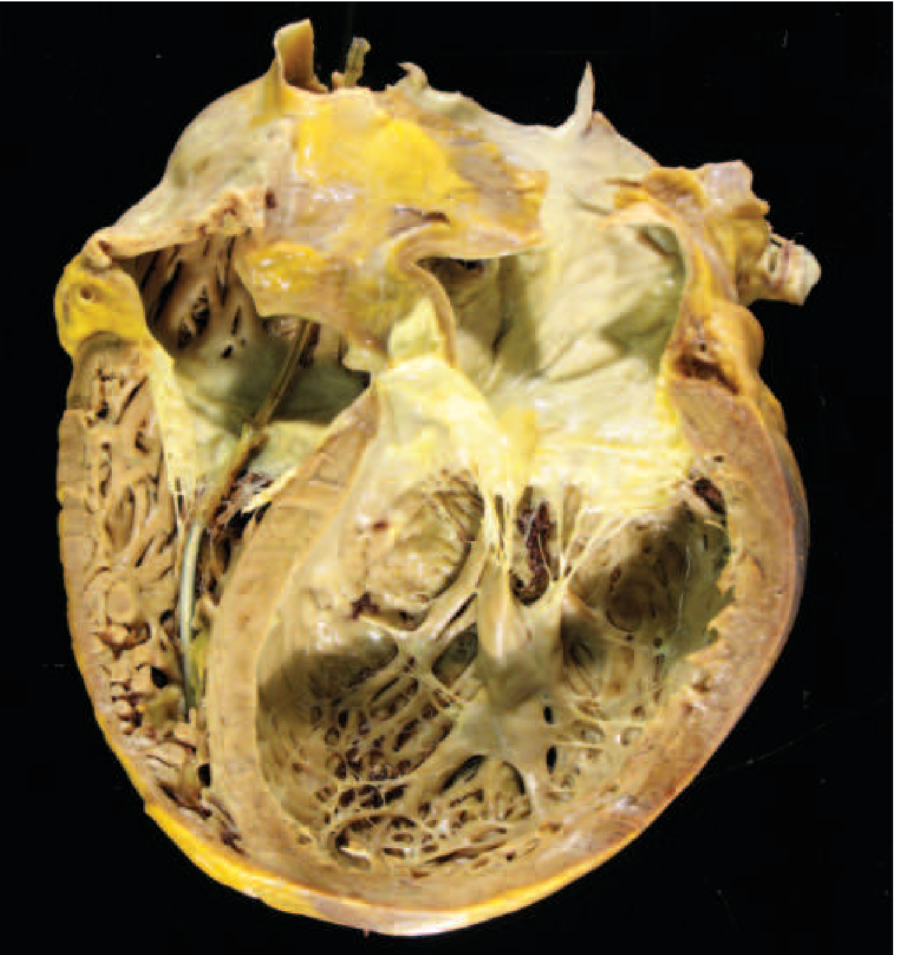
- Gross: Four-chamber dilation and hypertrophy; flabby, poorly contractile walls; mural thrombus (LV apex). Heart weight often >800 g (normal upper limit ~360 g)
- Histology: Nonspecific - myocyte hypertrophy + interstitial fibrosis (Masson trichrome shows blue collagen). No diagnostic features at end-stage
DCM gross specimen: massive biventricular dilation; apparent wall thinning despite overall hypertrophy (>800 g). Harrison's 22E, Fig. 267-6
Diagnosis
- TTE (transthoracic echo): first line; LV dilation, reduced LVEF, wall motion abnormalities
- CMR: gold standard for tissue characterization; mid-wall late gadolinium enhancement (LGE) is a key prognostic marker and predicts VT; 2023 ESC endorses CMR as primary diagnostic modality
- Biomarkers: BNP/NT-proBNP elevated; high-sensitivity troponin (consensus across all guidelines)
- ECG: LBBB, conduction defects, arrhythmias; LBBB + DCM = indication for CRT evaluation
- Genetic testing: Recommended when diagnostic phenotype established; essential for cascade family screening
Management: DCM
Heart Failure with Reduced EF (HFrEF) - Guideline-Directed Medical Therapy (GDMT):
All four pillars of the 2022 AHA/ACC/HFSA HF guideline apply:
| Drug Class | Example | Key Points |
|---|
| ACE inhibitor / ARB / ARNI | Sacubitril/valsartan (preferred over ACEi if tolerated) | Reduce mortality; sacubitril/valsartan superior to enalapril (PARADIGM-HF) |
| Beta-blocker | Carvedilol, bisoprolol, metoprolol succinate | Reduce SCD; carvedilol start 6.25 mg BID, titrate to 25 mg BID |
| MRA | Spironolactone, eplerenone | Reduce mortality and hospitalizations |
| SGLT2 inhibitor | Empagliflozin, dapagliflozin | Reduce HF hospitalizations/CV death (HFrEF + HFpEF); now standard of care |
2023 ESC novel recommendation: HF therapies may be initiated in asymptomatic patients with subclinical DCM/NDLVC phenotype to prevent adverse ventricular remodeling (Class IIb C).
Device therapy:
- CRT (cardiac resynchronization therapy): LVEF ≤35% + LBBB + QRS ≥150 ms (Class I) or QRS 120-149 ms (Class IIa)
- ICD for primary prevention: Standard threshold LVEF ≤35% despite 3+ months of GDMT
- Genotype-driven ICD (earlier implantation): LMNA or FLNC variants - ICD before LVEF <35% if ≥2 risk factors (male sex, NSVT, >10% fibrosis on CMR, non-missense mutations). 2023 ESC recommends SCD risk stratification by genotype even when LVEF >35% (Class IIa B)
Anticoagulation: In presence of LV thrombus, AF, or severe ventricular dilation
(Goldman-Cecil Medicine; Harrison's 22E; 2025 systematic review [PMID: 39674807])
PART 3: HYPERTROPHIC CARDIOMYOPATHY (HCM)
Definition & Epidemiology
- Unexplained LV hypertrophy in absence of abnormal loading conditions (valve disease, HTN, congenital defects)
- Prevalence: 0.2-0.5% worldwide; all racial groups
- Most common inherited cardiac disease
Genetics & Pathogenesis
- Autosomal dominant, gain-of-function mutations in sarcomeric contractile proteins
-
1,400 mutations in >9 genes identified; 50-60% of cases have identifiable sarcomere variants
- The Big 3 genes (account for 70-80%):
- MYH7 (beta-myosin heavy chain) - most common
- MYBPC3 (myosin-binding protein C)
- TNNT2 (troponin T)
- Mechanism: hypercontractility → increased energy use → net negative energy balance → compensatory hypertrophy + fibrosis
- Same genes (MYH7, etc.) cause DCM when loss-of-function; cause HCM when gain-of-function
Pathology
Gross:
- Massive hypertrophy WITHOUT ventricular dilation
- 90%: Asymmetric septal hypertrophy - septum disproportionately thicker than LV free wall
- 10%: concentric hypertrophy
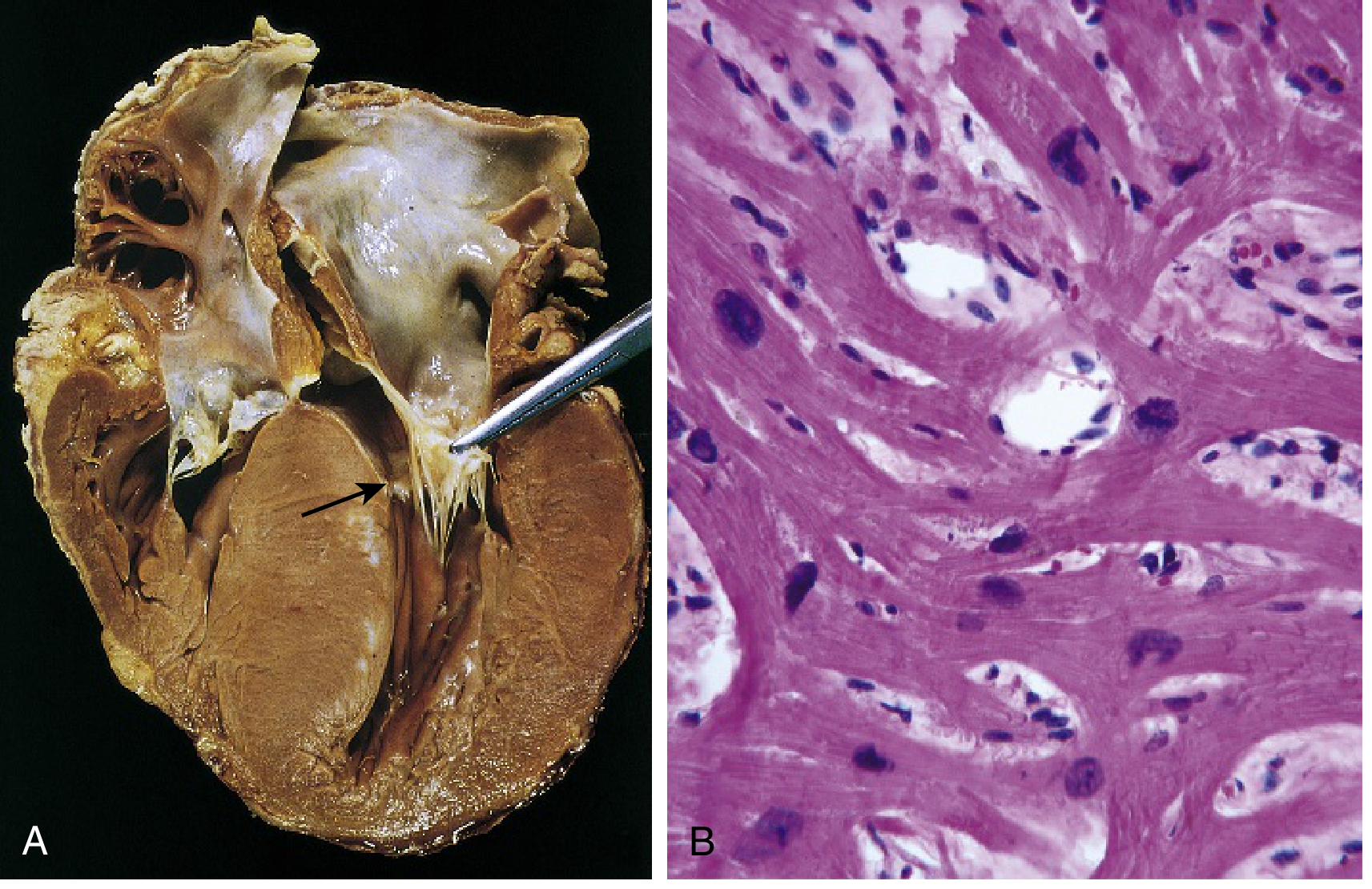
- LV cavity: "banana-shaped" on longitudinal section
- Fibrous endocardial plaque in LV outflow tract (from SAM contact with septum)
- Systolic anterior motion (SAM) of the mitral valve → LV outflow tract obstruction (LVOTO) in 1/3 of cases
Histology (hallmark triad):
- Extreme myocyte hypertrophy
- Myocyte disarray (haphazard, chaotic architecture) - the pathognomonic feature
- Interstitial fibrosis
(A) Septal muscle compressing LV outflow tract into a "banana" shape; LA enlarged; fibrous endocardial plaque visible (arrow). (B) Myocyte disarray and interstitial fibrosis on H&E. Robbins & Kumar Basic Pathology, Fig. 9.27
Clinical Features
- Often asymptomatic or mild; presents during postpubertal growth spurt or young adulthood
- Triad of symptoms: exertional dyspnea, chest pain (angina without CAD), syncope
- Harsh systolic ejection murmur at LLSB; increases with Valsalva, standing (decreased preload); decreases with squatting, leg raise
- Sudden cardiac death - most important complication; HCM is responsible for ~1/3 of SCD in athletes under 35
ECG findings in HCM
- LVH pattern
- Deep narrow Q waves in lateral and inferior leads (septal depolarization)
- T-wave abnormalities
- AF is common (20-25%)
2024 AHA/ACC HCM Guidelines - Key Updates (PMID: 38718139)
Diagnosis:
- TTE: first-line; LV wall thickness ≥15 mm (or ≥13 mm with family history/genetic variant)
- CMR: when TTE inconclusive; superior tissue characterization; LGE ≥15% of LV mass = major SCD risk factor
Medical management of obstructive HCM (oHCM):
| Step | Drug | Mechanism |
|---|
| 1st line | Beta-blockers (metoprolol, atenolol) | Reduce HR, prolong diastolic filling, reduce LVOT gradient |
| 2nd line | Verapamil or diltiazem | Improve diastolic relaxation |
| 3rd line | Disopyramide (+ beta-blocker) | Negative inotrope |
| NEW (2024) | Mavacamten (Camzyos) | Cardiac myosin inhibitor; reduces hypercontractility and LVOT gradient; first-in-class disease-modifying therapy |
Mavacamten (major 2024 guideline addition):
- Selective, reversible cardiac myosin ATPase inhibitor
- Approved by FDA; EXPLORER-HCM trial showed significant improvement in LVOT gradient, symptoms, and exercise capacity
- Monitoring: Echo every 4 weeks for first 12 weeks (FDA requirement); discontinue if LVEF drops <50%
- Contraindicated with strong CYP3A4 inhibitors; teratogenic
Septal reduction therapy (for refractory oHCM):
- Surgical septal myectomy (Morrow procedure) - gold standard; preferred for younger patients
- Alcohol septal ablation (ASA): catheter-based; intracoronary ethanol injection causes controlled septal MI; preferred for older/high surgical risk patients
- Indication: LVOT gradient ≥50 mmHg at rest or with provocation + refractory symptoms (NYHA III-IV) despite maximal medical therapy
SCD prevention / ICD indications (2024 ACC/AHA):
Primary prevention ICD - Class IIa if ≥1 major risk factor:
- Family history of SCD at young age (≤40 years)
- Unexplained syncope (especially within 6 months)
- LVEF <50%
- Apical aneurysm with transmural scar
- Extensive LGE on CMR (≥15% of LV mass)
- NSVT on ambulatory monitoring
Use the HCM Risk-SCD calculator to estimate 5-year SCD risk - includes LVOT gradient and LA diameter (2024 addition).
AF management in HCM (2024 guidelines):
- Anticoagulation recommended in ALL HCM patients with AF regardless of CHA2DS2-VASc score
- DOACs (direct oral anticoagulants) = first-line
- Vitamin K antagonists = second-line
- Rate control: beta-blockers, verapamil, diltiazem
- Rhythm control: amiodarone (effective in HCM); catheter ablation (higher relapse rate vs non-HCM)
Physical activity (2024 guidelines - paradigm shift):
- HCM patients should engage in mild to moderate recreational exercise for cardiovascular health
- Competitive/vigorous sports: individualized shared decision-making with HCM expert (no longer blanket prohibition)
- 2025 ACC/AHA Sports Participation Guidelines: athletes with confirmed HCM - "Reasonable to consider participation" (vs 2015 guideline: "Should not participate")
Family screening (2024 ACC/AHA):
- First-degree relatives of HCM proband: ECG + TTE every 12-18 months during adolescence; every 3-5 years in adults
- Genetic testing: recommended when clinical diagnosis established; valuable for cascade screening
- Genotype positive/phenotype negative: annual clinical evaluation
PART 4: RESTRICTIVE CARDIOMYOPATHY (RCM)
Definition
Decreased ventricular compliance → impaired diastolic filling. Systolic function usually preserved. Least common primary cardiomyopathy.
Pathology
- Ventricles normal or slightly enlarged (NOT dilated)
- Both atria markedly dilated (consequence of chronic elevated filling pressures)
- Myocardium is firm
- Mimics constrictive pericarditis clinically (must distinguish - important exam distinction!)
| Feature | Restrictive CMP | Constrictive Pericarditis |
|---|
| Pericardium | Normal | Thickened/calcified |
| Kussmaul sign | Present | Present |
| Pericardial knock | Absent | Present |
| Systemic JVD equalization | Less common | Characteristic |
| Treatment | Cause-specific | Pericardiectomy |
Causes
1. Cardiac amyloidosis (most clinically important)
| Type | Protein | Key Features |
|---|
| AL amyloid (primary) | Immunoglobulin light chains | Plasma cell dyscrasia (MM); light chains also directly cardiotoxic to myocytes |
| ATTR-wild type (senile systemic amyloidosis) | Normal transthyretin (TTR) | Older men (>60); carpal tunnel syndrome, spinal stenosis, biceps tendon rupture - classic clues |
| ATTR-hereditary | Mutant TTR | 4% of African Americans carry the Val122Ile mutation → 4-fold increased risk; younger onset |
Clues to cardiac amyloidosis on exam:
- "Sparkling" myocardium on echo (granular appearance)
- Low-voltage ECG despite LV hypertrophy on echo (pseudo-LVH)
- Bilateral carpal tunnel syndrome + HF
- Spinal stenosis + HF
- Macroglossia (AL type)
- "Cherry blossoms" (HCC-like) on CMR with specific LGE pattern
Diagnosis of ATTR:
- Pyrophosphate (PYP/DPD) bone scintigraphy - can diagnose ATTR noninvasively (Perugini Grade 2-3) if monoclonal protein screen is negative (SPEP, UPEP, serum FLC)
- If monoclonal protein present → biopsy needed to exclude AL
Treatment of ATTR cardiomyopathy (2025 ACC Concise Clinical Guidance, PMID: 41171219):
- Tafamidis (Vyndaqel) - TTR stabilizer; FDA-approved; reduces mortality (ATTR-ACT trial); first-line
- Acoramidis - TTR stabilizer; newer, approved 2023-2024
- Vutrisiran (siRNA) - TTR silencer; reduces TTR production; alternative approach
- SGLT2 inhibitors and MRAs: now endorsed as broadly effective HF therapies in ATTR
- Standard HF therapy: diuretics (symptom relief); digoxin relatively contraindicated (binds amyloid fibrils); calcium channel blockers generally avoided
2. Endomyocardial fibrosis
- Most common RCM worldwide (children/young adults, sub-Saharan Africa, tropical regions)
- Diffuse fibrosis of ventricular endocardium and subendocardium
- Often involves tricuspid and mitral valves
- Linked to nutritional deficiencies and helminthic infections
3. Loeffler endomyocarditis
- No geographic predilection
- Peripheral hypereosinophilia + eosinophilic tissue infiltrates
- Eosinophil major basic protein → endocardial necrosis → mural thrombus → organization → fibrosis
- Stages: necrotic → thrombotic → fibrotic
4. Other causes: Radiation fibrosis, sarcoidosis, storage diseases (mucopolysaccharidoses, Gaucher, Fabry), hemochromatosis, idiopathic
PART 5: ARRHYTHMOGENIC CARDIOMYOPATHY (ACM/ARVC)
Definition
Genetically determined structural cardiomyopathy with prominent arrhythmic presentation; fibro-fatty replacement of myocardium - predominantly RV but may involve LV (ALVC) or be biventricular.
- Responsible for up to 22% of SCD in athletes
- Prevalence: ~1 in 5,000
Genetics
- Desmosomal protein mutations (cell-cell adhesion):
- PKP2 (plakophilin-2) - most common
- DSP (desmoplakin)
- DSG2 (desmoglein-2)
- DSC2 (desmocollin-2)
- JUP (junction plakoglobin)
- Non-desmosomal: LMNA, desmin, others
- Naxos disease: autosomal recessive; PKP2 mutation; clinical triad = ARVC + woolly hair + palmoplantar keratoses (first described on Greek island of Naxos)
- Carvajal syndrome: DSP mutation; ARVC + woolly hair + palmoplantar keratosis + DILATED (not just RV) cardiomyopathy
Pathology
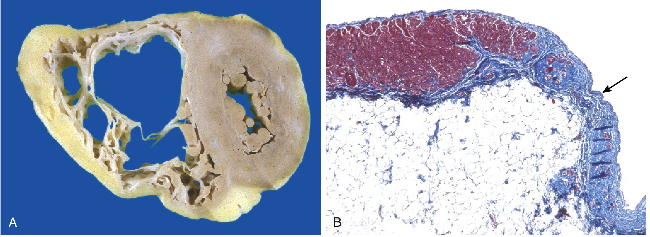
- RV markedly dilated; fibro-fatty replacement of RV free wall (almost transmural)
- LV may be involved to lesser extent
- "Triangle of dysplasia": RV inflow, outflow, and apex
(A) RV dilated with fibro-fatty replacement of free wall. (B) Masson trichrome: RV myocardium (red) replaced by fibrosis (blue) and fat (arrow). Robbins & Kumar Basic Pathology, Fig. 9.26
Diagnosis: Task Force Criteria (Major and Minor)
Diagnosis requires 2 major, 1 major + 2 minor, or 4 minor criteria across 6 categories:
| Category | Major | Minor |
|---|
| Structural/functional (RV) | RVEF ≤40% or RV dilation on echo/CMR/angiography | RVEF 41-45% or milder dilation |
| Tissue characterization | Fibro-fatty replacement on biopsy | |
| Repolarization | T-wave inversion V1-V3 (age >14, no RBBB) | T-wave inversion V1-V2 |
| Depolarization | Epsilon wave (V1-V3); terminal QRS >55 ms in V1-V3 | Late potentials on signal-averaged ECG |
| Arrhythmia | Sustained or NSVT with LBBB morphology, superior axis | LBBB-type VT from RVOT; >500 PVCs/24 hrs |
| Family history | ARVC in first-degree relative; pathogenic desmosomal mutation | |
Key ECG features:
- Epsilon wave (classic) - small high-frequency deflection after QRS in V1-V3
- T-wave inversions V1-V3 (most sensitive)
- Terminal QRS prolongation >55 ms in V1-V3 (distinct from RBBB)
- VT with LBBB morphology + superior or inferior axis = arises from RV
Management
- Lifestyle: Avoid strenuous exercise/competitive sports (especially endurance); exercise accelerates disease progression by increasing RV wall stress
- Beta-blockers: first-line for arrhythmia prevention
- Antiarrhythmics: sotalol (160-240 mg/day), amiodarone (200 mg/day maintenance); flecainide (endorsed by 2023 ESC for atrial and ventricular arrhythmias in ARVC)
- ICD indications:
- Definite: prior SCA, sustained VT, LVEF ≤35%
- Primary prevention (Class IIa): LVEF ≤50% + ≥1 major risk factor (NSVT, inducible VT, RV dysfunction, male sex, proband, >1 desmosomal mutation)
- Catheter ablation: for drug-refractory VT or frequent ICD shocks
- Heart failure therapy (ACEi, beta-blocker, diuretics) if progresses to severe HF
- Anticoagulation: if AF, marked ventricular dilation, or ventricular aneurysms
- Cardiac transplantation: for refractory end-stage disease
2025 ACC/AHA Sports Participation update:
- PKP2 ACM: risks may outweigh benefits (sports caution)
- Non-PKP2 ACM: can consider participation with shared decision-making
- Genotype positive/phenotype negative: reasonable to consider participation
(Goldman-Cecil Medicine; Fuster and Hurst's The Heart 15E; Harrison's 22E)
PART 6: PERIPARTUM CARDIOMYOPATHY (PPCM)
Definition & Diagnostic Criteria
DCM occurring in the last trimester of pregnancy OR within months of delivery, in the absence of prior heart disease, with:
- LVEF <45%
- No other identifiable cause of HF
Epidemiology
- US: 1 per ~3,000 live births; Haiti/Africa: 1 per ~300 live births (much higher in Black women)
- Risk factors: older maternal age, hypertension, preeclampsia, African-American race, multifetal pregnancies
Pathogenesis (multifactorial)
- Genetic: 15% have rare truncating variants (2/3 involve TTN); similar rate to idiopathic DCM → PPCM may be an unmasked familial DCM
- Prolactin cleavage: Oxidative stress → prolactin cleaved into a pro-inflammatory, vasotoxic 16-kDa fragment → endothelial and cardiomyocyte dysfunction
- Anti-angiogenic: Placenta releases soluble fms-like tyrosine kinase-1 (sFlt-1) → angiogenic imbalance
- Overlap with preeclampsia (~25% of PPCM vs 5% population)
Prognosis
- ~40-70% have normalization of LVEF within 5 years (better than idiopathic DCM)
- Patients with persistent LV dysfunction at baseline or African-American race: worse outcomes
- Subsequent pregnancy: Risk of recurrence in all patients, even those who normalize LV function
- Normalized LV function: HF recurs in 20% of subsequent pregnancies (mortality 0% in this group)
- Persistent LV dysfunction: HF recurs in 40%; maternal mortality 19%
Treatment
| Drug | Comment |
|---|
| Hydralazine + isosorbide dinitrate | Vasodilators safe in pregnancy (ACEi/ARBs/ARNI contraindicated - teratogenic) |
| Beta-blockers | Safe in pregnancy (carvedilol, metoprolol) |
| Diuretics | Judicious use for symptom relief |
| Bromocriptine (dopamine agonist) | Inhibits prolactin; small studies show improvement in LVEF; still controversial |
| Anticoagulation | If severe LV dysfunction; LMWH preferred (not warfarin - teratogenic in first trimester) |
| Digoxin | Safe in pregnancy; used for rate control |
(Creasy & Resnik's Maternal-Fetal Medicine; Goldman-Cecil Medicine)
PART 7: STRESS (TAKOTSUBO) CARDIOMYOPATHY
Overview
- Triggered by acute severe emotional, psychological, or physical stress
- Characterized by transient apical LV ballooning and dysfunction that is usually reversible
- "Takotsubo" = Japanese octopus trap (the shape of the LV during systole)
Epidemiology
- Predominantly postmenopausal women (>90% of cases)
- Accounts for 2-3% of all suspected ACS presentations
- COVID-19 increased incidence: 7.8% of ACS presentations during pandemic vs 1.5-1.8% pre-pandemic
Key Features (how to distinguish from STEMI)
| Feature | Takotsubo | STEMI |
|---|
| Troponin elevation | Mild (low) | Marked |
| ST elevation | May be present, diffuse | Focal, territory-matching |
| Coronary angiography | Normal (no culprit lesion) | Culprit stenosis/occlusion |
| Wall motion abnormality | Extends beyond single coronary territory (apical ballooning) | Territory of one coronary artery |
| Resolution | Complete in days-weeks | Permanent (infarct) |
| LVEF | Acutely reduced (~30%), recovers | Depends on infarct size |
Management
- Supportive care: beta-blockers, ACEi, diuretics for HF
- Avoid catecholamines (worsens condition)
- If cardiogenic shock: IABP or Impella; avoid vasopressors if possible
- Anticoagulation if LV thrombus suspected
- Prognosis: generally excellent with full recovery; mortality ~4-5% in hospitalized patients (usually comorbid conditions)
PART 8: OTHER / SECONDARY CARDIOMYOPATHIES
Infiltrative/Storage Diseases Mimicking HCM ("Pseudo-hypertrophy")
| Disease | Key Feature | Treatment |
|---|
| Fabry disease | X-linked; alpha-galactosidase A deficiency; glycolipid accumulation; classic: angiokeratomas, acroparesthesias, corneal opacities | Enzyme replacement therapy (agalsidase); oral chaperone migalastat |
| Pompe disease | Glycogen storage (acid maltase deficiency); massive hypertrophy in infants; short PR interval | Enzyme replacement |
| Anderson-Fabry | Above; also causes HCM phenotype | |
| Danon disease | LAMP2 mutation; X-linked; HCM + Wolff-Parkinson-White + skeletal myopathy | Supportive; transplant |
| LEOPARD/Noonan syndrome | RAS/MAPK pathway mutations; HCM phenotype + lentigines, ECG abnormalities, hypertelorism | Supportive |
| Cardiac sarcoidosis | Granulomatous infiltration; heart block, VT, DCM phenotype; MRI shows patchy LGE | Steroids; ICD |
Chemotherapy-Induced Cardiomyopathy (Cardio-oncology)
| Agent | Mechanism | Management |
|---|
| Anthracyclines (doxorubicin) | Oxidative damage, topoisomerase II inhibition; dose-dependent; overt HF at >450 mg/m² | Dexrazoxane (cardioprotective); carvedilol or enalapril prophylaxis |
| Trastuzumab | HER2 pathway; up to 11% DCM; reversible with withdrawal | Withdraw + standard HFrEF therapy |
| Immune checkpoint inhibitors | T-cell off-target myocarditis; rare but potentially fatal | High-dose steroids; stop immunotherapy |
| Tyrosine kinase inhibitors (sunitinib) | Reduce LVEF; more with CAD | Withdraw; standard HFrEF therapy |
PART 9: MYOCARDITIS (Related Entity)
Overview
Myocardial damage due to inflammatory infiltrates. Important because:
- Can cause acute HF
- Can progress to DCM (DCM is considered a potential sequela of viral myocarditis)
Causes
| Category | Examples |
|---|
| Viral (most common in US) | Coxsackievirus A and B, enteroviruses; parvovirus B19, HHV-6; CMV, HIV, influenza |
| Bacterial | Diphtheria (Corynebacterium diphtheriae), Lyme disease (Borrelia) |
| Parasitic | Chagas disease (Trypanosoma cruzi) - most important worldwide cause of cardiomyopathy in Latin America |
| Autoimmune | SLE, polymyositis; giant cell myocarditis (rare but fatal) |
| Toxic | Anthracyclines, cocaine |
| Eosinophilic | Drug hypersensitivity, hypereosinophilic syndrome |
Giant Cell Myocarditis
- Rapidly fatal without treatment
- Diagnosis: endomyocardial biopsy (multinucleated giant cells)
- Treatment: prolonged immunosuppression (cyclosporine + steroids); transplant if refractory
Chagas Cardiomyopathy
- Trypanosoma cruzi - vector: Reduviid (kissing) bug
- Affects 8-10 million in Latin America; leading cause of cardiomyopathy in endemic regions
- Chronic phase: dilated cardiomyopathy + right bundle branch block + left anterior fascicular block (classic ECG pattern) + apical aneurysm + thromboembolic events
- Treatment: Benznidazole/nifurtimox (antiparasitic); standard HFrEF therapy; ICD for VT/VF
PART 10: SUMMARY COMPARISON TABLE (HIGH-YIELD EXAM REFERENCE)
| Feature | DCM | HCM | RCM | ARVC |
|---|
| LV size | Dilated (4-chamber) | Normal-enlarged (thick-walled) | Normal/small | RV dilated |
| Wall thickness | Thin (relative) | Massively thick | Variable | RV thin (fibro-fatty) |
| Systolic function | Severely impaired (LVEF <40%) | Preserved (LVEF 50-80%) | Preserved | RV impaired |
| Diastolic function | Secondarily impaired | Primarily impaired (stiff) | Severely impaired | - |
| Pathognomonic finding | 4-chamber dilation | Myocyte disarray + septal hypertrophy | Biatrial dilation + firm myocardium | Fibro-fatty RV replacement |
| Key histology | Myocyte hypertrophy + fibrosis | Disarray + fibrosis | Amyloid/fibrosis | Fibroadipose tissue |
| Murmur | Functional MR | Harsh systolic ejection murmur (LLSB); increases with Valsalva | None/S3-S4 | None |
| Top genetic causes | TTN, LMNA, DMD (loss-of-function) | MYH7, MYBPC3, TNNT2 (gain-of-function) | TTR, J chain | PKP2, DSP, DSG2 (desmosomal) |
| Key drug (new) | SGLT2i, sacubitril/valsartan | Mavacamten | Tafamidis/acoramidis (ATTR) | Beta-blocker + sotalol |
| SCD risk | Moderate (LMNA/FLNC high) | High (young athletes) | Low-moderate | Very high (athletes) |
| ICD threshold | LVEF ≤35% (earlier for LMNA/FLNC) | ≥1 major risk factor | Cause-specific | Per Task Force criteria |
| Sports restriction | Shared decision-making (2025) | Shared decision-making (2024) | Low-moderate activity | Avoid strenuous exercise |
PART 11: GUIDELINE SUMMARY REFERENCE
| Guideline | Year | Key Highlights |
|---|
| 2023 ESC Guidelines on Cardiomyopathies | 2023 | New NDLVC entity; CMR as primary diagnostic modality; genotype-driven SCD risk stratification; HF therapy in asymptomatic subclinical phenotype (IIb C); flecainide endorsed in ARVC; anticoagulation in HCM/amyloid regardless of risk score |
| 2024 AHA/ACC HCM Guidelines | 2024 | Mavacamten introduced (cardiac myosin inhibitor); shared decision-making for sports; DOACs first-line in HCM+AF regardless of CHA2DS2-VASc; updated SCD risk factors (LGE ≥15%, apical aneurysm); HCM Risk-SCD calculator updated |
| 2025 ACC ATTR Guidance | 2025-2026 | Tafamidis and acoramidis (TTR stabilizers) + vutrisiran (siRNA silencer) now endorsed; SGLT2i and MRA for all ATTR-CM patients; PYP scintigraphy for noninvasive diagnosis |
| 2025 ACC/AHA Sports Participation | 2025 | Paradigm shift - cardiomyopathy no longer uniformly excludes athletes; gene+/phenotype- can participate; confirmed HCM = "reasonable to consider participation" |
| 2022 AHA/ACC/HFSA HF Guidelines | 2022 | Four pillars of GDMT (ACEi/ARB/ARNI + BB + MRA + SGLT2i) for ALL HFrEF including DCM |
| 2025 ESC Myocarditis Guidelines | 2025 | Updated diagnostic criteria; genetic screening for family members; new CMR Lake Louise criteria |
Sources: Goldman-Cecil Medicine; Harrison's Principles of Internal Medicine, 22E; Robbins & Kumar Basic Pathology (9th ed.); Robbins, Cotran & Kumar Pathologic Basis of Disease; Creasy & Resnik's Maternal-Fetal Medicine; Fuster & Hurst's The Heart, 15E; Tintinalli's Emergency Medicine;
2024 AHA/ACC HCM Guideline (PMID: 38718139);
DCM Guidelines Systematic Review 2025 (PMID: 39674807);
2025 ACC ATTR Guidance (PMID: 41171219); 2023 ESC Cardiomyopathy Guidelines (Eur Heart J 44:3503-3626)