
Expansile medullary glioma with mass effects on cm cistern ,adjacent cerebellar hemispheres & pontomedullary junction understanding with images & treatment plan
medullary glioma MRI brainstem expansile mass
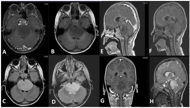
This diagnostic imaging panel consists of eight MRI slices (axial, sagittal, and coronal planes) demonstrating an expansile intra-axial mass in the brainstem, characteristic of a Diffuse Midline Glioma (DMG). The mass is primarily centered in the pons and exhibits a classic radiological profile: hypointense signal on T1-weighted pre-contrast (B, F) and post-contrast (A, E, G) sequences, indicating a lack of significant enhancement. T2-weighted (D, H) and FLAIR (C) sequences show the lesion as uniformly hyperintense. Anatomically, the tumor causes significant expansion of the pons, leading to the obliteration of the prepontine cistern and encasement of the basilar artery. Posteriorly, the mass compresses the fourth ventricle, though no proximal obstructive hydrocephalus is currently evident. Sagittal and coronal views (E, F, G, H) illustrate the longitudinal extent of the lesion, stretching from the pontomesencephalic junction superiorly to the pontomedullary junction inferiorly, with characteristically ill-defined, infiltrative borders. This series serves as a pedagogical example of the typical imaging features and local mass effect of pontine gliomas in neuro-oncology.
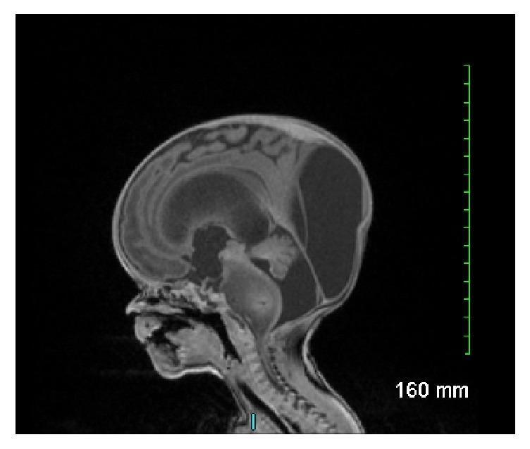
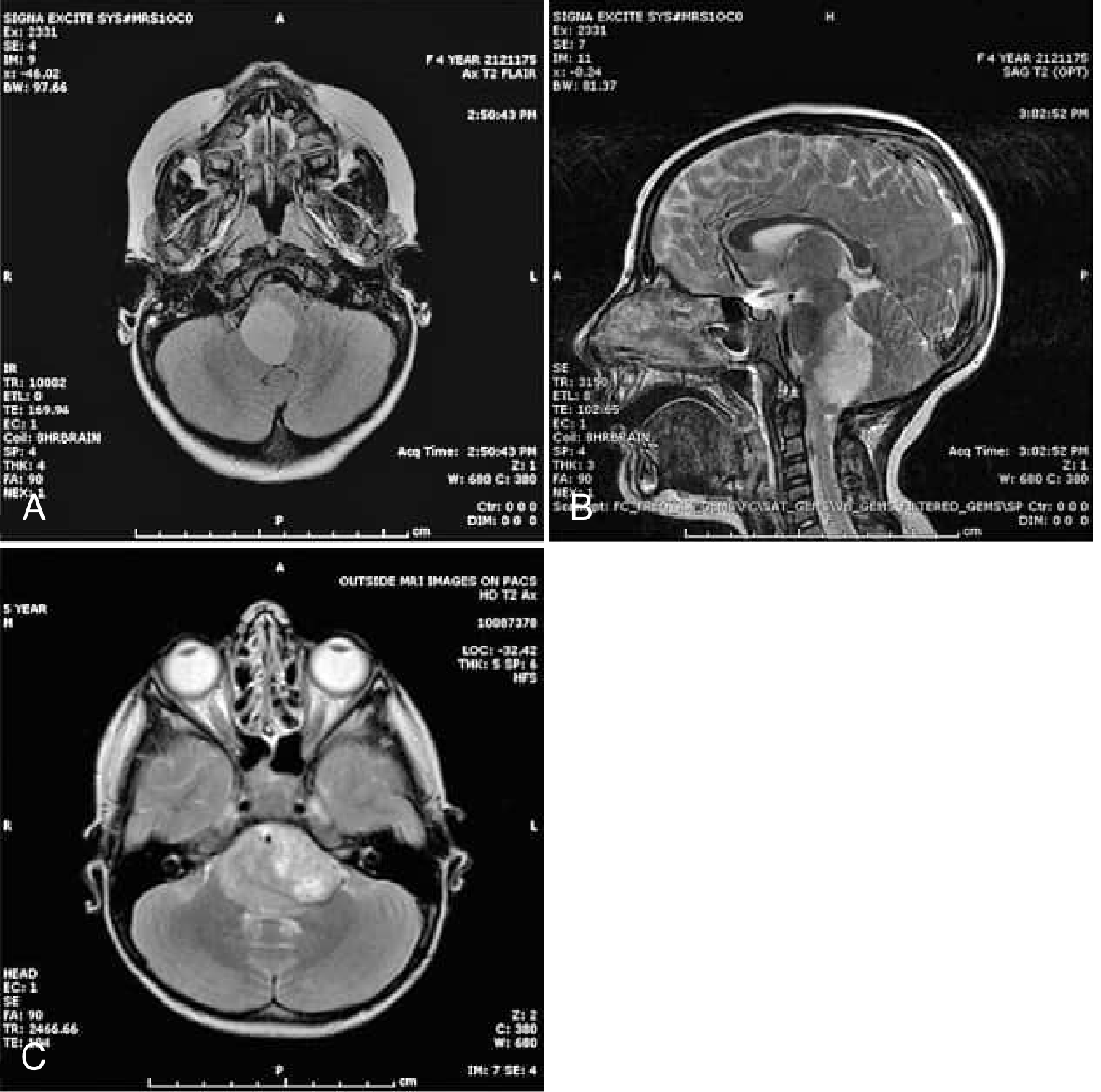
This sagittal T1-weighted fetal MRI scan demonstrates significant intracranial pathology in a developing fetus. The primary finding is a large, expansile, and poorly demarcated mass involving the brainstem, centered in the pons. The mass appears heterogeneously signal-intense and significantly expands the diameter of the pons, extending superiorly towards the midbrain and inferiorly toward the medulla. Secondary to this mass, there is evidence of severe obstructive hydrocephalus, characterized by marked dilation of the lateral ventricles and the third ventricle, with associated thinning of the overlying cerebral mantle and macrocephaly. The posterior fossa shows displacement of the cerebellar structures. This visual presentation is highly characteristic of a congenital brainstem glioma, specifically a diffuse intrinsic pontine glioma (DIPG) or similar anaplastic neoplasm, illustrating the typical obstructive complications and mass effect associated with neonatal neuro-oncology.
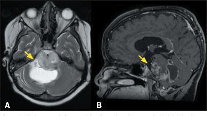
This diagnostic imaging composite displays two magnetic resonance imaging (MRI) sequences of the brain in a pediatric patient, illustrating a diffuse midline glioma affecting the brainstem. Image A is an axial T2-weighted sequence showing an expansile, infiltrative lesion located in the pons. The lesion demonstrates a heterogeneous signal intensity, with hyperintense focal areas (yellow arrow) indicative of cystic components or internal necrosis. The mass effect is evident as it expands the pontine contours. Image B is a sagittal, fat-saturated, T1-weighted sequence with gadolinium contrast enhancement. It reveals the solid portions of the tumor exhibit irregular, heterogeneous enhancement (yellow arrow), suggesting varying vascularity and blood-brain barrier disruption within the mass. The clinical presentation and imaging features are characteristic of a high-grade pediatric-type diffuse midline glioma, specifically the H3K27-altered molecular subtype. This case highlights the typical neuroradiological appearance of aggressive midline gliomas in children, focusing on expansile growth, signal heterogeneity, and variable contrast uptake.
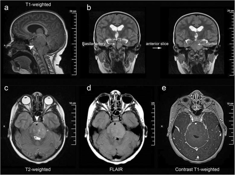
Multi-modal MRI series of the brain demonstrating a Diffuse Intrinsic Pontine Glioma (DIPG). (a) T1-weighted sagittal view shows an infiltrative, expansile mass centered within the pons. (b) Coronal T2-weighted images reveal the exophytic tumor portion extending into the prepontine and suprasellar cisterns, notably encasing the basilar artery. (c-e) Axial sequences highlight characteristic signal patterns: the lesion is hyperintense on T2-weighted (c) and Fluid-Attenuated Inversion Recovery (FLAIR) (d) sequences, indicating significant vasogenic edema or tumor infiltration. (e) Post-gadolinium T1-weighted axial sequence shows a conspicuous absence of enhancement, a typical feature of DIPG indicating an intact blood-brain barrier within the tumor. Associated secondary findings include obstructive hydrocephalus, evidenced by dilated lateral ventricles in the coronal views. This imaging series illustrates the classic neuroradiological criteria for pediatric brainstem gliomas, emphasizing anatomical distortion of the brainstem, cisternal extension, and specific signal characteristics across varying MRI sequences.
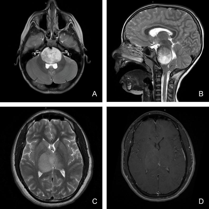
This composite of four magnetic resonance imaging (MRI) scans illustrates the diagnostic features of pediatric diffuse midline glioma in different anatomical locations. Panels A (axial) and B (sagittal) display T2-weighted sequences showing a large, hyperintense, and expansile mass involving the brainstem (pons), characteristic of a diffuse intrinsic pontine glioma (DIPG). The lesion demonstrates an infiltrative growth pattern, causing significant local mass effect and compression of adjacent structures. Panels C and D focus on a thalamic presentation. Panel C is an axial T2-weighted image showing a homogenous, hyperintense lesion within the right thalamus with poorly defined margins extending into the surrounding white matter tracts. Panel D shows the corresponding axial T1-weighted post-gadolinium sequence, revealing minimal to heterogeneous contrast enhancement within the thalamic mass. These images highlight key radiological hallmarks including the tumor's midline location, infiltrative nature, perilesional edema, and variable vascular permeability as indicated by enhancement patterns, which are critical for the clinical classification of these high-grade pediatric gliomas.
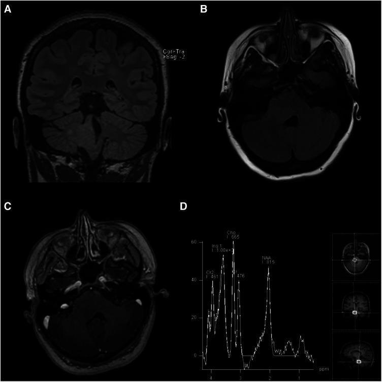
This composite of four images (A-D) presents a multi-modal MRI evaluation of a brainstem lesion. Panels (A) Coronal T2-weighted FLAIR and (B) Axial T2-weighted FLAIR demonstrate an ill-defined, hyperintense, and expansile mass within the right pons. The lesion extends into the right middle cerebellar peduncle and the right posterolateral medulla, causing partial effacement of the fourth ventricle. Panel (C) represents an axial post-gadolinium contrast T1-weighted image, which shows no contrast enhancement, indicating a preserved blood-brain barrier within the lesion. Panel (D) provides Magnetic Resonance Spectroscopy (MRS) data, showing a markedly increased choline (Cho) peak and a significantly decreased N-acetylaspartate (NAA) peak. This metabolic profile (high Cho:NAA ratio) is characteristic of increased cellular turnover and loss of neuronal integrity, typically seen in infiltrating gliomas such as Diffuse Intrinsic Pontine Glioma (DIPG). The findings are essential for neurosurgical and oncological education regarding the diagnostic imaging and metabolic characteristics of pediatric-type diffuse low-grade gliomas in adult patients.
medullary glioma brainstem treatment
cisterna magna cerebellomedullary cistern anatomy posterior fossa
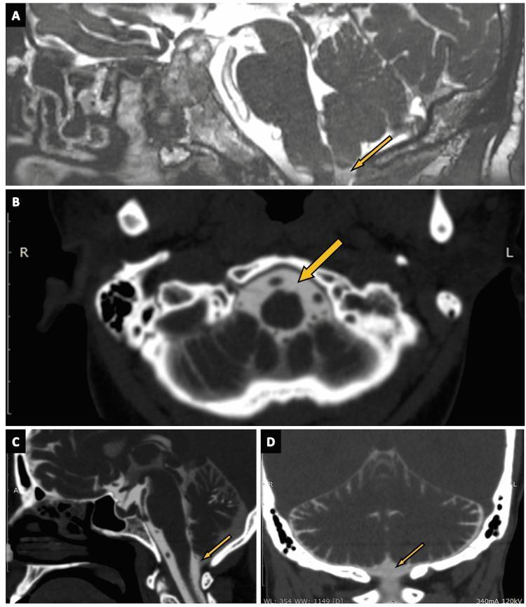
This composite diagnostic image illustrates the cisterna magna across multiple modalities and planes. Panel A shows a sagittal view using 3D constructive interference in steady state (CISS) MRI, where the cistern appears as a dark, hypointense fluid-filled space. Panels B, C, and D utilize CT cisternography in axial, sagittal, and coronal planes, respectively, to visualize the cistern as a region of low attenuation enhanced by contrast material. Anatomically, the cisterna magna is located in the posterior cranial fossa, situated inferior to the cerebellum and posterior to the medulla oblongata. It is identified by arrows in each panel. The images demonstrate the cistern's relationship to surrounding structures, including the brainstem, cerebellar vermis, and the foramen magnum. This visual guide serves as a clinical reference for identifying normal subarachnoid cistern anatomy, which is essential for diagnosing conditions such as CSF rhinorrhea or posterior fossa malformations. The target audience includes medical students and radiology residents specializing in neuroimaging.
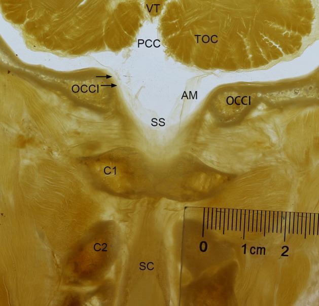
A frontal P45 plastinated anatomical section of the human occipito-cervical junction (OCJ) passing through the posterior arch of the atlas (C1). The specimen demonstrates the morphological relationship between the cerebellum and the upper cervical spine. Key structures labeled include the tuber of vermis (VT) and tonsil of cerebellum (TOC) superiorly. The posterior cerebellomedullary cistern (PCC) is visible above the foramen magnum. A prominent, rhombic-shaped subarachnoid space (SS) is observed between the cerebellum and the atlas (C1), bordered by the arachnoid membrane (AM) and the spinal dura mater (indicated by arrows). Bony landmarks identified include the occipital bone (OCCI), the atlas (C1), and the axis (C2). The spinal cord (SC) is visible descending through the vertebral canal. The image illustrates the continuity of the subarachnoid space from the posterior cranial fossa into the upper cervical vertebral canal, highlighting the anatomy of the cisterna magna and the dural sac at the craniocervical transition. A metric ruler is included for scale.
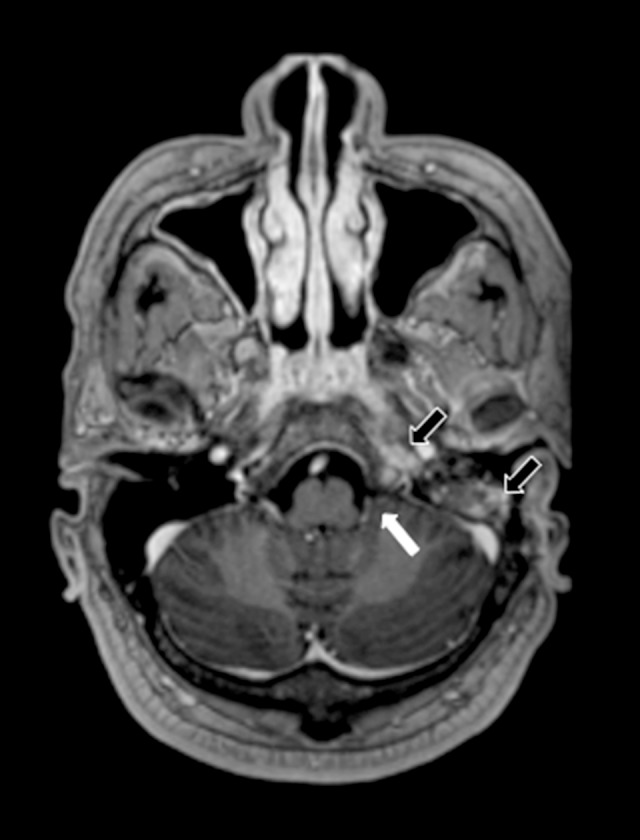
**Imaging Modality:** Magnetic Resonance Imaging (MRI), Axial plane. **Sequence:** Post-contrast T1-weighted image. **Anatomical Region:** Posterior fossa and skull base, specifically focusing on the left cerebellomedullary cistern (cisterna magna), petrous apex, and mastoid air cells. **Observed Findings:** * **Cisterna Magna:** A white arrow indicates the left cerebellomedullary cistern. There is a notable absence of a peripherally enhancing collection, signifying the resolution of a previously documented abscess. * **Skull Base and Mastoid:** Black arrows highlight persistent, though reduced, contrast enhancement within the left petrous apex and the mastoid air cells. These features are consistent with improving petrositis and mastoiditis. * **Cranial Nerves:** Normalization of contrast uptake in the vicinity of the lower cranial nerves is observed compared to previous involvement. **Diagnostic Context:** Post-treatment follow-up of a skull base infection (likely secondary to otomastoiditis) with secondary intracranial extension. The image demonstrates therapeutic response characterized by the resolution of a focal infectious collection (abscess) while showing residual inflammatory changes in the adjacent osseous structures.
diffuse midline glioma H3K27M radiation ONC201
PMID: 42317168
PMID: 39941789
pontomedullary junction anatomy cerebellar hemispheres fourth ventricle diagram
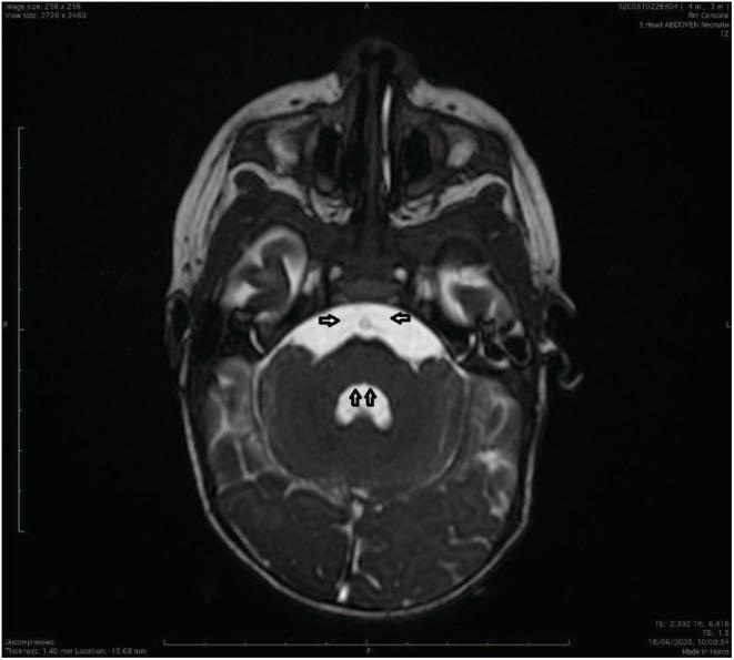
This diagnostic image is an axial T2-weighted MRI of the brain at the level of the pons and fourth ventricle, illustrating neuroanatomical abnormalities characteristic of Möbius syndrome. The image reveals a bilateral absence of the facial colliculi in the dorsal pontine tegmentum, indicated by vertical arrows, leading to an atypical 'inverted V' shape of the fourth ventricle. Furthermore, there is a radiologic absence of the abducens nerves (Cranial Nerve VI) along their expected cisternal trajectory from the pontomedullary junction, marked by horizontal arrows. The surrounding middle cerebellar peduncles and cerebellar hemispheres appear otherwise intact. These findings—the absence of the facial colliculus (formed by facial nerve fibers looping around the abducens nucleus) and the lack of visible abducens nerves—are pathognomonic radiological signs of brainstem hypoplasia involving the VIth and VIIth cranial nerve nuclei, correlating with clinical symptoms of congenital facial paralysis and impaired lateral eye movement.
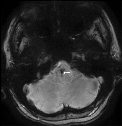
**Imaging Modality:** Magnetic Resonance Imaging (MRI), Susceptibility-Weighted Imaging (SWI). **Anatomical Region:** Axial section of the posterior fossa, specifically localized to the pontomedullary junction of the brainstem. **Observed Pathology:** Capillary telangiectasia. **Characteristic Visual Features:** The image demonstrates a focal area of signal loss (hypointensity) within the brainstem parenchyma, indicated by a white arrow. This "blooming" effect is characteristic of paramagnetic substances, such as deoxyhemoglobin or hemosiderin, within slow-flow vascular channels. The lesion exhibits a subtle, radiating "caput medusa" morphology, characterized by fine, brush-like peripheral vessels converging toward a central point. There is no evidence of surrounding edema or mass effect on the adjacent cerebellar hemispheres or fourth ventricle. **Key Diagnostic Features:** The marked hypointensity on susceptibility-weighted sequences in an asymptomatic location is highly suggestive of a capillary telangiectasia. The lack of signal void on T1 or T2 sequences (not shown) and the presence of fine radiating vasculature help differentiate this from other vascular malformations like cavernous malformations or developmental venous anomalies (DVA).
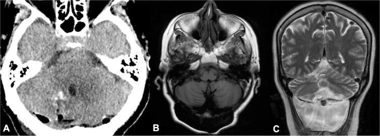
This composite figure displays postoperative and follow-up neuroimaging of a 66-year-old patient following surgical resection of neurocysticercosis. Panel (A) is an axial non-contrast head CT demonstrating the posterior fossa. Key features include the brainstem, cerebellum, and a centrally located fourth ventricle, with postoperative changes visible in the right cerebellar hemisphere indicating successful cyst removal. Panel (B) shows an axial T1-weighted brain MRI at the 2-year follow-up, confirming the absence of parasitic cyst recurrence. Panel (C) is a coronal T2-weighted brain MRI illustrating the posterior fossa anatomy and supratentorial brain parenchyma. This sequence highlights the cerebrospinal fluid (CSF) spaces, including the lateral and fourth ventricles. A hyperintense signal area at the pontomedullary junction suggests a CSF fistula, a known postoperative complication. The images collectively demonstrate the radiological assessment of treatment success for neurocysticercosis and the identification of long-term postoperative sequelae in a clinical neurosurgical context.
Expansile Medullary Glioma: Mass Effect on CM Cistern, Cerebellar Hemispheres & Pontomedullary Junction
1. Anatomical Context - Understanding the Region
The Cerebellomedullary (CM) Cistern (Cisterna Magna)
The Pontomedullary Junction (PMJ)
- Cranial nerve VI nuclei (abducens) just above
- Cranial nerve VII (facial) looping around CN VI in the facial colliculus
- Cranial nerve IX, X, XI, XII originating from the medulla
- Pyramidal tracts and medial lemniscus passing through
- Middle and inferior cerebellar peduncles connecting brainstem to cerebellar hemispheres
2. Pathology and Classification
What is a Medullary Glioma?
| Classification | Key Features |
|---|---|
| Diffuse Midline Glioma, H3K27-altered | H3K27M mutation in histone 3; most aggressive; includes DIPG and medullary variants |
| Diffuse Astrocytoma (IDH-mutant, grade 2-3) | Rare in medulla; IDH1 mutations in ~80% of adult DAs |
| Focal/Nodular Pilocytic Astrocytoma | Dorsal exophytic type; better prognosis; often enhancing cystic mural nodule |
| Cervicomedullary glioma | Straddles medulla and upper cervical cord; often low-grade in children |
Expansile Growth Pattern - Two Subtypes
- Asymmetric enlargement of the medulla/pons
- Hypointense T1, heterogeneously hyperintense T2/FLAIR
- Poor margins; engulfs cranial nerve nuclei and tracts
- Minimal or no gadolinium enhancement (intact blood-brain barrier in low-grade)
- Displaces and encroaches on cisterns and adjacent structures
- H3K27M mutation common
- Grows out of the dorsal medullary surface into the CM cistern
- May look like an extra-axial mass on imaging
- Often pilocytic histology; may enhance with contrast
- Surgical resection sometimes feasible
- Long-term survivors documented
Adams & Victor's Principles of Neurology, 12th Ed. distinguishes these subtypes, noting that "focal or nodular tumors tend to occur in the dorsal brainstem and often protrude in an exophytic manner" with a better prognosis than diffusely infiltrating tumors.
3. MRI Imaging - Features & Mass Effect
Characteristic MRI Findings
T2/FLAIR: Hyperintense signal reflecting tumor infiltration and edema
Gadolinium (T1+C): Variable - diffuse type typically non-enhancing; exophytic/focal type may show ring or nodular enhancement
DWI: Usually restricted diffusion in high-grade variants
MR Spectroscopy: Elevated Cho:NAA ratio - high choline (increased cell turnover), reduced NAA (neuronal loss)
MRI Spectrum of Mass Effect on Specific Structures
| Structure | Mass Effect Observed |
|---|---|
| CM cistern | Obliteration of posterior CSF space; dorsal exophytic component may fill/displace cistern |
| Cerebellar hemispheres | Direct compression via inferior/middle cerebellar peduncles; displaced laterally and posteriorly |
| Pontomedullary junction | Loss of normal brainstem contours; engulfment of CN nuclei VI, VII; trapped basilar artery |
| 4th ventricle | Compression from below/anteriorly → obstructive hydrocephalus |
| Foramen magnum | Inferior extension may cause cervicomedullary syndrome |
4. Clinical Presentation
From Medullary Involvement
- Dysphagia (CN IX, X nuclei)
- Hoarseness and dysphonia (nucleus ambiguus)
- Hiccups, nausea, early satiety - tegmental medullary involvement near the dorsal vagal nucleus and area postrema
- Respiratory irregularities - pre-Botzinger complex
From Pontomedullary Junction Mass Effect
- Ipsilateral CN VI palsy (diplopia, lateral gaze failure)
- Ipsilateral CN VII palsy (peripheral facial weakness)
- Contralateral hemiparesis (corticospinal tract compression - crossed signs)
- Hemisensory deficits (medial lemniscus)
- Nystagmus, vertigo (vestibular nuclei)
From Cerebellar Hemisphere Compression
- Ipsilateral limb ataxia (dentate-thalamic pathway disruption)
- Dysmetria, dysdiadochokinesia
- Wide-based unsteady gait
From CM Cistern Obliteration / 4th Ventricle Compromise
- Raised intracranial pressure: Headache (morning, positional), vomiting, papilledema
- Obstructive hydrocephalus (late sign)
- Downward Herniation Risk if craniocervical mass extends inferiorly
Adams & Victor's Neurology notes: "Patients with medullary tumors may present with a long history of dysphagia, hoarseness, ataxia, and hemiparesis. Cervicomedullary tumors may cause medullary or upper cervical symptoms, neck discomfort, weakness or numbness of the hands, and asymmetrical quadriparesis."
5. Molecular Biology - The H3K27M Mutation
- Inhibits Polycomb Repressive Complex 2 (PRC2), reducing global H3K27 trimethylation
- Leads to widespread epigenetic dysregulation and transcriptional activation of oncogenes
- Confers WHO Grade 4 behavior regardless of histological appearance
- Strongly associated with poor prognosis (median OS ~9-11 months from diagnosis)
- Diagnostic: H3K27M IHC on biopsy confirms Diffuse Midline Glioma (DMG) classification
6. Treatment Plan
Step 1: Diagnosis Confirmation
- MRI brain + spine with gadolinium (full neuraxis staging)
- Stereotactic biopsy - now standard even for posterior fossa/brainstem lesions; yields molecular diagnosis (H3K27M, IDH1/2, BRAF, FGFR status)
- LP for CSF cytology if leptomeningeal spread suspected
- Ophthalmology for papilledema assessment
Step 2: Management of Mass Effect / Raised ICP
| Situation | Intervention |
|---|---|
| Obstructive hydrocephalus | Endoscopic 3rd ventriculostomy (ETV) or VP shunt |
| Acute neurological deterioration | IV dexamethasone (0.5-1 mg/kg/day); osmotic therapy if needed |
| Dorsally exophytic component accessible | Surgical debulking/resection of exophytic portion (subtotal resection possible) |
| Diffuse infiltrating type | Surgery NOT indicated as primary treatment; biopsy only |
Adams & Victor's: "Treatment of the diffuse infiltrative type has generally been radiation, and if increased ICP develops as a result of hydrocephalus, ventricular shunting of CSF becomes necessary."
Step 3: Radiation Therapy - The Primary Treatment
- Dose: 54 Gy in 30 fractions (1.8 Gy/fraction) for standard fractionation
- Alternative: Hypofractionated RT (39 Gy in 13 fractions) for very young children (<3 years) or palliation
- Field: Involved field RT (IFRT); whole-brain RT only if leptomeningeal disease
- Response: ~80-90% of patients show temporary clinical improvement; median duration 6-9 months
- Re-irradiation: At recurrence, re-RT (24-30 Gy) provides meaningful palliation in selected patients; craniospinal irradiation for leptomeningeal disease
Step 4: Systemic Therapy
A. Dordaviprone (ONC201) - FDA Approved 2024 (Recurrent H3K27M DMG)
- Mechanism: Dopamine receptor D2/D3 antagonist + ClpP mitochondrial protease activator; inhibits Akt/ERK kinases; epigenetically reprograms tumor microenvironment
- FDA approval: Accelerated approval for recurrent/progressive H3K27M DMG in adults and children ≥6 months (2024)
- Evidence: Phase 1/2 trials - ORR 20%, disease control rate 40%, median duration of response 11.2 months
- Dosing: 375 mg once weekly orally
- Side effects: Fatigue, headache, vomiting, nausea (generally well tolerated)
- Current status: NOT a substitute for radiation; used at recurrence. The ACTION trial is evaluating it in newly diagnosed H3K27M DMG
B. Other Systemic Options
| Agent | Class | Status |
|---|---|---|
| Panobinostat | HDAC inhibitor (epigenetic) | Phase I/II trials; promising in H3K27M |
| Nimotuzumab + Vinorelbine | Anti-EGFR + vinca alkaloid | Phase II evidence; combination used post-RT |
| BRAF/MEK inhibitors (dabrafenib + trametinib) | Targeted therapy | For BRAF V600E-mutant pilocytic/focal gliomas |
| BRAF + MEK inhibitors | Targeted | BRAF-mutant DA with early malignant transformation |
| CAR-T cell therapy | Immunotherapy | GD2-directed CAR-T (H3K27M): early phase trials; CSF delivery |
| Convection-enhanced delivery (CED) | Local drug delivery | Direct brainstem infusion bypassing BBB; experimental |
| Oncolytic viruses (DNX-2401) | Virotherapy | Preliminary series show possible prolonged survival |
C. Conventional Chemotherapy
- Adjuvant chemotherapy has generally not been helpful for diffuse infiltrating brainstem gliomas
- Temozolomide: No survival benefit in DIPG/medullary diffuse type (multiple failed trials)
- May be considered for IDH-mutant variants in adults (similar to supratentorial DAs)
Step 5: Supportive & Palliative Care
- Dexamethasone: Titrate to lowest effective dose; long-term side effects (Cushingoid features, hyperglycemia, myopathy)
- Antiepileptics: If seizures develop
- Nutritional support: Dysphagia management; PEG tube if aspiration risk
- Speech and swallowing therapy
- Physical and occupational therapy: Manage ataxia and hemiparesis
- Palliative care integration: Early referral; goals-of-care discussions given poor prognosis
7. Prognosis
| Tumor Type | Median OS | Notes |
|---|---|---|
| Diffuse infiltrating medullary/DIPG (H3K27M) | 9-15 months | Worst prognosis group |
| Diffuse infiltrating (H3K27 wild-type) | 15-24 months | Slightly better |
| Focal dorsally exophytic (pilocytic) | 5-10 years | Best prognosis; surgical candidates |
| IDH-mutant diffuse astrocytoma (adult) | 5-10+ years | Slower progression |
| Cervicomedullary (low-grade) | 3-7 years | Depends on extent of resection |
8. Summary Algorithm
Posterior fossa mass on imaging
        ↓
Medullary location? → MRI with gadolinium (brain + spine)
        ↓
Morphology assessment
    ├── Diffuse/infiltrating? → Biopsy only → Molecular profiling
    │           ↓
    │   H3K27M+ → Diffuse Midline Glioma (WHO Grade 4)
    │   → Focal RT 54 Gy + Dordaviprone at recurrence
    │
    └── Focal/exophytic? → Surgical exploration
                ↓
        Resection of exophytic mural nodule
        → RT if residual/progressive
        → Targeted therapy if BRAF-mutant
        ↓
Hydrocephalus? → ETV or VP shunt
Raised ICP? → Dexamethasone ± osmotherapy
Leptomeningeal? → Consider craniospinal RT
- Adams & Victor's Principles of Neurology, 12th Ed. - Glioma of the Brainstem (Ch. 30)
- Bradley & Daroff's Neurology in Clinical Practice - Diffuse Intrinsic Pontine Glioma/Diffuse Midline Glioma (Ch. 75)
- Lo Greco et al. H3K27M DIPG management narrative review, Cancers 2025 (PMID 39941789)
- Nuss et al. Dordaviprone pharmacotherapy review, J Oncol Pharm Pract 2026 (PMID 42317168)
- Akdemir et al. H3K27M DMG diagnostic/therapeutic challenges, Pract Radiat Oncol 2024 (PMID 38704025)