
Additional ECG placement methods apart from 12 lead ECG placement.
Reading File
Reading File
Searching the Web
alternative ECG lead placement methods 15 lead 18 lead right-sided posterior leads guideline
Searching PubMed
electrocardiography lead placement
Finding Sources
Searching Images
diagram posterior ECG lead placement V7 V8 V9 and right-sided V4R ECG
Reading File
Apart from the standard 12-lead ECG, common additional or modified lead-placement methods include:
-
15-lead ECG
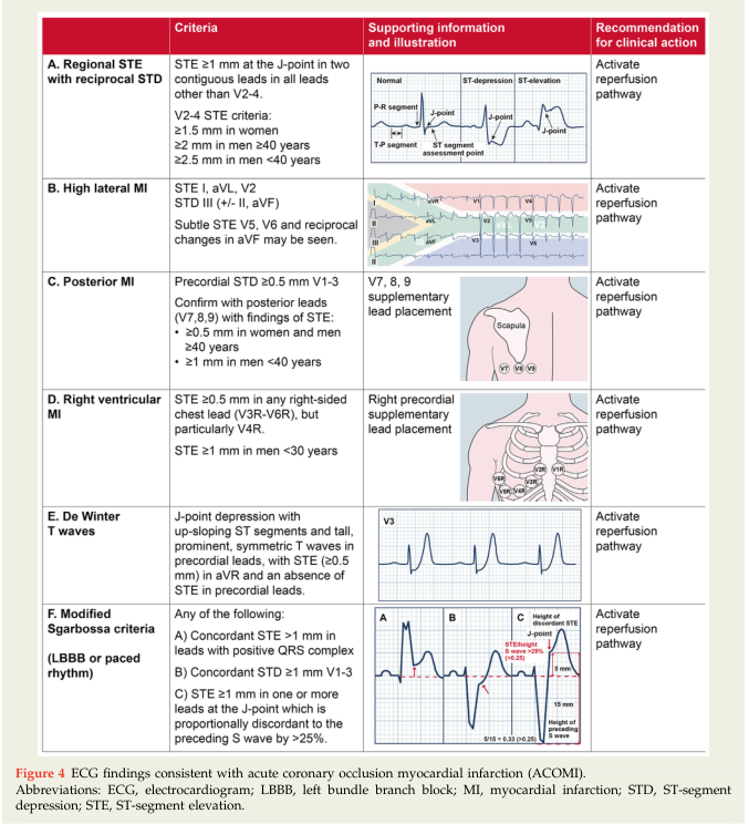
- Adds V4R for the right ventricle and V7-V9 for the posterior wall.
- Used especially in inferior STEMI, suspected right-ventricular infarction, hypotension with ACS, or ST depression in V1-V3.
- V4R: 5th right intercostal space, right mid-clavicular line.
- V7: left posterior axillary line, at the same horizontal level as V6.
- V8: left mid-scapular line, same level.
- V9: left paraspinal line, same level.
Rosen's notes that V4R is the preferred right-sided lead in a 15-lead tracing and that posterior ST elevation may be subtle. [Rosen's Emergency Medicine, p. 2110]
-
Right-sided precordial ECG
- Records V1R-V6R or sometimes V1R-V8R as mirror images across the right chest.
- Assesses right ventricular ischemia/infarction, commonly with inferior MI.
- V4R is the most useful single right-sided lead.
-
Posterior ECG
- Uses V7, V8, V9 to detect posterior myocardial infarction that may be missed on routine leads.
- Particularly useful when V1-V3 show horizontal ST depression with tall R waves.
-
18-lead ECG
- Combines the standard 12 leads with posterior leads V7-V9 and right-sided leads, usually V3R-V5R or V4R.
- Offers broader coverage of posterior and right-ventricular injury. A recent systematic review compared 18-lead with 12-lead ECG in acute coronary syndrome (PMID 41338016).
-
24-lead ECG / body-surface mapping
- Adds more chest electrodes for detailed spatial mapping of cardiac electrical activity.
- Used mainly in specialist cardiology, electrophysiology, research, or selected diagnostic settings, rather than routine emergency assessment.
-
Modified limb-lead placement
- Limb electrodes may be placed on the torso rather than wrists and ankles in emergency, surgical, exercise, or monitoring settings.
- This reduces movement artifact and is practical when limb access is difficult.
- Important: label the tracing, because torso limb placement can alter frontal-plane wave amplitudes and axis, so it is not fully interchangeable with standard diagnostic placement.
-
Mason-Likar modification
- A common torso-limb placement used for exercise ECG/stress testing and telemetry:
- Arm electrodes below the clavicles
- Leg electrodes on lower torso/iliac areas
- It improves stability during movement but changes the ECG appearance somewhat.
- A common torso-limb placement used for exercise ECG/stress testing and telemetry:
-
Lewis lead
- A modified lead configuration designed to make atrial activity, especially P waves, easier to see.
- Helpful when distinguishing atrial activity from ventricular tachycardia or clarifying AV dissociation.
-
Fontaine bipolar precordial leads
- Specialized right-precordial configuration, mainly used in arrhythmia evaluation, including arrhythmogenic right ventricular cardiomyopathy.
- Can help display epsilon waves or low-amplitude atrial activity.
Key clinical point: Additional leads are not replacements for a correctly recorded standard 12-lead ECG. They are targeted extensions, most often for suspected posterior or right-ventricular MI. ECG lead positioning overview
This is a shared conversation. Sign in to Orris to start your own chat.