
# THE ELECTRON TRANSPORT CHAIN (ETC) — A COMPREHENSIVE TREATISE --- ## TABLE OF CONTENTS 1. Introduction & Overview 2. Location and Structural Organization 3. Historical Background 4. Thermodynamic Basis 5. Components of the ETC 6. Complex I (NADH: Ubiquinone Oxidoreductase) 7. Complex II (Succinate: Ubiquinone Oxidoreductase) 8. Ubiquinone (Coenzyme Q) 9. Complex III (Ubiquinol: Cytochrome c Oxidoreductase) 10. Cytochrome c 11. Complex IV (Cytochrome c Oxidase) 12. Complex V (ATP Synthase) — Oxidative Phosphorylation 13. The Proton Motive Force 14. Chemiosmotic Hypothesis 15. P/O Ratios and ATP Yield 16. Shuttle Systems 17. Inhibitors of ETC 18. Uncouplers of ETC 19. Regulation of ETC 20. Reactive Oxygen Species (ROS) 21. Supercomplex Organization (Respirasomes) 22. Clinical Correlations (Comprehensive) 23. Summary Tables --- ## 1. INTRODUCTION & OVERVIEW The **Electron Transport Chain** (also called the **respiratory chain** or **mitochondrial electron transport system**) is the final common pathway through which electrons derived from fuel molecules (carbohydrates, fats, amino acids) are transferred to molecular oxygen (O₂), the terminal electron acceptor. This process is coupled with the generation of a **proton gradient** across the inner mitochondrial membrane, which drives **ATP synthesis** via **oxidative phosphorylation**. ### Key Concepts: - **Oxidation** = loss of electrons - **Reduction** = gain of electrons - Electrons flow from carriers with **more negative** (lower) reduction potential to carriers with **more positive** (higher) reduction potential - The overall reaction: > **NADH + H⁺ + ½O₂ → NAD⁺ + H₂O** (ΔG°' = −220 kJ/mol = −52.6 kcal/mol) > **FADH₂ + ½O₂ → FAD + H₂O** (ΔG°' = −152 kJ/mol = −36.4 kcal/mol) This large free energy change is released in a **stepwise manner** through the chain, preventing explosive release of energy and allowing efficient capture in the form of ATP. --- ## 2. LOCATION AND STRUCTURAL ORGANIZATION ### 2.1 Mitochondrial Anatomy The ETC is located in the **inner mitochondrial membrane (IMM)**. **Mitochondria** are **double-membrane** organelles: | Structure | Features | |-----------|----------| | **Outer Mitochondrial Membrane (OMM)** | Permeable to molecules <5 kDa via porins (VDAC — Voltage-Dependent Anion Channel); contains monoamine oxidase (MAO) | | **Intermembrane Space (IMS)** | ~pH 7.0 (slightly acidic relative to matrix); contains cytochrome c, adenylate kinase | | **Inner Mitochondrial Membrane (IMM)** | Highly folded into **cristae**; IMPERMEABLE to most ions and molecules; contains ETC complexes, ATP synthase, cardiolipin; protein:lipid ratio ≈ 75:25 | | **Mitochondrial Matrix** | ~pH 7.8 (slightly alkaline); contains TCA cycle enzymes, β-oxidation enzymes, mtDNA, mitoribosomes, PDH complex | ### 2.2 Cardiolipin - **Diphosphatidylglycerol** — a unique phospholipid found almost exclusively in the IMM - Contains **4 fatty acid chains** (usually linoleic acid) - Essential for the function of Complex III, Complex IV, and ATP synthase - Provides structural support and maintains the impermeability of the IMM to protons > **🔴 CLINICAL: Barth Syndrome** > - X-linked recessive disorder > - Mutation in the **tafazzin gene (TAZ)** on Xq28 > - Tafazzin is a transacylase required for **cardiolipin remodeling** > - Results in abnormal cardiolipin → impaired ETC function > - Features: **dilated cardiomyopathy, skeletal myopathy, neutropenia, 3-methylglutaconic aciduria, growth retardation** > - Predominantly affects males ### 2.3 Mitochondrial DNA (mtDNA) - **Circular, double-stranded DNA** — 16,569 base pairs - Encodes **37 genes**: 13 polypeptides (all ETC/ATP synthase subunits), 22 tRNAs, 2 rRNAs - **Maternal inheritance** (mitochondria come from the ovum) - No histones, limited repair mechanisms → high mutation rate (~10x nuclear DNA) - **Heteroplasmy**: a cell can contain a mixture of normal and mutant mtDNA - **Threshold effect**: disease manifests when the proportion of mutant mtDNA exceeds a critical threshold **Subunits encoded by mtDNA:** | Complex | mtDNA-encoded subunits | |---------|----------------------| | Complex I | ND1, ND2, ND3, ND4, ND4L, ND5, ND6 (7 subunits) | | Complex II | **None** (all 4 subunits nuclear-encoded) | | Complex III | Cytochrome b (1 subunit) | | Complex IV | COX I, COX II, COX III (3 subunits) | | Complex V | ATPase 6, ATPase 8 (2 subunits) | | **Total** | **13 subunits** | > **🔴 CLINICAL: Mitochondrial inheritance patterns** > - All children of an affected mother may be affected (but variable expressivity due to heteroplasmy) > - An affected father **CANNOT** transmit the disease > - Tissues with high energy demands (brain, heart, skeletal muscle, retina, kidney) are most affected → explains the clinical phenotype of mitochondrial diseases --- ## 3. HISTORICAL BACKGROUND | Year | Contribution | |------|-------------| | 1897 | **Buchner** — cell-free fermentation | | 1900 | **Warburg** — identified "Atmungsferment" (respiratory enzyme), later identified as cytochrome oxidase | | 1925 | **Keilin** — rediscovered cytochromes (a, b, c) using spectroscopy | | 1937 | **Kalckar** — linked oxidation to phosphorylation | | 1948 | **Kennedy & Lehninger** — localized oxidative phosphorylation to mitochondria | | 1961 | **Peter Mitchell** — proposed the **Chemiosmotic Hypothesis** (Nobel Prize 1978) | | 1964 | **Hatefi** — isolated the four respiratory complexes | | 1979 | **Anderson** — sequenced human mtDNA | | 1994 | **John Walker** — determined the crystal structure of ATP synthase (Nobel Prize 1997 with Paul Boyer) | --- ## 4. THERMODYNAMIC BASIS ### 4.1 Standard Reduction Potential (E°') Electrons flow from a **more negative** E°' to a **more positive** E°'. | Redox Pair | E°' (Volts) | |------------|-------------| | NAD⁺/NADH | −0.32 V | | FAD/FADH₂ (free) | −0.22 V | | FAD/FADH₂ (in Complex II) | +0.03 V | | CoQ/CoQH₂ | +0.04 V | | Cytochrome b (Fe³⁺/Fe²⁺) | +0.07 V | | Cytochrome c₁ (Fe³⁺/Fe²⁺) | +0.22 V | | Cytochrome c (Fe³⁺/Fe²⁺) | +0.25 V | | Cytochrome a (Fe³⁺/Fe²⁺) | +0.29 V | | Cytochrome a₃ (Fe³⁺/Fe²⁺) | +0.55 V | | ½O₂/H₂O | +0.82 V | ### 4.2 Free Energy Relationship $$\Delta G°' = -nF\Delta E°'$$ Where: - n = number of electrons transferred - F = Faraday constant (96,485 J/V·mol = 23.06 kcal/V·mol) - ΔE°' = E°'(acceptor) − E°'(donor) **For NADH oxidation:** ΔE°' = +0.82 − (−0.32) = +1.14 V ΔG°' = −2 × 96.485 × 1.14 = −220 kJ/mol **For FADH₂ oxidation:** ΔE°' = +0.82 − (−0.03 for bound FAD) = +0.79 V ΔG°' ≈ −152 kJ/mol ### 4.3 Sites of Sufficient Free Energy for ATP Synthesis The ΔG°' for ATP synthesis = +30.5 kJ/mol (minimum required) Three sites have sufficient ΔG drop to drive proton pumping → ATP synthesis: 1. **Complex I**: NADH → CoQ (ΔE = 0.36 V → ΔG = −69.5 kJ/mol) 2. **Complex III**: CoQH₂ → Cytochrome c (ΔE = 0.21 V → ΔG = −40.5 kJ/mol) 3. **Complex IV**: Cytochrome c → O₂ (ΔE = 0.57 V → ΔG = −110 kJ/mol) Complex II has **insufficient** free energy change to pump protons. --- ## 5. COMPONENTS OF THE ETC — ELECTRON CARRIERS ### 5.1 Types of Electron Carriers | Carrier Type | Carries | Examples | |-------------|---------|----------| | **NAD⁺/NADH** | 2 electrons + 1 H⁺ (hydride ion) | Soluble in matrix | | **FAD/FADH₂** | 2 electrons + 2 H⁺ | Prosthetic group (tightly bound) | | **FMN/FMNH₂** | 2 electrons + 2 H⁺ | Prosthetic group in Complex I | | **Coenzyme Q (Ubiquinone)** | 2 electrons + 2 H⁺ | Mobile carrier in lipid bilayer | | **Iron-Sulfur Centers (Fe-S)** | 1 electron | Multiple types: [2Fe-2S], [3Fe-4S], [4Fe-4S] | | **Cytochromes (heme iron)** | 1 electron (Fe²⁺ ↔ Fe³⁺) | Cyt b, c₁, c, a, a₃ | | **Copper Centers** | 1 electron (Cu⁺ ↔ Cu²⁺) | CuA, CuB in Complex IV | ### 5.2 Flavin Nucleotides (FMN and FAD) - Derived from **Riboflavin (Vitamin B₂)** - Can accept **1 or 2 electrons** (can form a semiquinone radical intermediate) - FMN is the **first electron acceptor** in Complex I - FAD is the prosthetic group in Complex II (succinate dehydrogenase) ### 5.3 Iron-Sulfur Clusters - Also called **non-heme iron** proteins - Contain iron atoms coordinated with **inorganic sulfide (S²⁻)** and/or **cysteine sulfur** from the protein - Types: - **[2Fe-2S]** — Rieske center in Complex III (unique: one Fe coordinated by 2 histidines) - **[4Fe-4S]** — most common, found in Complexes I, II, III - **[3Fe-4S]** — found in Complex II - Transfer **only 1 electron** at a time despite having multiple iron atoms - The most complex arrangement is in **Complex I**: contains **8 Fe-S clusters** forming an electron "wire" ~95 Å long ### 5.4 Heme Groups and Cytochromes Cytochromes are proteins with **heme prosthetic groups** containing iron that undergoes reversible Fe²⁺/Fe³⁺ transitions. | Cytochrome | Heme Type | Location | Unique Features | |-----------|-----------|----------|-----------------| | Cyt b(bL and bH) | Heme B (protoporphyrin IX) | Complex III | Two b-type cytochromes in Q cycle | | Cyt c₁ | Heme C (covalently attached) | Complex III | Part of the complex | | Cyt c | Heme C | IMS (mobile carrier) | Small, water-soluble, peripheral protein; also involved in apoptosis | | Cyt a | Heme A | Complex IV | Contains formyl group and hydroxyethylfarnesyl side chain | | Cyt a₃ | Heme A | Complex IV | Contains the binuclear center (with CuB) where O₂ binds | ### 5.5 Copper Centers - **CuA** in Complex IV: dinuclear copper center; first electron acceptor from cytochrome c - **CuB** in Complex IV: paired with heme a₃ in the binuclear center; directly involved in O₂ binding and reduction --- ## 6. COMPLEX I — NADH:UBIQUINONE OXIDOREDUCTASE (EC 1.6.5.3) ### 6.1 Structure - **Largest** respiratory complex - **~1,000 kDa** (1 MDa) in mammals - **45 subunits** in mammals (7 encoded by mtDNA: ND1–ND6, ND4L; 38 nuclear-encoded) - Shaped like an **"L"** or a **boot**: - **Hydrophilic arm** (peripheral arm): projects into the matrix; contains FMN and all Fe-S clusters - **Hydrophobic arm** (membrane arm): embedded in the IMM; contains the proton-pumping machinery - Contains: - **1 FMN** (flavin mononucleotide) - **8 iron-sulfur clusters**: N1a [2Fe-2S], N1b [2Fe-2S], N2 [4Fe-4S], N3 [4Fe-4S], N4 [4Fe-4S], N5 [4Fe-4S], N6a [4Fe-4S], N6b [4Fe-4S] - The Fe-S clusters form a **~95 Å electron transfer wire** ### 6.2 Mechanism 1. **NADH** binds to the hydrophilic arm and donates **2 electrons as a hydride ion (H⁻)** to **FMN** → **FMNH₂** 2. FMNH₂ passes electrons **one at a time** through the chain of **8 Fe-S clusters** 3. The terminal Fe-S cluster **N2** (highest potential in the chain) donates electrons to **ubiquinone (CoQ)** bound at the ubiquinone-binding site at the junction of the two arms 4. CoQ is reduced: **Q → QH⁻ (semiquinone) → QH₂ (ubiquinol)** 5. The energy released drives **conformational changes** in the membrane arm that translocate **4 H⁺** from the matrix to the IMS ### 6.3 Proton Pumping Mechanism - The membrane arm contains **three antiporter-like subunits** (ND2, ND4, ND5) each pumping **1 H⁺** - A **fourth proton** is pumped at the junction/interface near the ubiquinone-binding site (ND1/ND6 region) - Total: **4 H⁺ pumped per NADH** (per 2 electrons) - The mechanism involves a **long-range conformational coupling** — a "piston-like" mechanism transmitted through a long amphipathic helix connecting the antiporter subunits ### 6.4 Overall Reaction > **NADH + H⁺ + Q + 4H⁺(matrix) → NAD⁺ + QH₂ + 4H⁺(IMS)** ### 6.5 Inhibitors of Complex I | Inhibitor | Source/Type | Mechanism | |-----------|-----------|-----------| | **Rotenone** | Plant insecticide (from Derris plant roots) | Blocks electron transfer from Fe-S cluster N2 to ubiquinone | | **Piericidin A** | Streptomyces antibiotic | CoQ analog; competes at Q-binding site | | **Barbiturates (Amobarbital/Amytal)** | Pharmaceutical | Block at the same site as rotenone | | **MPP⁺ (1-methyl-4-phenylpyridinium)** | Active metabolite of MPTP | Inhibits Complex I | > **🔴 CLINICAL: Rotenone and Parkinson's Disease** > - Chronic rotenone exposure in animal models produces **Parkinsonism** including selective dopaminergic neuron death and **Lewy body-like inclusions** > - Dopaminergic neurons in the **substantia nigra pars compacta** are especially vulnerable due to high energy demands and high oxidative stress > - Epidemiological studies link pesticide exposure (rotenone, paraquat) to increased Parkinson's disease risk > **🔴 CLINICAL: MPTP Toxicity (Drug-induced Parkinsonism)** > - **MPTP** (1-methyl-4-phenyl-1,2,3,6-tetrahydropyridine) is a contaminant of synthetic heroin ("designer drug") > - MPTP crosses the blood-brain barrier → taken up by astrocytes → converted to **MPP⁺** by **MAO-B (monoamine oxidase B)** > - MPP⁺ is selectively taken up by dopaminergic neurons via the **dopamine transporter (DAT)** > - MPP⁺ inhibits **Complex I** → ATP depletion, ROS generation → selective death of dopaminergic neurons > - Produces **acute, irreversible Parkinsonism** > - This discovery was pivotal in understanding Parkinson's disease pathogenesis > - Treatment: L-DOPA, dopamine agonists (same as idiopathic Parkinson's) > - Prevention: MAO-B inhibitors (selegiline/rasagiline) can block MPTP → MPP⁺ conversion > **🔴 CLINICAL: Leber's Hereditary Optic Neuropathy (LHON)** > - **Mitochondrial inheritance** (maternal) > - Caused by point mutations in mtDNA genes encoding **Complex I subunits** > - Most common mutations: **m.11778G>A** (ND4, ~70% of cases), **m.3460G>A** (ND1), **m.14484T>C** (ND6) > - Presents in **young adults (15-35 years)**, more commonly in **males** (male:female = 4:1, suggesting nuclear modifier genes like X-linked factors) > - **Acute or subacute painless bilateral central vision loss** (sequential, one eye then the other within weeks to months) > - Loss of **central vision** with **centrocecal scotoma** (central blind spot) > - **Optic disc pseudoedema**, peripapillary telangiectatic microangiopathy (does NOT leak on fluorescein angiography) > - Eventually → **optic atrophy** > - Retinal ganglion cells (especially the papillomacular bundle) are selectively vulnerable > - No consistently effective treatment; **idebenone** (CoQ analog) may have some benefit, especially in early stages > - Variable penetrance due to **heteroplasmy** > - Smoking and alcohol use may trigger/worsen the disease (environmental factors) > **🔴 CLINICAL: Complex I Deficiency** > - Most common cause of **mitochondrial disease** in children > - Can be caused by mutations in any of the 45 structural genes (7 mtDNA, 38 nuclear) or assembly factor genes > - Clinical spectrum: > - **Leigh syndrome** (subacute necrotizing encephalomyelopathy) — most severe > - **MELAS-like syndrome** > - **Neonatal lactic acidosis** > - **Hypertrophic cardiomyopathy** > - **Leukodystrophy** > - Lab findings: elevated **lactate**, elevated **lactate:pyruvate ratio** (>20:1), elevated **alanine** --- ## 7. COMPLEX II — SUCCINATE:UBIQUINONE OXIDOREDUCTASE (EC 1.3.5.1) ### 7.1 Structure - **Smallest** respiratory complex: ~124 kDa - **4 subunits** — **ALL nuclear-encoded** (the ONLY ETC complex entirely nuclear-encoded) - **SDHA**: flavoprotein subunit; contains covalently bound **FAD** and the succinate-binding site - **SDHB**: iron-sulfur protein; contains three Fe-S clusters: [2Fe-2S], [4Fe-4S], [3Fe-4S] - **SDHC** and **SDHD**: membrane-anchor subunits; contain **heme b** (not involved in electron transfer — may serve in ROS protection) and the ubiquinone-binding site - Part of **both** the TCA cycle (as succinate dehydrogenase, enzyme #6) **and** the ETC ### 7.2 Mechanism 1. **Succinate** is oxidized to **fumarate** (trans double bond) at the SDHA subunit 2. **FAD** is reduced to **FADH₂** (covalently bound, does NOT dissociate) 3. Electrons pass through the three Fe-S clusters in SDHB: [2Fe-2S] → [4Fe-4S] → [3Fe-4S] 4. Electrons are donated to **ubiquinone (CoQ)** at the Q-binding site in SDHC/SDHD → **ubiquinol (QH₂)** ### 7.3 Key Points - **Does NOT pump protons** — insufficient free energy change (ΔG ≈ −6 kJ/mol for FAD → CoQ transfer) - FADH₂ generated here enters the chain at the level of CoQ, bypassing Complex I - Therefore, FADH₂ from Complex II generates **fewer ATP** than NADH (1.5 vs. 2.5 ATP) ### 7.4 Overall Reaction > **Succinate + Q → Fumarate + QH₂** > (No proton pumping) ### 7.5 Inhibitors of Complex II | Inhibitor | Mechanism | |-----------|-----------| | **Malonate** | Competitive inhibitor of succinate (structural analog — dicarboxylic acid) | | **Carboxin** | Blocks Q-binding site | | **TTFA (thenoyltrifluoroacetone)** | Blocks Q-binding site | | **3-Nitropropionic acid (3-NPA)** | Irreversible inhibitor; causes striatal necrosis | > **🔴 CLINICAL: Hereditary Paraganglioma-Pheochromocytoma Syndrome** > - Germline mutations in **SDHB, SDHC, SDHD** (and also **SDHA**, **SDHAF2**) > - These are **tumor suppressor genes** (loss-of-function mutations) > - SDH deficiency → accumulation of **succinate** → inhibits **prolyl hydroxylases (PHDs)** → stabilization of **HIF-1α (Hypoxia-Inducible Factor)** → **pseudohypoxic signaling** → angiogenesis, proliferation > - This is an example of an **oncometabolite** mechanism > - Clinical manifestations: > - **Paragangliomas** (especially head and neck — carotid body tumors) > - **Pheochromocytomas** (adrenal medulla) > - **Gastrointestinal stromal tumors (GISTs)** > - **Renal cell carcinoma** > - **SDHB** mutations: highest malignancy risk (~30-40% metastatic) > - **SDHD** mutations: predominantly **paternally inherited** expression (genomic imprinting) > - Screening: plasma/urine **metanephrines and catecholamines** > - Immunohistochemistry: loss of **SDHB** staining in tumor tissue is a marker > **🔴 CLINICAL: Carney-Stratakis Syndrome** > - Dyad of **paraganglioma** and **gastrointestinal stromal tumor (GIST)** > - Due to germline **SDH subunit mutations** > - Distinct from Carney triad (paraganglioma, GIST, pulmonary chondroma — usually sporadic) > **🔴 CLINICAL: 3-Nitropropionic Acid (3-NPA) Toxicity** > - Found in **moldy sugarcane** (Arthrinium saccharicola fungus) > - Irreversibly inhibits Complex II (SDH) > - Causes **acute encephalopathy** with selective **bilateral striatal (basal ganglia) necrosis** > - Mimics **Huntington's disease** pathology > - Used as an experimental model for Huntington's disease ### 7.6 Other FADH₂-Linked Dehydrogenases Several other enzymes feed electrons to CoQ via FADH₂ without going through Complex I or Complex II: | Enzyme | Pathway | Location | |--------|---------|----------| | **Electron-transferring flavoprotein (ETF) dehydrogenase** / ETF:QO | β-oxidation of fatty acids | IMM | | **Glycerol-3-phosphate dehydrogenase (mitochondrial)** | Glycerol-3-phosphate shuttle | Outer surface of IMM | | **Dihydroorotate dehydrogenase** | Pyrimidine biosynthesis | IMM | | **Choline dehydrogenase** | Choline metabolism | IMM | | **Proline dehydrogenase** | Proline metabolism | IMM | These all reduce CoQ to CoQH₂, entering the ETC at the same point as Complex II (bypassing Complex I → therefore ~1.5 ATP per FADH₂). > **🔴 CLINICAL: Multiple Acyl-CoA Dehydrogenase Deficiency (MADD) / Glutaric Aciduria Type II** > - Deficiency of **ETF** or **ETF:ubiquinone oxidoreductase (ETF-QO)** > - Impairs electron transfer from multiple FAD-linked dehydrogenases → impaired β-oxidation, branched-chain amino acid oxidation, and others > - Biochemical: elevated multiple acylcarnitines, organic acids (glutaric, ethylmalonic, adipic, suberic acids) > - Clinical: severe neonatal form (with or without congenital anomalies — renal cysts, facial dysmorphism) or milder late-onset form (lipid storage myopathy) > - Treatment: **riboflavin** (B₂) — many cases are riboflavin-responsive (especially late-onset) --- ## 8. UBIQUINONE (COENZYME Q / CoQ₁₀) ### 8.1 Structure - **2,3-dimethoxy-5-methyl-1,4-benzoquinone** with an **isoprenoid side chain** - In humans: **10 isoprenoid units** (CoQ₁₀ or ubiquinone-50) - The long hydrophobic tail allows it to dissolve freely in the **lipid bilayer** of the IMM - **Small, lipid-soluble, mobile electron carrier** — NOT a protein ### 8.2 Redox States CoQ can exist in **three redox states**: 1. **Ubiquinone (Q)** — fully oxidized 2. **Semiquinone radical (QH• or Q•⁻)** — partially reduced (one electron) 3. **Ubiquinol (QH₂)** — fully reduced (two electrons) This ability to accept 1 or 2 electrons is crucial for the **Q cycle** in Complex III. ### 8.3 Function - **Collects electrons** from Complexes I and II (and other FADH₂-linked dehydrogenases) - **Shuttles electrons** to Complex III - Acts as a **mobile electron carrier** in the membrane (unlike Fe-S clusters and cytochromes which are protein-bound) - The **CoQ pool** contains ~10× more CoQ than Complex III → functions as a mobile electron buffer ### 8.4 Biosynthesis - Synthesized endogenously via the **mevalonate pathway** (same pathway as cholesterol synthesis) - The benzoquinone ring is derived from **tyrosine** (or phenylalanine) - The isoprenoid tail is from the **mevalonate pathway** (farnesyl pyrophosphate → decaprenyl-PP) > **🔴 CLINICAL: CoQ₁₀ Deficiency** > - **Primary CoQ₁₀ deficiency**: autosomal recessive mutations in genes of CoQ₁₀ biosynthetic pathway (COQ2, COQ4, COQ6, COQ7, COQ8A, COQ8B, COQ9, PDSS1, PDSS2) > - Clinical phenotypes: > - **Cerebellar ataxia** (most common, often with cerebellar atrophy) > - **Steroid-resistant nephrotic syndrome** (especially COQ2, COQ6, COQ8B mutations) > - **Encephalomyopathy** > - **Isolated myopathy** with ragged red fibers > - **Multisystem infantile form** (severe) > - **Secondary CoQ₁₀ deficiency**: seen in mitochondrial diseases, aging, statin therapy > - Treatment: **oral CoQ₁₀ supplementation** (one of the few treatable mitochondrial disorders, especially the nephrotic and ataxic forms if treated early) > **🔴 CLINICAL: Statins and CoQ₁₀** > - Statins (HMG-CoA reductase inhibitors) inhibit the mevalonate pathway > - This reduces not only **cholesterol** synthesis but also **CoQ₁₀** and **dolichol** synthesis > - May contribute to **statin-associated myopathy/myalgia** (controversial but biologically plausible) > - Some clinicians recommend **CoQ₁₀ supplementation** with statin therapy, though evidence is mixed ### 8.5 CoQ₁₀ as an Antioxidant - Ubiquinol (QH₂) is a potent **lipid-soluble antioxidant** - Protects membrane lipids from peroxidation - Regenerates **vitamin E** (α-tocopherol) from its radical form --- ## 9. COMPLEX III — UBIQUINOL:CYTOCHROME c OXIDOREDUCTASE (EC 1.10.2.2) Also called: **Cytochrome bc₁ complex** ### 9.1 Structure - **~240 kDa** as a functional **homodimer** (each monomer ~11 subunits in mammals) - **11 subunits** per monomer: **1 mtDNA-encoded** (cytochrome b) + **10 nuclear-encoded** - Key catalytic subunits: 1. **Cytochrome b**: contains **two heme b groups** — heme bL (low potential, +0.07 V) and heme bH (high potential, +0.03 V → but higher in the context of electron flow) 2. **Rieske iron-sulfur protein**: contains [2Fe-2S] cluster — unique because one Fe is coordinated by **2 histidines** instead of cysteines (higher reduction potential: +0.28 V) 3. **Cytochrome c₁**: contains heme c₁ ### 9.2 The Q Cycle (Mitchell's Q Cycle) The Q cycle is an elegant mechanism that explains how Complex III: - Transfers electrons from a **2-electron carrier** (QH₂) to a **1-electron carrier** (cytochrome c) - Pumps **protons** across the membrane - Effectively doubles the number of protons translocated per QH₂ **Two binding sites for CoQ:** - **Qp site** (also called Qo site) — on the **P-side** (IMS side / positive side) — where QH₂ is **oxidized** - **Qn site** (also called Qi site) — on the **N-side** (matrix side / negative side) — where Q is **reduced** #### First Half of the Q Cycle: 1. **QH₂** binds at the **Qp site** 2. First electron → **Rieske [2Fe-2S]** → **Cytochrome c₁** → **Cytochrome c** (which departs as a reduced, mobile carrier) 3. 2 H⁺ are released into the **IMS** (from QH₂ oxidation) 4. The remaining electron goes to the **low-potential pathway**: QH• → **heme bL** → **heme bH** → reduces **Q** to **Q•⁻ (semiquinone)** at the **Qn site** 5. QH₂ at Qp is now fully oxidized → Q, which leaves #### Second Half of the Q Cycle: 1. A **second QH₂** binds at the **Qp site** 2. Again: first electron → Rieske → cyt c₁ → **second cytochrome c** (reduced and departs) 3. 2 more H⁺ released into IMS 4. Second electron → heme bL → heme bH → reduces the **semiquinone (Q•⁻)** at Qn to **QH₂** (picking up **2 H⁺ from the matrix**) 5. This regenerated QH₂ can re-enter the cycle #### Net Q Cycle (per pair of electrons reaching cytochrome c): > **QH₂ + 2 cyt c (ox) + 2H⁺(matrix) → Q + 2 cyt c (red) + 4H⁺(IMS)** **In one complete Q cycle** (processing 2 QH₂): - 2 QH₂ oxidized at Qp - 1 Q reduced to QH₂ at Qn (net consumption: 1 QH₂) - **4 H⁺ released to IMS** (2 from each QH₂ oxidation at Qp) - **2 H⁺ consumed from matrix** (for QH₂ formation at Qn) - **Net proton translocation: ~4 H⁺ per 2 electrons** reaching O₂ via cyt c (but conventionally counted as **4 H⁺ per pair of electrons from QH₂ entering Complex III**, which equals 2 H⁺ per electron delivered to cyt c) **Revised consensus: 4 H⁺ translocated per 2 electrons through Complex III** (per QH₂ oxidized net). ### 9.3 Inhibitors of Complex III | Inhibitor | Site of Action | Mechanism | |-----------|---------------|-----------| | **Antimycin A** | **Qn (Qi) site** | Blocks electron transfer from heme bH to Q at the Qn site; prevents the second half of Q cycle | | **Myxothiazol** | **Qp (Qo) site** | Blocks QH₂ oxidation at the Qp site | | **Stigmatellin** | **Qp (Qo) site** | Blocks electron transfer to Rieske center | > **🔴 CLINICAL: Antimycin A and ROS** > - When Antimycin A blocks the Qn site, the **semiquinone radical (Q•⁻)** at the Qp site accumulates > - This semiquinone can donate its electron directly to O₂ → forming **superoxide (O₂•⁻)** > - Complex III (Qp site) is a major site of **mitochondrial ROS production** > - Relevant to aging, ischemia-reperfusion injury, and neurodegenerative diseases > **🔴 CLINICAL: Complex III Deficiency** > - Rare; can be caused by mutations in: > - **Cytochrome b** (mtDNA — MT-CYB) > - **BCS1L** (assembly factor for Rieske protein) > - **BCS1L mutations** cause: > - **GRACILE syndrome** (Growth Retardation, Aminoaciduria, Cholestasis, Iron overload, Lactic acidosis, Early death) — Finnish heritage > - **Björnstad syndrome** (sensorineural deafness + pili torti — twisted hair) > - Isolated Complex III deficiency with encephalopathy > - **MT-CYB mutations**: exercise intolerance, myopathy, cardiomyopathy --- ## 10. CYTOCHROME c ### 10.1 Structure and Function - Small (~12.4 kDa), highly conserved, water-soluble protein - Contains **one heme c** (covalently attached via thioether bonds to two cysteine residues — -CXXCH- motif) - Located in the **intermembrane space** - **Peripheral membrane protein** — loosely associated with the outer surface of the IMM (bound by electrostatic interactions with cardiolipin) - Transfers **one electron** from Complex III (cyt c₁) → Complex IV (CuA) ### 10.2 Role in Apoptosis > **🔴 CLINICAL: Cytochrome c and Apoptosis (Intrinsic Pathway)** > - During **intrinsic (mitochondrial) apoptosis**: > 1. Pro-apoptotic stimuli (DNA damage, oxidative stress, growth factor withdrawal) activate BH3-only proteins (BID, BIM, BAD, etc.) > 2. These activate **BAX** and **BAK** (pro-apoptotic BCL-2 family members) > 3. BAX/BAK oligomerize and form pores in the **outer mitochondrial membrane** → **MOMP (Mitochondrial Outer Membrane Permeabilization)** > 4. **Cytochrome c is released** from the IMS into the cytoplasm > 5. Cytochrome c binds **APAF-1** (Apoptotic Protease-Activating Factor 1) → forms the **apoptosome** (wheel-of-death) > 6. The apoptosome activates **Caspase-9** (initiator caspase) > 7. Caspase-9 activates **Caspase-3 and Caspase-7** (executioner caspases) → cell death > - **Anti-apoptotic proteins** (BCL-2, BCL-XL, MCL-1) prevent BAX/BAK activation → prevent cytochrome c release > - **Cancer cells** often overexpress BCL-2 → resistance to apoptosis > - **Venetoclax**: BCL-2 inhibitor used in CLL and AML — restores apoptosis by allowing cytochrome c release > - Other proteins released from mitochondria during apoptosis: **Smac/DIABLO** (inhibits IAPs), **AIF** (apoptosis-inducing factor — caspase-independent death), **endonuclease G**, **Omi/HtrA2** ### 10.3 Cytochrome c in Evolution - One of the most conserved proteins in evolution - Comparing cytochrome c amino acid sequences across species has been used to construct **phylogenetic trees** - Human and chimpanzee cytochrome c are **identical** (100% homology) - Human and yeast share ~60% identity --- ## 11. COMPLEX IV — CYTOCHROME c OXIDASE (EC 1.9.3.1) ### 11.1 Structure - **~200 kDa** as a functional **dimer** (each monomer: 13 subunits in mammals) - **3 mtDNA-encoded** subunits: **COX I, COX II, COX III** (catalytic core) - **10 nuclear-encoded** subunits (regulatory/structural) - Contains **4 redox-active metal centers**: - **CuA center** (in COX II): dinuclear copper; first to accept electron from cytochrome c - **Heme a** (in COX I): transfers electron to the binuclear center - **Heme a₃** (in COX I): part of the binuclear center - **CuB** (in COX I): part of the binuclear center; paired with heme a₃ - Also contains **Zn** and **Mg** ions (structural roles) ### 11.2 Mechanism 1. **Cytochrome c** (reduced) binds to the IMS face of Complex IV and donates **1 electron** to **CuA** 2. CuA → **heme a** → **binuclear center (heme a₃–CuB)** 3. At the binuclear center, **O₂ binds** (between Fe of heme a₃ and CuB) 4. **Four electrons** are required to fully reduce O₂: > **O₂ + 4 e⁻ + 4 H⁺ (matrix) → 2 H₂O** 5. This occurs in a carefully controlled sequence to avoid release of partially reduced oxygen species (superoxide, peroxide) ### 11.3 Proton Pumping Complex IV pumps **protons** across the IMM through **two proton channels** (D-channel and K-channel): - **4 "chemical" protons**: consumed in the matrix to make H₂O (scalar protons) - **4 "pumped" protons**: translocated from matrix to IMS (vectorial protons) - Total: **8 H⁺ removed from the matrix** per O₂ (4 used for H₂O + 4 pumped) - But per 2 electrons (from 1 NADH): **2 H⁺ pumped** + 2 H⁺ consumed for 1 H₂O ### 11.4 Overall Reaction > **4 cyt c (red) + O₂ + 8H⁺(matrix) → 4 cyt c (ox) + 2H₂O + 4H⁺(IMS)** (4 H⁺ consumed to make H₂O, 4 H⁺ pumped — from the matrix perspective, 8 H⁺ disappear) **Per pair of electrons (per NADH):** > 2 cyt c (red) + ½O₂ + 4H⁺(matrix) → 2 cyt c (ox) + H₂O + 2H⁺(IMS) ### 11.5 Inhibitors of Complex IV | Inhibitor | Mechanism | |-----------|-----------| | **Cyanide (CN⁻)** | Binds to **Fe³⁺ of heme a₃** → blocks O₂ binding | | **Carbon monoxide (CO)** | Binds to **Fe²⁺ of heme a₃** → blocks O₂ binding | | **Hydrogen sulfide (H₂S)** | Binds to heme a₃ → blocks O₂ binding | | **Azide (N₃⁻)** | Binds to Fe³⁺ of heme a₃ | | **Nitric oxide (NO)** | Reversible inhibitor; binds to heme a₃ and CuB | | **Phosphine (PH₃)** | Insecticide; inhibits Complex IV | | **Formate** (from methanol metabolism) | Inhibits Complex IV | > **🔴 CLINICAL: Cyanide Poisoning** > - Sources: industrial chemicals (electroplating, mining), **smoke inhalation** (burning of plastics, wool, silk releases HCN), **cyanogenic glycosides** (bitter almonds, cassava, apple seeds, cherry pits — contain amygdalin), sodium nitroprusside (releases CN⁻ during prolonged infusion) > - Mechanism: CN⁻ binds Fe³⁺ of cytochrome a₃ → **complete inhibition of Complex IV** → cells cannot use O₂ → **histotoxic/cytotoxic hypoxia** > - Classic features: > - **Bright cherry-red skin** (venous blood remains oxygenated because cells can't extract O₂) > - **"Bitter almond" breath odor** (only 40% of people can detect this — genetically determined) > - High **venous PO₂** (venous blood is almost as oxygenated as arterial → narrow **A-V O₂ difference**) > - **Lactic acidosis** (severe, from anaerobic glycolysis) > - Rapidly: headache, confusion, seizures, coma, death > - PaO₂ and O₂ saturation may be **normal** (pulse oximetry and ABG can be misleadingly normal) > - Lab: **elevated lactate**, **elevated venous O₂ saturation**, cyanide levels >0.5 mg/L are toxic, >3 mg/L lethal > - Treatment: > - **100% oxygen** > - **Hydroxocobalamin (Cyanokit®)**: binds CN⁻ → cyanocobalamin (vitamin B₁₂) → renally excreted. **Preferred antidote**, especially in smoke inhalation (safe in combined CO/CN poisoning) > - **Sodium thiosulfate**: provides sulfur donor for **rhodanese** (thiosulfate sulfurtransferase) → converts CN⁻ → **thiocyanate (SCN⁻)** → renally excreted. Slow onset. > - **Sodium nitrite** (or amyl nitrite): induces **methemoglobinemia** → methemoglobin (Fe³⁺) binds CN⁻ → cyanomethemoglobin → draws CN away from cytochrome oxidase. **Caution in smoke inhalation** (may already have carboxyhemoglobin → further reduces O₂ carrying capacity) > - **Dicobalt edetate (Kelocyanor)**: used in UK — cobalt directly chelates CN⁻ > - Traditional "Cyanide Antidote Kit": amyl nitrite + sodium nitrite + sodium thiosulfate > **🔴 CLINICAL: Carbon Monoxide (CO) Poisoning** > - CO binds to: > 1. **Hemoglobin** (240× greater affinity than O₂) → **carboxyhemoglobin (COHb)** → leftward shift of O₂-Hb dissociation curve → impaired O₂ delivery > 2. **Cytochrome c oxidase (Complex IV)** → Fe²⁺ of heme a₃ → histotoxic hypoxia > 3. **Myoglobin** → impaired muscle O₂ utilization > - Sources: incomplete combustion, house fires, car exhaust, charcoal grills, furnaces > - Findings: > - **Cherry-red skin** (due to COHb, but this is a LATE finding — often patients appear pale/cyanotic) > - Headache (most common early symptom), nausea, confusion, syncope, seizures, coma > - **PaO₂ is NORMAL** (dissolved O₂ is unaffected) > - **O₂ saturation by pulse oximetry is FALSELY NORMAL** (pulse ox can't distinguish COHb from OxyHb) > - **CO-oximetry** is the diagnostic test → directly measures COHb% > - Normal COHb <3% (non-smokers), <10% (smokers); symptomatic >15-20%; lethal >50-60% > - Treatment: > - **100% O₂ via non-rebreather mask** (reduces COHb half-life from 5 hours → 90 minutes) > - **Hyperbaric oxygen (HBO₂)**: reduces half-life to ~30 minutes; indicated for COHb >25%, loss of consciousness, neurological symptoms, cardiac ischemia, pregnancy > - Delayed neuropsychiatric sequelae (DNS): cognitive impairment, personality changes weeks later → may be prevented by HBO₂ > **🔴 CLINICAL: Hydrogen Sulfide (H₂S) Poisoning** > - "Knockdown gas" — toxic in sewer gas, volcanic emissions, petroleum industry > - Mechanism: like cyanide, inhibits Complex IV > - Can cause sudden collapse ("sewer gas asphyxia") > - Rotten egg smell at low concentrations; at high concentrations → **olfactory nerve paralysis** (can no longer smell it — extremely dangerous) > - Treatment: similar to cyanide → nitrites (to form sulfmethemoglobin), 100% O₂, supportive > **🔴 CLINICAL: Nitric Oxide (NO) and Complex IV** > - NO is a physiological signaling molecule (vasodilator, neurotransmitter) > - At physiological concentrations: **reversibly inhibits Complex IV** → may regulate mitochondrial respiration and O₂ utilization > - In excess (sepsis, inflammation → iNOS activation): excessive NO → mitochondrial dysfunction → contributes to **septic shock** pathophysiology > - NO competes with O₂ at the binuclear center → when O₂ is low, NO inhibition is enhanced → may help redistribute O₂ in tissues (metabolic signaling) > **🔴 CLINICAL: Methanol and Formate Toxicity** > - Methanol → formaldehyde → **formic acid/formate** (via alcohol dehydrogenase and aldehyde dehydrogenase) > - Formate inhibits **cytochrome c oxidase (Complex IV)** > - Causes: **metabolic acidosis (high anion gap)**, **blindness** (retinal/optic nerve toxicity — retinal ganglion cells are especially vulnerable) > - Treatment: **fomepizole** (4-methylpyrazole — ADH inhibitor) or ethanol (competitive substrate for ADH), dialysis, folate (enhances formate metabolism to CO₂ and H₂O) --- ## 12. COMPLEX V — ATP SYNTHASE (F₁F₀-ATP SYNTHASE) (EC 7.1.2.2) ### 12.1 Overview - Also called: **F₁F₀-ATPase**, **Complex V** - Not a component of the electron transport chain per se, but is the **site of oxidative phosphorylation** - Uses the **proton motive force** (PMF) generated by Complexes I, III, IV to synthesize ATP from ADP + Pi - **~600 kDa** - **16 subunits** in mammals (2 mtDNA-encoded: ATPase 6 = subunit a, ATPase 8 = subunit A6L) ### 12.2 Structure ATP synthase has two main components: #### F₁ Component (Soluble, Matrix side — "Factor 1") - **Knob-like structure** projecting into the matrix - Composition: **α₃β₃γδε** - **3 α subunits** and **3 β subunits**: arranged alternately like segments of an orange - Each **β subunit** contains a **catalytic site** for ATP synthesis - The **3 β subunits** exist in 3 different conformations (see binding change mechanism) - **γ subunit**: central stalk/axle; rotates within the α₃β₃ ring; asymmetric → drives conformational changes in β subunits - **ε subunit**: attached to γ; part of the rotor - **δ subunit**: part of the peripheral stalk (in bacterial nomenclature; in mitochondria, the OSCP — Oligomycin Sensitivity Conferring Protein) #### F₀ Component (Membrane-embedded — "Factor 0" — "oligomycin-sensitive factor") - **Proton channel** - Composition (in mammals): - **c-ring**: ring of **8-15 c subunits** (8 in mammals, 10 in yeast); each c subunit has a proton-binding carboxylate (aspartate or glutamate) - **a subunit** (subunit 6): has two half-channels for proton translocation; stationary - **b subunit**: part of the peripheral/lateral stalk connecting F₀ to F₁ (stator) - **Additional subunits**: d, F6, A6L, e, f, g (structural and regulatory) #### Peripheral Stalk (Stator) - Connects F₁ to F₀ without rotating - Composed of: **b, d, F6, OSCP** subunits - Prevents co-rotation of α₃β₃ with γ → allows the conformational changes in β subunits ### 12.3 The Rotary Catalysis — Boyer's Binding Change Mechanism (Paul Boyer — Nobel Prize 1997) The γ subunit acts as a **rotating cam shaft**. As protons flow through F₀, the c-ring rotates, and the attached γ subunit rotates within the α₃β₃ hexamer, sequentially changing the conformation of each β subunit: #### Three Conformations of β Subunits: 1. **O (Open)**: low affinity for substrates; releases ATP 2. **L (Loose)**: binds ADP + Pi loosely 3. **T (Tight)**: catalyzes ATP formation; binds ATP very tightly #### Catalytic Cycle (per 120° rotation of γ): - **Step 1**: ADP + Pi bind to the **L-site** (Loose) - **Step 2**: γ rotates 120° → L-site converts to **T-site** (Tight) → ATP is synthesized (essentially a spontaneous condensation reaction when substrates are properly positioned) - **Step 3**: The previous T-site converts to **O-site** (Open) → ATP is released - **Step 4**: The previous O-site converts to L-site → ready for new ADP + Pi **One full rotation (360°)** of γ = **3 ATP molecules** synthesized ### 12.4 Proton Translocation Through F₀ 1. H⁺ from IMS enters the **half-channel** in the **a subunit** (inlet half-channel) 2. H⁺ binds to the **carboxylate** (Asp/Glu) on a **c subunit** 3. This neutralization allows the c subunit to move into the **hydrophobic lipid bilayer** environment 4. As the c-ring rotates (driven by the proton gradient), each c subunit eventually reaches the **outlet half-channel** on the matrix side 5. The H⁺ is released into the matrix **Number of protons per ATP:** - Depends on the number of **c subunits** in the ring - In mammals (8 c subunits): 360°/3 = 120° per ATP; 8 H⁺ per 360° → **8/3 ≈ 2.67 H⁺ per ATP** - Plus **1 H⁺** for the phosphate carrier (Pi/H⁺ symport) → **~3.67 H⁺ per ATP** - Often approximated as **~4 H⁺ per ATP** (including phosphate transport) ### 12.5 ATP/ADP Translocase (Adenine Nucleotide Translocator — ANT) - Located in IMM - **Antiporter**: exchanges **ATP⁴⁻ (out to IMS)** for **ADP³⁻ (in to matrix)** - This exchange is **electrogenic** — net export of 1 negative charge → driven by the membrane potential (Δψ) - Consumes energy from the proton gradient - Accounts for ~25% of the energy of the PMF **Inhibitors of ANT:** | Inhibitor | Effect | |-----------|--------| | **Atractyloside** (from thistle plant) | Binds to cytoplasmic face of ANT → blocks ADP import | | **Bongkrekic acid** (from contaminated coconut/corn fermentation — Burkholderia gladioli) | Binds to matrix face → locks ANT in "m-state" | > **🔴 CLINICAL: Bongkrekic Acid Poisoning** > - From fermented coconut or corn products contaminated with **Burkholderia gladioli pathovar cocovenenans** > - Outbreaks in Southeast Asia, China > - Inhibits ANT → ATP depletion → multiorgan failure > - Very high mortality rate (40-100%) ### 12.6 Phosphate Carrier (PiC) - **Symporter**: transports **Pi (H₂PO₄⁻)** with **H⁺** into the matrix - Driven by the ΔpH component of the PMF - Total cost of ATP synthesis and transport: - 3-4 H⁺ through ATP synthase - 1 H⁺ for phosphate import - Electrical potential used by ANT ### 12.7 Inhibitors of ATP Synthase (Complex V) | Inhibitor | Mechanism | |-----------|-----------| | **Oligomycin** | Blocks the **proton channel** in F₀ (binds between a and c subunits) → prevents H⁺ flow → stops both ATP synthesis AND electron transport (because the PMF builds up and back-pressure stops proton pumping by Complexes I, III, IV) | | **DCCD (Dicyclohexylcarbodiimide)** | Reacts with the carboxylate (Asp/Glu) on **c subunits** → blocks proton binding | | **Aurovertin** | Binds to **β subunit** of F₁ → blocks catalytic activity | | **Efrapeptin** | Blocks the F₁ catalytic site | > **🔴 CLINICAL: Oligomycin Effect** > - When oligomycin blocks ATP synthase → protons accumulate in IMS → PMF increases → proton pumping by Complexes I, III, IV stops → electron transport stops → O₂ consumption stops > - This is called **"respiratory control"** — the tight coupling between electron transport and ATP synthesis > - Shows that under normal conditions, ETC activity is directly coupled to ATP synthesis > **🔴 CLINICAL: ATP Synthase Deficiency** > - Mutations in **MT-ATP6** (subunit a): > - **NARP syndrome** (Neuropathy, Ataxia, Retinitis Pigmentosa): when mutant mtDNA load is 70-90% > - **Maternally Inherited Leigh Syndrome (MILS)**: when mutant load >90% > - Most common mutation: **m.8993T>G** or **m.8993T>C** (both in MT-ATP6) > - **MT-ATP8** mutations: rare; cardiomyopathy > - Nuclear-encoded assembly factors: **TMEM70** mutations — most common cause of nuclear-encoded ATP synthase deficiency; 3-methylglutaconic aciduria, cardiomyopathy, lactic acidosis --- ## 13. THE PROTON MOTIVE FORCE (PMF) ### 13.1 Definition The energy stored in the electrochemical proton gradient across the IMM. **Two components:** $$\Delta p = \Delta \psi - \frac{2.3RT}{F} \Delta pH$$ Where: - **Δψ** = membrane potential (voltage gradient) — inside (matrix) is **negative**; ~150-180 mV in mammals - **ΔpH** = pH gradient — matrix is more **alkaline** (~0.5-1.0 pH units) - Under physiological conditions: ~80% from Δψ, ~20% from ΔpH ### 13.2 Total Protons Pumped per NADH | Complex | H⁺ pumped (per 2 electrons from NADH) | |---------|---------------------------------------| | Complex I | 4 H⁺ | | Complex III | 4 H⁺ (via Q cycle) | | Complex IV | 2 H⁺ (pumped) + 2 H⁺ (consumed for H₂O) | | **Total vectorial H⁺** | **10 H⁺** per NADH | For FADH₂ (enters at CoQ, bypasses Complex I): | Complex | H⁺ pumped (per 2 electrons from FADH₂) | |---------|----------------------------------------| | Complex III | 4 H⁺ | | Complex IV | 2 H⁺ (pumped) + 2 H⁺ (consumed) | | **Total vectorial H⁺** | **6 H⁺** per FADH₂ | --- ## 14. CHEMIOSMOTIC HYPOTHESIS ### Peter Mitchell (1961) — Nobel Prize in Chemistry (1978) ### 14.1 Key Postulates: 1. The ETC complexes are **vectorially arranged** in the IMM, pumping protons from matrix → IMS 2. The IMM is **impermeable to protons** (and most ions) — the proton gradient is maintained 3. The electrochemical proton gradient (**PMF**) stores energy 4. Protons flow back into the matrix **only through ATP synthase** → this drives ATP synthesis 5. The processes of electron transport and ATP synthesis are **coupled** through the PMF (not by chemical intermediates) ### 14.2 Evidence Supporting the Hypothesis: 1. **Intact IMM is required** for oxidative phosphorylation (disrupted membranes → no ATP synthesis) 2. **Uncouplers** (which dissipate the proton gradient) abolish ATP synthesis without stopping electron transport 3. Artificially created pH gradients across vesicles can drive ATP synthesis 4. ATP synthase reconstituted in artificial liposomes produces ATP when a pH gradient is applied 5. Ionophores that collapse the gradient inhibit ATP synthesis --- ## 15. P/O RATIOS AND ATP YIELD ### 15.1 Definition **P/O ratio** = moles of ATP produced per atom of oxygen consumed (per pair of electrons reaching O₂) ### 15.2 Revised Values (Current Consensus) Based on: - ~4 H⁺ needed per ATP (including transport costs) - 10 H⁺ pumped per NADH; 6 H⁺ pumped per FADH₂ | Substrate | H⁺ pumped | ATP per molecule (H⁺/~4) | P/O ratio | |-----------|-----------|---------------------------|-----------| | NADH | 10 | **~2.5** (10/4) | 2.5 | | FADH₂ | 6 | **~1.5** (6/4) | 1.5 | ### 15.3 Total ATP Yield from Complete Oxidation of Glucose | Reaction | NADH | FADH₂ | GTP/ATP | ATP equivalent | |----------|------|-------|---------|----------------| | Glycolysis: Glucose → 2 Pyruvate | 2 | — | 2 ATP (substrate level) | 2 | | 2 Pyruvate → 2 Acetyl-CoA (PDH) | 2 | — | — | — | | 2 turns TCA cycle | 6 | 2 | 2 GTP | 2 | | **Totals** | **10 NADH** | **2 FADH₂** | **4 ATP/GTP** | | **Calculation:** - 10 NADH × 2.5 = 25 ATP - 2 FADH₂ × 1.5 = 3 ATP - 4 ATP/GTP (substrate level) - **But**: glycolytic NADH (2) is in the cytoplasm and must be shuttled into mitochondria **Shuttle systems affect the yield:** - **Malate-Aspartate Shuttle**: delivers electrons as NADH → 2.5 ATP each → 5 ATP - **Glycerol-3-Phosphate Shuttle**: delivers electrons as FADH₂ → 1.5 ATP each → 3 ATP | Using Malate-Aspartate Shuttle | Using Glycerol-3-Phosphate Shuttle | |-------------------------------|--------------------------------------| | 25 + 3 + 4 + (2 × 2.5) = **30-32 ATP** | 25 + 3 + 4 + (2 × 1.5) = **30 ATP** | **Most quoted: ~30-32 ATP per glucose** (Older textbooks used 36-38 ATP based on P/O ratios of 3 and 2) --- ## 16. SHUTTLE SYSTEMS The IMM is **impermeable to NADH**. Cytoplasmic NADH (from glycolysis) cannot directly enter mitochondria. Two shuttle systems transfer reducing equivalents: ### 16.1 Malate-Aspartate Shuttle **Location**: primarily in **liver, heart, kidney** **Mechanism**: 1. Cytoplasmic **oxaloacetate + NADH** → **malate + NAD⁺** (cytoplasmic malate dehydrogenase) 2. **Malate** crosses IMM via the **malate-α-ketoglutarate antiporter** 3. Matrix **malate** → **oxaloacetate + NADH** (mitochondrial malate dehydrogenase) 4. Matrix **oxaloacetate** cannot cross IMM → transaminated to **aspartate** (by aspartate aminotransferase, using glutamate → α-ketoglutarate) 5. **Aspartate** exits via the **glutamate-aspartate antiporter** (electrogenic — driven by Δψ) 6. Cytoplasmic aspartate → oxaloacetate (by cytoplasmic aspartate aminotransferase) 7. α-ketoglutarate returns to matrix via the malate-αKG antiporter **Net result**: cytoplasmic NADH → mitochondrial NADH → **2.5 ATP** (most energetically efficient) **Enzymes used**: malate dehydrogenase (both sides), aspartate aminotransferase (both sides) ### 16.2 Glycerol-3-Phosphate Shuttle **Location**: primarily in **skeletal muscle, brain** **Mechanism**: 1. Cytoplasmic **DHAP + NADH** → **glycerol-3-phosphate + NAD⁺** (cytoplasmic glycerol-3-phosphate dehydrogenase — NAD⁺-linked) 2. **Glycerol-3-phosphate** → **DHAP + FADH₂** (mitochondrial glycerol-3-phosphate dehydrogenase — FAD-linked, located on **outer surface of IMM**, has active site facing IMS) 3. FADH₂ directly reduces **CoQ** in the IMM **Net result**: cytoplasmic NADH → mitochondrial FADH₂ → **1.5 ATP** (less efficient) **Key difference**: this shuttle is **irreversible** (the mitochondrial enzyme is FAD-linked with a more positive E°') and **faster** than the malate-aspartate shuttle > **🔴 CLINICAL: Malate-Aspartate Shuttle and Aralar1/AGC1 Deficiency** > - **SLC25A12** mutations (encoding mitochondrial aspartate-glutamate carrier isoform 1 — AGC1/Aralar1) > - Causes **global cerebral hypomyelination**, developmental delay, seizures > - Impaired transfer of reducing equivalents into brain mitochondria --- ## 17. INHIBITORS OF THE ETC — COMPREHENSIVE SUMMARY ### 17.1 Classification by Site | Complex | Inhibitors | |---------|-----------| | **Complex I** | Rotenone, Piericidin A, Barbiturates (amytal/amobarbital), MPP⁺ | | **Complex II** | Malonate, Carboxin, TTFA, 3-Nitropropionic acid | | **Complex III** | Antimycin A (Qi/Qn site), Myxothiazol (Qo/Qp site), Stigmatellin (Qo/Qp site) | | **Complex IV** | Cyanide (CN⁻), Carbon monoxide (CO), Hydrogen sulfide (H₂S), Azide (N₃⁻), Nitric oxide (NO), Phosphine, Formate | | **Complex V** | Oligomycin, DCCD, Aurovertin, Efrapeptin | | **ANT** | Atractyloside, Bongkrekic acid | ### 17.2 Effects of ETC Inhibitors When an inhibitor blocks at any site: 1. **Upstream** of the block: carriers become **more reduced** (can't pass electrons forward) 2. **Downstream** of the block: carriers become **more oxidized** (no electrons coming in) 3. **O₂ consumption decreases** (except with uncouplers) 4. **ATP synthesis decreases** 5. **NADH/NAD⁺ ratio increases** → inhibits TCA cycle and other NAD⁺-dependent reactions 6. **Lactate production increases** → **lactic acidosis** --- ## 18. UNCOUPLERS OF OXIDATIVE PHOSPHORYLATION ### 18.1 Definition Uncouplers **dissipate the proton gradient** without generating ATP. They allow electron transport to continue (actually at an **accelerated rate**) but without ATP production. The energy is released as **heat**. ### 18.2 Mechanism Uncouplers are **lipophilic weak acids** that: 1. Pick up H⁺ on the IMS side (protonated form is lipid-soluble) 2. Cross the IMM in their protonated form 3. Release H⁺ on the matrix side 4. Return across the membrane in their anionic form 5. This short-circuits the proton gradient → collapses Δp ### 18.3 Effects of Uncoupling | Parameter | Effect | |-----------|--------| | Electron transport | ↑ (accelerated — no back-pressure from PMF) | | O₂ consumption | ↑↑ | | ATP synthesis | ↓↓ (abolished) | | NADH/NAD⁺ ratio | ↓ (more NAD⁺ generated — TCA cycle accelerated) | | Heat production | ↑↑ (**thermogenesis**) | | Proton gradient | ↓↓ (dissipated) | | Fuel oxidation | ↑↑ | ### 18.4 Examples of Uncouplers | Uncoupler | Notes | |-----------|-------| | **2,4-dinitrophenol (DNP)** | Classical uncoupler; used as a weight-loss drug in 1930s (banned due to toxicity) | | **FCCP (carbonyl cyanide-p-trifluoromethoxyphenylhydrazone)** | Research tool | | **CCCP (carbonyl cyanide m-chlorophenylhydrazone)** | Research tool | | **Thermogenin (UCP1)** | Natural uncoupling protein in brown adipose tissue | | **Valinomycin** | K⁺ ionophore; dissipates Δψ component | | **Nigericin** | K⁺/H⁺ antiporter ionophore; dissipates ΔpH | | **Salicylates** (high dose aspirin) | Weak uncoupling activity | | **Bilirubin** (high levels) | Mild uncoupler | | **Calcium ions** (high levels) | Uncouple via mitochondrial permeability transition pore (mPTP) | | **Long-chain fatty acids** | Can act as mild uncouplers | > **🔴 CLINICAL: 2,4-Dinitrophenol (DNP) Toxicity** > - Used in the 1930s as a **weight-loss agent** due to uncoupling → increased metabolic rate → fat burning > - Banned by FDA in 1938 due to severe side effects > - Still available illegally online → causes deaths in body-builders/dieters > - Symptoms: **profuse sweating, tachycardia, tachypnea, hyperthermia** (potentially fatal **malignant hyperthermia-like syndrome**) > - Cataracts (with chronic use) > - **No specific antidote** — treatment is supportive: cooling, dantrolene may help > - Narrow therapeutic index → small dose increase can be lethal > **🔴 CLINICAL: Aspirin (Salicylate) Overdose** > - High-dose salicylates are: > 1. **Uncouplers of oxidative phosphorylation** (weak acid, lipid-soluble) > 2. Direct stimulants of the respiratory center (medullary) > - Clinical toxicity: > - **Respiratory alkalosis** (early — direct stimulation of respiratory center) > - Followed by **metabolic acidosis** (high anion gap — from uncoupling → lactate, ketoacids + salicylate itself) > - **Mixed respiratory alkalosis and metabolic acidosis** is characteristic > - Hyperthermia, tinnitus, diaphoresis, nausea/vomiting > - Severe: pulmonary edema, cerebral edema, seizures, death > - Treatment: sodium bicarbonate (alkalinize urine and blood → traps salicylate in ionized form in urine), hemodialysis for severe cases, cooling ### 18.5 Thermogenin (UCP1) and Brown Adipose Tissue (BAT) > **🔴 CLINICAL: Non-Shivering Thermogenesis** > - **Brown adipose tissue (BAT)** contains abundant mitochondria (brown color from cytochromes) > - Contains **UCP1 (Uncoupling Protein 1 / Thermogenin)** — a natural proton channel in the IMM > - UCP1 allows H⁺ to re-enter the matrix without passing through ATP synthase → energy released as **HEAT** > - Important in: > - **Neonates** (who cannot shiver) — major source of heat production > - **Hibernating animals** > - Adults (recently discovered to have metabolically active BAT, especially in supraclavicular, paravertebral, perirenal regions) > - **Regulation of UCP1**: > - Activated by: **free fatty acids**, **norepinephrine** (via β₃-adrenergic receptors → cAMP → PKA → hormone-sensitive lipase → FFA release → FFA activates UCP1) > - Inhibited by: **purine nucleotides (GDP, GTP, ADP, ATP)** — bind and block the channel > - **Cold exposure** → sympathetic nervous system → norepinephrine → β₃ receptors on BAT → thermogenesis > - **Thyroid hormones** (T₃) upregulate UCP1 expression → explains heat intolerance in hyperthyroidism > - **BAT and obesity**: less active BAT is associated with obesity; activating BAT is a potential anti-obesity strategy ### 18.6 Other Uncoupling Proteins | UCP | Location | Proposed Function | |-----|----------|-------------------| | UCP1 | Brown adipose tissue | Thermogenesis | | UCP2 | Widely expressed (pancreatic β-cells, brain, etc.) | ROS regulation, β-cell function | | UCP3 | Skeletal muscle, heart | Fatty acid metabolism, ROS protection | | UCP4 | Brain | Neuroprotection | | UCP5 (BMCP1) | Brain | Neuroprotection | > **🔴 CLINICAL: UCP2 and Type 2 Diabetes** > - UCP2 in **pancreatic β-cells** may reduce ATP production → decreased ATP/ADP ratio → impaired glucose-stimulated insulin secretion > - UCP2 polymorphisms have been associated with **type 2 diabetes** risk and **obesity** (though controversial) > - UCP2 knockout mice show improved insulin secretion --- ## 19. TIGHT COUPLING, RESPIRATORY CONTROL, AND REGULATION ### 19.1 Respiratory Control - Under normal conditions, electron transport is **tightly coupled** to ATP synthesis - The rate of O₂ consumption (electron transport) is determined by the **availability of ADP** - When ADP levels are low (cell has enough ATP) → no ATP synthesis needed → proton gradient builds up → back-pressure inhibits ETC → O₂ consumption decreases - When ADP levels are high (ATP is being used) → ADP enters mitochondria → ATP synthase becomes active → proton gradient is consumed → ETC speeds up → O₂ consumption increases ### 19.2 Respiratory States (Chance & Williams) | State | Substrate | ADP | O₂ | Rate of respiration | |-------|-----------|-----|-----|---------------------| | **State 1** | Low | Low | Adequate | Slow | | **State 2** | Added | Low | Adequate | Slow | | **State 3** (active) | Adequate | Added (high) | Adequate | **Fast** | | **State 4** (resting/controlled) | Adequate | Depleted (low) | Adequate | **Slow** | | **State 5** | Adequate | Adequate | **Absent** | **Zero** | - **Respiratory Control Ratio (RCR)** = State 3 rate / State 4 rate - Normal RCR = 5-10 (indicates tight coupling) - Low RCR suggests **uncoupling** or **damaged mitochondria** ### 19.3 Regulation of ETC | Regulatory Factor | Effect | |------------------|--------| | **[ADP]** | Primary regulator — high ADP → increased respiration | | **[ATP]** | High ATP → inhibits Complex IV (allosteric) | | **[NADH]/[NAD⁺] ratio** | Controls substrate supply to Complex I | | **[O₂]** | Must be adequate (Km of Complex IV for O₂ is very low ~1 μM — efficient even at low O₂) | | **Thyroid hormones** | Increase mitochondrial biogenesis, ETC expression, UCP expression → increase BMR | | **Ca²⁺** | Activates PDH, isocitrate DH, α-KG DH → increases NADH supply; also stimulates Complex IV and ATP synthase | | **NO** | Reversible inhibition of Complex IV — helps regulate O₂ distribution | | **Allosteric regulation** | ATP inhibits cytochrome c oxidase; ADP relieves inhibition | --- ## 20. REACTIVE OXYGEN SPECIES (ROS) ### 20.1 Overview The ETC is the **major source of intracellular ROS production** (~1-2% of O₂ consumed under normal conditions leads to superoxide formation instead of water). ### 20.2 Sites of ROS Production | Site | ROS produced | Mechanism | |------|-------------|-----------| | **Complex I** (FMN site and N1a cluster) | **O₂•⁻** (superoxide) | Especially during **reverse electron transfer (RET)** from succinate when Δp is high | | **Complex III** (Qp/Qo site) | **O₂•⁻** | Semiquinone radical at Qp site donates electron to O₂ | | **Complex II** (flavin site) | O₂•⁻ | Minor source | | **Other sources** | | Monoamine oxidase (OMM), p66^shc, xanthine oxidase, NADPH oxidase | ### 20.3 Types of ROS | Species | Formula | Formation | |---------|---------|-----------| | **Superoxide** | O₂•⁻ | 1-electron reduction of O₂ | | **Hydrogen peroxide** | H₂O₂ | By SOD from superoxide; or 2-electron reduction of O₂ | | **Hydroxyl radical** | •OH | **Fenton reaction**: Fe²⁺ + H₂O₂ → Fe³⁺ + •OH + OH⁻ | | **Peroxynitrite** | ONOO⁻ | O₂•⁻ + NO• → ONOO⁻ | ### 20.4 Antioxidant Defense Systems | Defense | Mechanism | |---------|-----------| | **SOD (Superoxide Dismutase)** | O₂•⁻ → H₂O₂ + O₂; **MnSOD (SOD2)** in matrix, **Cu/ZnSOD (SOD1)** in cytoplasm and IMS | | **Catalase** | 2 H₂O₂ → 2 H₂O + O₂ (in peroxisomes) | | **Glutathione peroxidase (GPx)** | H₂O₂ + 2 GSH → 2 H₂O + GSSG (requires **selenium**) | | **Glutathione reductase** | GSSG + NADPH → 2 GSH (requires NADPH from pentose phosphate pathway) | | **Thioredoxin / Peroxiredoxin system** | Reduces H₂O₂ and organic peroxides | | **Vitamin E (α-tocopherol)** | Lipid-soluble radical scavenger; breaks chain of lipid peroxidation | | **Vitamin C (ascorbate)** | Water-soluble radical scavenger; regenerates vitamin E | | **CoQ₁₀ (ubiquinol)** | Lipid-soluble antioxidant in membranes | ### 20.5 ROS-Mediated Damage | Target | Damage | |--------|--------| | **DNA** | Oxidation of bases (8-oxo-guanine), strand breaks → mutations, cancer | | **Proteins** | Carbonylation, disulfide bond formation, loss of function | | **Lipids** | Lipid peroxidation → malondialdehyde (MDA), 4-hydroxynonenal (4-HNE) → membrane damage | | **mtDNA** | Especially vulnerable (no histones, near ROS source, limited repair) → vicious cycle of mitochondrial dysfunction | > **🔴 CLINICAL: ROS and Disease** > - **Aging**: "Mitochondrial free radical theory of aging" — cumulative mtDNA damage → declining ETC function → more ROS → more damage (vicious cycle) > - **Ischemia-Reperfusion Injury**: during reperfusion, sudden O₂ reintroduction → massive ROS burst from dysfunctional ETC → cell death (relevant in **MI, stroke, organ transplantation**) > - **Neurodegenerative diseases**: Parkinson's (Complex I deficiency, ROS in substantia nigra), Alzheimer's, ALS (SOD1 mutations in familial ALS) > - **Cancer**: ROS cause DNA mutations → oncogenic transformation; paradoxically, many cancer cells have increased ROS which promotes proliferation and genomic instability > - **Diabetes**: Hyperglycemia → increased ETC substrate → ROS overproduction → diabetic complications (retinopathy, nephropathy, neuropathy) > **🔴 CLINICAL: Superoxide Dismutase and ALS** > - ~20% of familial ALS (amyotrophic lateral sclerosis) cases involve mutations in **SOD1 (Cu/ZnSOD)** > - Not a loss-of-function → **gain of toxic function** (misfolded SOD1 aggregates → motor neuron death) > - Autosomal dominant > **🔴 CLINICAL: Friedreich's Ataxia and Iron-Sulfur Clusters** > - Autosomal recessive; most common hereditary ataxia > - GAA trinucleotide repeat expansion in **FXN gene** (frataxin) > - **Frataxin**: mitochondrial protein involved in **iron-sulfur cluster assembly** and iron homeostasis > - Frataxin deficiency → impaired Fe-S cluster synthesis → **dysfunctional Complexes I, II, III** and **aconitase** > - Mitochondrial iron accumulation → **Fenton reaction** → ROS damage > - Clinical: progressive **gait and limb ataxia** (cerebellar and spinocerebellar), **dysarthria**, **hypertrophic cardiomyopathy** (major cause of death), diabetes mellitus, **absent deep tendon reflexes** (peripheral neuropathy), **pes cavus**, **scoliosis**, upgoing plantars (corticospinal tract involvement) > - Treatment: **idebenone** (CoQ analog, antioxidant — limited benefit), **omaveloxolone** (Nrf2 activator — FDA approved 2023) --- ## 21. SUPERCOMPLEXES (RESPIRASOMES) ### 21.1 Concept Traditionally, ETC components were thought to diffuse freely in the IMM ("fluid model" or "random collision model"). Current evidence supports the **"solid-state" model** or **"plasticity model"** — complexes form **supramolecular assemblies**: | Supercomplex | Composition | Name | |-------------|-------------|------| | **I + III₂** | Complex I + dimer of Complex III | Supercomplex | | **I + III₂ + IV₁₋₄** | Complex I + Complex III dimer + 1-4 copies of Complex IV | **Respirasome** | | **III₂ + IV** | Complex III dimer + Complex IV | Supercomplex | ### 21.2 Functional Significance - **Substrate channeling**: CoQ and cytochrome c may be channeled between complexes → faster electron transfer → reduced ROS production - **Enhanced efficiency**: reduces the distance electrons must travel - **Reduced ROS**: shorter transit time → less chance of electron leak to O₂ - **Stabilization**: individual complexes are stabilized by supercomplex formation (Complex I is unstable without Complex III) - **Cardiolipin** is essential for supercomplex assembly > **🔴 CLINICAL: Supercomplex Dysfunction** > - **Heart failure**: decreased supercomplex assembly → increased ROS, decreased ATP production > - **Barth syndrome**: cardiolipin deficiency → impaired supercomplex formation > - **Aging**: decreased supercomplex levels in aged tissues --- ## 22. COMPREHENSIVE CLINICAL CORRELATIONS ### 22.1 Mitochondrial Diseases — Overview **General Features** (Mnemonic: mitochondrial diseases preferentially affect tissues with high ATP demand): - **Brain**: encephalopathy, seizures, stroke-like episodes, cognitive decline, ataxia - **Muscle**: myopathy, exercise intolerance, ophthalmoplegia - **Heart**: cardiomyopathy, conduction defects - **Eye**: optic neuropathy, retinitis pigmentosa, ptosis - **Ear**: sensorineural hearing loss - **Endocrine**: diabetes mellitus, growth hormone deficiency - **GI**: pseudo-obstruction - **Kidney**: tubular dysfunction, Fanconi syndrome - **Liver**: hepatopathy **Diagnostic clues**: - **Elevated serum lactate** (most consistent finding) - **Elevated lactate:pyruvate ratio** (>20:1, indicating NADH/NAD⁺ imbalance) - **Elevated alanine** (transamination of pyruvate) - **Ragged red fibers (RRF)** on **Gomori trichrome stain** of muscle biopsy (subsarcolemmal accumulation of abnormal mitochondria) - **COX-negative fibers** (cytochrome c oxidase staining absent in affected fibers) - **Strongly SDH-reactive fibers** ("ragged blue fibers") - **MRI**: stroke-like lesions not conforming to vascular territories, bilateral basal ganglia lesions (Leigh syndrome), cerebellar atrophy - **Genetic testing**: mtDNA sequencing, nuclear gene panels, whole exome/genome sequencing - **Enzyme analysis**: measurement of individual complex activities in muscle tissue ### 22.2 Specific Mitochondrial Syndromes #### MELAS (Mitochondrial Encephalomyopathy, Lactic Acidosis, Stroke-like episodes) - **Mutation**: **m.3243A>G** in MT-TL1 (tRNA^Leu(UUR)) — ~80% of cases - Also affects Complex I function predominantly - **Onset**: usually childhood-young adulthood (onset before age 40) - **Features**: - **Stroke-like episodes** (NOT true strokes — do not follow vascular territories; often occipital/parietal) - Lactic acidosis - Seizures (often associated with stroke-like episodes) - Myopathy, exercise intolerance - Sensorineural hearing loss - **Diabetes mellitus** (m.3243A>G is a common cause of **maternally inherited diabetes and deafness — MIDD**) - Short stature - Migraine headaches - Cognitive decline - **Ragged red fibers** on muscle biopsy - Important: the same m.3243A>G mutation can cause **MELAS**, **MIDD**, or **progressive external ophthalmoplegia (PEO)** — variable phenotype due to heteroplasmy #### MERRF (Myoclonic Epilepsy with Ragged Red Fibers) - **Mutation**: **m.8344A>G** in MT-TK (tRNA^Lys) — ~80% of cases - **Features**: - **Myoclonus** (stimulus-sensitive, progressive) - **Generalized epilepsy** - Myopathy with **ragged red fibers** - Cerebellar **ataxia** - **Lipomas** (multiple, symmetric — especially cervical) - **Sensorineural hearing loss** - Short stature, optic atrophy, dementia - Lactic acidosis #### Leigh Syndrome (Subacute Necrotizing Encephalomyelopathy) - Most common mitochondrial disease in infancy/childhood - Can be caused by mutations in >75 different genes (both mtDNA and nuclear) - Complex I (most common), Complex II, Complex III, Complex IV, Complex V, PDH, CoQ₁₀ biosynthesis - Common mtDNA mutations: MT-ATP6 (m.8993T>G/C), MT-ND genes - Common nuclear gene mutations: SURF1 (Complex IV assembly), NDUFS4 (Complex I), SDHA - **Pathology**: bilateral, symmetric **necrotizing lesions** in basal ganglia (putamen, caudate), **brainstem**, thalamus, spinal cord - **Features**: - Developmental regression - Hypotonia → spasticity - Respiratory abnormalities (central hypoventilation — brainstem involvement) - Eye movement abnormalities (nystagmus, ophthalmoplegia) - Dysphagia - Lactic acidosis - Usually fatal in early childhood (but variable) - **MRI**: bilateral symmetric T2-hyperintense lesions in basal ganglia and brainstem > **🔴 CLINICAL: SURF1 Mutations and Leigh Syndrome** > - **SURF1** is a **Complex IV assembly factor** (involved in heme a insertion) > - Autosomal recessive > - Most common nuclear gene cause of **COX-deficient Leigh syndrome** > - Muscle biopsy: **COX-negative fibers** with **SDH-positive** fibers #### Kearns-Sayre Syndrome (KSS) - Caused by **large-scale mtDNA deletions** (typically 1.3-10 kb; most common: "common deletion" — 4,977 bp deletion removing genes from ATPase8 to ND5) - Usually **sporadic** (not maternally inherited — deletions arise de novo in oocyte or early embryogenesis) - **Diagnostic triad** (all required): 1. **Progressive External Ophthalmoplegia (PEO)** — ptosis + restricted eye movements 2. **Pigmentary retinopathy** ("salt and pepper" fundus) 3. **Onset before age 20** - **Plus at least one of**: cardiac conduction defect, cerebellar ataxia, CSF protein >100 mg/dL - Other features: short stature, hearing loss, endocrinopathies (diabetes, hypoparathyroidism, growth hormone deficiency), cognitive decline - **Cardiac conduction defects** are life-threatening → may require pacemaker (heart block can cause sudden death) - **Ragged red fibers** and **COX-negative fibers** on muscle biopsy #### Pearson Syndrome - Also caused by **large-scale mtDNA deletions** (same deletions as KSS) - Presents in **infancy** (much earlier than KSS) - **Sideroblastic anemia** + **pancreatic exocrine insufficiency** - Bone marrow: **vacuolization of precursors**, **ringed sideroblasts** - Many die in infancy; survivors may develop **KSS** in later years - Same deletion → different phenotype due to tissue distribution of mutant mtDNA #### Chronic Progressive External Ophthalmoplegia (CPEO) - **PEO** without the full features of KSS - Can be caused by: - Single mtDNA deletions (sporadic) - Nuclear gene mutations affecting mtDNA maintenance → multiple mtDNA deletions: - **POLG** (mitochondrial DNA polymerase γ) - **Twinkle/TWNK** (mitochondrial helicase) - **ANT1/SLC25A4** (adenine nucleotide translocator) - **RRM2B** (ribonucleotide reductase) - Features: progressive ptosis, ophthalmoplegia, exercise intolerance > **🔴 CLINICAL: POLG Mutations** > - **POLG** encodes the catalytic subunit of **mitochondrial DNA polymerase γ** (responsible for mtDNA replication) > - Autosomal recessive (or dominant) > - Can cause a wide spectrum: > - **Alpers-Huttenlocher syndrome**: childhood; progressive encephalopathy, intractable seizures, hepatopathy → **liver failure** (especially with valproate — **VALPROATE IS CONTRAINDICATED in POLG mutations** as it precipitates fatal hepatotoxicity) > - **CPEO** (with multiple mtDNA deletions) > - **Ataxia-neuropathy spectrum** (MIRAS — Mitochondrial Recessive Ataxia Syndrome, SANDO — Sensory Ataxic Neuropathy, Dysarthria, Ophthalmoparesis) > - **Myocerebrohepatopathy spectrum** #### NARP (Neuropathy, Ataxia, Retinitis Pigmentosa) - **Mutation**: **m.8993T>G** (or T>C) in **MT-ATP6** - When heteroplasmy level is **70-90%** → NARP - When >90% → **Leigh syndrome** (maternally inherited Leigh syndrome — MILS) - Features: peripheral neuropathy (sensorimotor), cerebellar ataxia, retinitis pigmentosa, proximal weakness, developmental delay #### Aminoglycoside-Induced Ototoxicity - **Mutation**: **m.1555A>G** in MT-RNR1 (12S rRNA) - This mutation makes the mitochondrial ribosome more "bacterial-like" → **aminoglycosides** (which target bacterial ribosomes) also damage mitochondrial translation - Individuals with this mutation: even a **single dose** of aminoglycosides can cause **permanent bilateral sensorineural hearing loss** - Important genetic counseling point; some hospitals screen for this mutation before aminoglycoside use ### 22.3 Acquired Conditions Affecting the ETC #### Ischemia-Reperfusion Injury - During **ischemia**: O₂ deprived → ETC cannot function → NADH/FADH₂ accumulate → succinate accumulates (via reversal of Complex II) → ATP depleted → anaerobic glycolysis → acidosis → Ca²⁺ accumulation → cell damage - During **reperfusion**: O₂ reintroduced → accumulated succinate is rapidly oxidized by Complex II → **reverse electron transport (RET)** through Complex I → massive superoxide production → oxidative damage, cell death, inflammation - **Clinical relevance**: myocardial infarction reperfusion, stroke thrombolysis, organ transplantation - **Therapeutic targets**: preventing succinate accumulation, inhibiting RET, antioxidants #### Sepsis - NO overproduction (iNOS) → Complex IV inhibition - ROS-mediated ETC damage - Mitochondrial dysfunction → bioenergetic failure → organ failure #### Cancer (Warburg Effect) - Many cancer cells rely on **aerobic glycolysis** ("Warburg effect") despite functional mitochondria - Some cancers have **ETC mutations**: - **SDH mutations** (paraganglioma, pheochromocytoma — discussed above) - **Fumarate hydratase (FH)** mutations → hereditary leiomyomatosis and renal cell cancer (HLRCC) - **Isocitrate dehydrogenase (IDH)** mutations → gliomas, AML (oncometabolite 2-hydroxyglutarate) #### Drug-Induced Mitochondrial Toxicity | Drug | Mechanism | Clinical Effect | |------|-----------|-----------------| | **NRTIs** (zidovudine, stavudine, didanosine) | Inhibit **mitochondrial DNA polymerase γ (POLG)** | Myopathy, lactic acidosis, lipodystrophy, neuropathy, hepatic steatosis | | **Metformin** | Mild Complex I inhibition | Lactic acidosis (rare, mainly in renal failure) | | **Doxorubicin** | ROS generation, Complex I inhibition, cardiolipin damage | Cardiomyopathy (dose-dependent, cumulative) | | **Linezolid** | Inhibits mitochondrial protein synthesis (targets 16S rRNA — similar to bacterial ribosome) | Lactic acidosis, peripheral neuropathy (with prolonged use >2 weeks) | | **Propofol** | Inhibits Complex I and Complex IV, impairs fatty acid oxidation | **Propofol infusion syndrome** (PRIS): cardiac failure, rhabdomyolysis, lactic acidosis, renal failure, lipemia; associated with high doses (>4 mg/kg/hr) for >48 hours, especially in critically ill children | | **Valproate** | Depletes CoA, carnitine; inhibits β-oxidation; inhibits Complex IV; toxic in POLG mutations | Hepatotoxicity, hyperammonemia, Reye-like syndrome | | **Chloramphenicol** | Inhibits mitochondrial ribosomes | Aplastic anemia | | **Amiodarone** | Inhibits Complexes I and II | Phospholipidosis, liver damage, thyroid dysfunction | | **Statins** | Decrease CoQ₁₀ synthesis | Myalgia/myopathy (mechanism debated) | ### 22.4 Malignant Hyperthermia > **🔴 CLINICAL: Malignant Hyperthermia** > - **Not a primary ETC disorder** but involves catastrophic thermogenesis > - Autosomal dominant; mutations in **RYR1 (Ryanodine Receptor 1)** — ~70% of cases — or CACNA1S (dihydropyridine receptor) > - Triggered by **volatile anesthetics** (halothane, sevoflurane, desflurane, isoflurane) and/or **succinylcholine** > - Mechanism: uncontrolled Ca²⁺ release from sarcoplasmic reticulum → sustained muscle contraction → massively increased ATP consumption → accelerated ETC activity → excessive heat production, CO₂ production > - Features: rapidly rising temperature, muscle rigidity (especially masseter/jaw rigidity), tachycardia, hypercarbia, metabolic acidosis, rhabdomyolysis, hyperkalemia > - Treatment: **dantrolene** (blocks RyR1 Ca²⁺ release), active cooling, treat hyperkalemia and acidosis > - Not the same mechanism as DNP uncoupling but shares the feature of excessive heat production ### 22.5 Thyroid Hormones and ETC - **Hyperthyroidism**: increased expression of ETC components + increased UCP expression → increased O₂ consumption, heat production → heat intolerance, weight loss, sweating, tachycardia, increased BMR - **Hypothyroidism**: decreased ETC activity → cold intolerance, weight gain, fatigue, decreased BMR --- ## 23. SUMMARY TABLES ### Table 1: Complete Summary of ETC Complexes | Feature | Complex I | Complex II | Complex III | Complex IV | Complex V | |---------|-----------|-----------|-------------|------------|-----------| | **Name** | NADH:CoQ oxidoreductase | Succinate:CoQ oxidoreductase | CoQH₂:cyt c oxidoreductase | Cytochrome c oxidase | ATP synthase | | **Subunits** | 45 | 4 | 11 (per monomer) | 13 | 16 | | **mtDNA-encoded** | 7 (ND1-6, ND4L) | 0 | 1 (cyt b) | 3 (COX I-III) | 2 (ATP6, ATP8) | | **Prosthetic groups** | FMN, 8 Fe-S clusters | FAD, 3 Fe-S clusters, heme b | Heme bL, heme bH, [2Fe-2S] Rieske, heme c₁ | Heme a, heme a₃, CuA, CuB | None (catalytic) | | **Electron donor** | NADH | Succinate (FADH₂) | CoQH₂ | Cytochrome c | — | | **Electron acceptor** | CoQ | CoQ | Cytochrome c | O₂ | — | | **H⁺ pumped** | 4 | 0 | 4 (via Q cycle) | 2 (+ 2 for H₂O) | ~4 H⁺ consumed per ATP | | **Inhibitors** | Rotenone, piericidin A, barbiturates, MPP⁺ | Malonate, carboxin, TTFA, 3-NPA | Antimycin A, myxothiazol, stigmatellin | CN⁻, CO, H₂S, N₃⁻, NO | Oligomycin, DCCD | | **Key diseases** | LHON, Leigh syndrome, Complex I deficiency | Paraganglioma-pheochromocytoma | GRACILE, Björnstad | Leigh (SURF1) | NARP, MILS | ### Table 2: Proton Accounting | Per NADH | Complex I | Complex III | Complex IV (pumped) | Total H⁺ pumped | |----------|-----------|-------------|--------------------|--------------------| | | 4 H⁺ | 4 H⁺ | 2 H⁺ | **10 H⁺** | | Per FADH₂ | Complex I | Complex III | Complex IV (pumped) | Total H⁺ pumped | |-----------|-----------|-------------|--------------------|--------------------| | | 0 (bypassed) | 4 H⁺ | 2 H⁺ | **6 H⁺** | ### Table 3: ATP Yield | | H⁺ per molecule | ATP per molecule | |---|-----------------|------------------| | NADH | 10 | **~2.5** | | FADH₂ | 6 | **~1.5** | ### Table 4: Vitamins and Minerals in ETC | Vitamin/Mineral | Role in ETC | |----------------|-------------| | **Riboflavin (B₂)** | Precursor of FMN (Complex I) and FAD (Complex II) | | **Niacin (B₃)** | Precursor of NAD⁺ | | **Pantothenic acid (B₅)** | Part of CoA (needed for acetyl-CoA — substrate for TCA) | | **Iron** | Fe-S clusters (Complexes I-III), heme groups (cytochromes) | | **Copper** | CuA and CuB in Complex IV | | **Sulfur** | Fe-S clusters | | **Coenzyme Q₁₀** | Mobile electron carrier (synthesized endogenously) | | **Ubiquinone biosynthesis** | Requires tyrosine, mevalonate pathway intermediates | ### Table 5: Inhibitors vs. Uncouplers | Parameter | Inhibitor | Uncoupler | |-----------|-----------|-----------| | Electron transport | ↓ Blocked | ↑ Accelerated | | O₂ consumption | ↓ | ↑↑ | | ATP synthesis | ↓ | ↓ | | Heat production | ↓ | ↑↑ | | Proton gradient | ↑ (builds up, if at Complex V) or normal | ↓↓ (dissipated) | | NADH/NAD⁺ | ↑ (upstream of block) | ↓ | --- ## FINAL INTEGRATIVE SUMMARY The Electron Transport Chain represents the culmination of aerobic metabolism. Through an elegant series of redox reactions catalyzed by four multi-subunit complexes embedded in the inner mitochondrial membrane, electrons derived from the oxidation of nutrients are passed in a thermodynamically favorable direction from NADH/FADH₂ to molecular oxygen. The free energy released is captured as a transmembrane electrochemical proton gradient (the proton motive force). ATP synthase, a remarkable molecular rotary motor, harnesses this gradient to synthesize ATP from ADP and Pi through Boyer's binding change mechanism. The system is subject to: - **Inhibition** (at any complex — leading to electron transport cessation, ATP depletion, and lactic acidosis) - **Uncoupling** (dissipation of the proton gradient — electron transport continues but ATP is not made; energy is released as heat) - **Genetic disorders** (mutations in either mtDNA or nuclear genes encoding ETC subunits, assembly factors, or mtDNA maintenance proteins — producing a vast spectrum of multisystem diseases preferentially affecting high-energy tissues) Understanding the ETC is essential for comprehending: - Normal energy metabolism - The pathophysiology of mitochondrial diseases - Mechanisms of toxicity (cyanide, CO, drug-induced mitochondrial toxicity) - The basis of thermogenesis - The role of ROS in aging, neurodegeneration, and ischemia-reperfusion injury - Emerging therapeutic targets in cancer, obesity, and metabolic disease --- *This document covers the essential biochemistry, molecular biology, physiology, pharmacology, and clinical medicine related to the Electron Transport Chain.* Rewrite the whole content as it is plus add maximum number of high yield clear images along the content from your side
electron transport chain diagram inner mitochondrial membrane complexes I II III IV V schematic
ATP synthase F1F0 structure rotary mechanism diagram
Q cycle Complex III ubiquinone cytochrome bc1 mechanism diagram
THE ELECTRON TRANSPORT CHAIN (ETC) — A COMPREHENSIVE TREATISE
TABLE OF CONTENTS
- Introduction & Overview
- Location and Structural Organization
- Historical Background
- Thermodynamic Basis
- Components of the ETC
- Complex I (NADH: Ubiquinone Oxidoreductase)
- Complex II (Succinate: Ubiquinone Oxidoreductase)
- Ubiquinone (Coenzyme Q)
- Complex III (Ubiquinol: Cytochrome c Oxidoreductase)
- Cytochrome c
- Complex IV (Cytochrome c Oxidase)
- Complex V (ATP Synthase) — Oxidative Phosphorylation
- The Proton Motive Force
- Chemiosmotic Hypothesis
- P/O Ratios and ATP Yield
- Shuttle Systems
- Inhibitors of ETC
- Uncouplers of ETC
- Regulation of ETC
- Reactive Oxygen Species (ROS)
- Supercomplex Organization (Respirasomes)
- Clinical Correlations (Comprehensive)
- Summary Tables
1. INTRODUCTION & OVERVIEW
Key Concepts:
- Oxidation = loss of electrons
- Reduction = gain of electrons
- Electrons flow from carriers with more negative (lower) reduction potential to carriers with more positive (higher) reduction potential
- The overall reaction:
NADH + H⁺ + ½O₂ → NAD⁺ + H₂O (ΔG°' = −220 kJ/mol = −52.6 kcal/mol)
FADH₂ + ½O₂ → FAD + H₂O (ΔG°' = −152 kJ/mol = −36.4 kcal/mol)

2. LOCATION AND STRUCTURAL ORGANIZATION
2.1 Mitochondrial Anatomy
| Structure | Features |
|---|---|
| Outer Mitochondrial Membrane (OMM) | Permeable to molecules <5 kDa via porins (VDAC — Voltage-Dependent Anion Channel); contains monoamine oxidase (MAO) |
| Intermembrane Space (IMS) | ~pH 7.0 (slightly acidic relative to matrix); contains cytochrome c, adenylate kinase |
| Inner Mitochondrial Membrane (IMM) | Highly folded into cristae; IMPERMEABLE to most ions and molecules; contains ETC complexes, ATP synthase, cardiolipin; protein:lipid ratio ≈ 75:25 |
| Mitochondrial Matrix | ~pH 7.8 (slightly alkaline); contains TCA cycle enzymes, β-oxidation enzymes, mtDNA, mitoribosomes, PDH complex |

2.2 Cardiolipin
- Diphosphatidylglycerol — a unique phospholipid found almost exclusively in the IMM
- Contains 4 fatty acid chains (usually linoleic acid)
- Essential for the function of Complex III, Complex IV, and ATP synthase
- Provides structural support and maintains the impermeability of the IMM to protons
🔴 CLINICAL: Barth Syndrome
- X-linked recessive disorder
- Mutation in the tafazzin gene (TAZ) on Xq28
- Tafazzin is a transacylase required for cardiolipin remodeling
- Results in abnormal cardiolipin → impaired ETC function
- Features: dilated cardiomyopathy, skeletal myopathy, neutropenia, 3-methylglutaconic aciduria, growth retardation
- Predominantly affects males
2.3 Mitochondrial DNA (mtDNA)
- Circular, double-stranded DNA — 16,569 base pairs
- Encodes 37 genes: 13 polypeptides (all ETC/ATP synthase subunits), 22 tRNAs, 2 rRNAs
- Maternal inheritance (mitochondria come from the ovum)
- No histones, limited repair mechanisms → high mutation rate (~10x nuclear DNA)
- Heteroplasmy: a cell can contain a mixture of normal and mutant mtDNA
- Threshold effect: disease manifests when the proportion of mutant mtDNA exceeds a critical threshold
| Complex | mtDNA-encoded subunits |
|---|---|
| Complex I | ND1, ND2, ND3, ND4, ND4L, ND5, ND6 (7 subunits) |
| Complex II | None (all 4 subunits nuclear-encoded) |
| Complex III | Cytochrome b (1 subunit) |
| Complex IV | COX I, COX II, COX III (3 subunits) |
| Complex V | ATPase 6, ATPase 8 (2 subunits) |
| Total | 13 subunits |
🔴 CLINICAL: Mitochondrial inheritance patterns
- All children of an affected mother may be affected (but variable expressivity due to heteroplasmy)
- An affected father CANNOT transmit the disease
- Tissues with high energy demands (brain, heart, skeletal muscle, retina, kidney) are most affected → explains the clinical phenotype of mitochondrial diseases
3. HISTORICAL BACKGROUND
| Year | Contribution |
|---|---|
| 1897 | Buchner — cell-free fermentation |
| 1900 | Warburg — identified "Atmungsferment" (respiratory enzyme), later identified as cytochrome oxidase |
| 1925 | Keilin — rediscovered cytochromes (a, b, c) using spectroscopy |
| 1937 | Kalckar — linked oxidation to phosphorylation |
| 1948 | Kennedy & Lehninger — localized oxidative phosphorylation to mitochondria |
| 1961 | Peter Mitchell — proposed the Chemiosmotic Hypothesis (Nobel Prize 1978) |
| 1964 | Hatefi — isolated the four respiratory complexes |
| 1979 | Anderson — sequenced human mtDNA |
| 1994 | John Walker — determined the crystal structure of ATP synthase (Nobel Prize 1997 with Paul Boyer) |
4. THERMODYNAMIC BASIS
4.1 Standard Reduction Potential (E°')
| Redox Pair | E°' (Volts) |
|---|---|
| NAD⁺/NADH | −0.32 V |
| FAD/FADH₂ (free) | −0.22 V |
| FAD/FADH₂ (in Complex II) | +0.03 V |
| CoQ/CoQH₂ | +0.04 V |
| Cytochrome b (Fe³⁺/Fe²⁺) | +0.07 V |
| Cytochrome c₁ (Fe³⁺/Fe²⁺) | +0.22 V |
| Cytochrome c (Fe³⁺/Fe²⁺) | +0.25 V |
| Cytochrome a (Fe³⁺/Fe²⁺) | +0.29 V |
| Cytochrome a₃ (Fe³⁺/Fe²⁺) | +0.55 V |
| ½O₂/H₂O | +0.82 V |
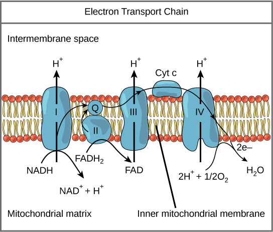
4.2 Free Energy Relationship
- n = number of electrons transferred
- F = Faraday constant (96,485 J/V·mol = 23.06 kcal/V·mol)
- ΔE°' = E°'(acceptor) − E°'(donor)
4.3 Sites of Sufficient Free Energy for ATP Synthesis
- Complex I: NADH → CoQ (ΔE = 0.36 V → ΔG = −69.5 kJ/mol)
- Complex III: CoQH₂ → Cytochrome c (ΔE = 0.21 V → ΔG = −40.5 kJ/mol)
- Complex IV: Cytochrome c → O₂ (ΔE = 0.57 V → ΔG = −110 kJ/mol)
5. COMPONENTS OF THE ETC — ELECTRON CARRIERS
5.1 Types of Electron Carriers
| Carrier Type | Carries | Examples |
|---|---|---|
| NAD⁺/NADH | 2 electrons + 1 H⁺ (hydride ion) | Soluble in matrix |
| FAD/FADH₂ | 2 electrons + 2 H⁺ | Prosthetic group (tightly bound) |
| FMN/FMNH₂ | 2 electrons + 2 H⁺ | Prosthetic group in Complex I |
| Coenzyme Q (Ubiquinone) | 2 electrons + 2 H⁺ | Mobile carrier in lipid bilayer |
| Iron-Sulfur Centers (Fe-S) | 1 electron | Multiple types: [2Fe-2S], [3Fe-4S], [4Fe-4S] |
| Cytochromes (heme iron) | 1 electron (Fe²⁺ ↔ Fe³⁺) | Cyt b, c₁, c, a, a₃ |
| Copper Centers | 1 electron (Cu⁺ ↔ Cu²⁺) | CuA, CuB in Complex IV |
5.2 Flavin Nucleotides (FMN and FAD)
- Derived from Riboflavin (Vitamin B₂)
- Can accept 1 or 2 electrons (can form a semiquinone radical intermediate)
- FMN is the first electron acceptor in Complex I
- FAD is the prosthetic group in Complex II (succinate dehydrogenase)
5.3 Iron-Sulfur Clusters
- Also called non-heme iron proteins
- Contain iron atoms coordinated with inorganic sulfide (S²⁻) and/or cysteine sulfur from the protein
- Types:
- [2Fe-2S] — Rieske center in Complex III (unique: one Fe coordinated by 2 histidines)
- [4Fe-4S] — most common, found in Complexes I, II, III
- [3Fe-4S] — found in Complex II
- Transfer only 1 electron at a time despite having multiple iron atoms
- The most complex arrangement is in Complex I: contains 8 Fe-S clusters forming an electron "wire" ~95 Å long
5.4 Heme Groups and Cytochromes
| Cytochrome | Heme Type | Location | Unique Features |
|---|---|---|---|
| Cyt b (bL and bH) | Heme B (protoporphyrin IX) | Complex III | Two b-type cytochromes in Q cycle |
| Cyt c₁ | Heme C (covalently attached) | Complex III | Part of the complex |
| Cyt c | Heme C | IMS (mobile carrier) | Small, water-soluble, peripheral protein; also involved in apoptosis |
| Cyt a | Heme A | Complex IV | Contains formyl group and hydroxyethylfarnesyl side chain |
| Cyt a₃ | Heme A | Complex IV | Contains the binuclear center (with CuB) where O₂ binds |
5.5 Copper Centers
- CuA in Complex IV: dinuclear copper center; first electron acceptor from cytochrome c
- CuB in Complex IV: paired with heme a₃ in the binuclear center; directly involved in O₂ binding and reduction
6. COMPLEX I — NADH:UBIQUINONE OXIDOREDUCTASE (EC 1.6.5.3)

6.1 Structure
- Largest respiratory complex
- ~1,000 kDa (1 MDa) in mammals
- 45 subunits in mammals (7 encoded by mtDNA: ND1–ND6, ND4L; 38 nuclear-encoded)
- Shaped like an "L" or a boot:
- Hydrophilic arm (peripheral arm): projects into the matrix; contains FMN and all Fe-S clusters
- Hydrophobic arm (membrane arm): embedded in the IMM; contains the proton-pumping machinery
- Contains:
- 1 FMN (flavin mononucleotide)
- 8 iron-sulfur clusters: N1a [2Fe-2S], N1b [2Fe-2S], N2 [4Fe-4S], N3 [4Fe-4S], N4 [4Fe-4S], N5 [4Fe-4S], N6a [4Fe-4S], N6b [4Fe-4S]
- The Fe-S clusters form a ~95 Å electron transfer wire
6.2 Mechanism
- NADH binds to the hydrophilic arm and donates 2 electrons as a hydride ion (H⁻) to FMN → FMNH₂
- FMNH₂ passes electrons one at a time through the chain of 8 Fe-S clusters
- The terminal Fe-S cluster N2 (highest potential in the chain) donates electrons to ubiquinone (CoQ) bound at the ubiquinone-binding site at the junction of the two arms
- CoQ is reduced: Q → QH⁻ (semiquinone) → QH₂ (ubiquinol)
- The energy released drives conformational changes in the membrane arm that translocate 4 H⁺ from the matrix to the IMS
6.3 Proton Pumping Mechanism
- The membrane arm contains three antiporter-like subunits (ND2, ND4, ND5) each pumping 1 H⁺
- A fourth proton is pumped at the junction/interface near the ubiquinone-binding site (ND1/ND6 region)
- Total: 4 H⁺ pumped per NADH (per 2 electrons)
- The mechanism involves a long-range conformational coupling — a "piston-like" mechanism transmitted through a long amphipathic helix connecting the antiporter subunits
6.4 Overall Reaction
NADH + H⁺ + Q + 4H⁺(matrix) → NAD⁺ + QH₂ + 4H⁺(IMS)
6.5 Inhibitors of Complex I
| Inhibitor | Source/Type | Mechanism |
|---|---|---|
| Rotenone | Plant insecticide (from Derris plant roots) | Blocks electron transfer from Fe-S cluster N2 to ubiquinone |
| Piericidin A | Streptomyces antibiotic | CoQ analog; competes at Q-binding site |
| Barbiturates (Amobarbital/Amytal) | Pharmaceutical | Block at the same site as rotenone |
| MPP⁺ (1-methyl-4-phenylpyridinium) | Active metabolite of MPTP | Inhibits Complex I |
🔴 CLINICAL: Rotenone and Parkinson's Disease
- Chronic rotenone exposure in animal models produces Parkinsonism including selective dopaminergic neuron death and Lewy body-like inclusions
- Dopaminergic neurons in the substantia nigra pars compacta are especially vulnerable due to high energy demands and high oxidative stress
- Epidemiological studies link pesticide exposure (rotenone, paraquat) to increased Parkinson's disease risk
🔴 CLINICAL: MPTP Toxicity (Drug-induced Parkinsonism)
- MPTP (1-methyl-4-phenyl-1,2,3,6-tetrahydropyridine) is a contaminant of synthetic heroin ("designer drug")
- MPTP crosses the blood-brain barrier → taken up by astrocytes → converted to MPP⁺ by MAO-B (monoamine oxidase B)
- MPP⁺ is selectively taken up by dopaminergic neurons via the dopamine transporter (DAT)
- MPP⁺ inhibits Complex I → ATP depletion, ROS generation → selective death of dopaminergic neurons
- Produces acute, irreversible Parkinsonism
- This discovery was pivotal in understanding Parkinson's disease pathogenesis
- Treatment: L-DOPA, dopamine agonists (same as idiopathic Parkinson's)
- Prevention: MAO-B inhibitors (selegiline/rasagiline) can block MPTP → MPP⁺ conversion
🔴 CLINICAL: Leber's Hereditary Optic Neuropathy (LHON)
- Mitochondrial inheritance (maternal)
- Caused by point mutations in mtDNA genes encoding Complex I subunits
- Most common mutations: m.11778G>A (ND4, ~70% of cases), m.3460G>A (ND1), m.14484T>C (ND6)
- Presents in young adults (15-35 years), more commonly in males (male:female = 4:1, suggesting nuclear modifier genes like X-linked factors)
- Acute or subacute painless bilateral central vision loss (sequential, one eye then the other within weeks to months)
- Loss of central vision with centrocecal scotoma (central blind spot)
- Optic disc pseudoedema, peripapillary telangiectatic microangiopathy (does NOT leak on fluorescein angiography)
- Eventually → optic atrophy
- Retinal ganglion cells (especially the papillomacular bundle) are selectively vulnerable
- No consistently effective treatment; idebenone (CoQ analog) may have some benefit, especially in early stages
- Variable penetrance due to heteroplasmy
- Smoking and alcohol use may trigger/worsen the disease (environmental factors)
🔴 CLINICAL: Complex I Deficiency
- Most common cause of mitochondrial disease in children
- Can be caused by mutations in any of the 45 structural genes (7 mtDNA, 38 nuclear) or assembly factor genes
- Clinical spectrum:
- Leigh syndrome (subacute necrotizing encephalomyelopathy) — most severe
- MELAS-like syndrome
- Neonatal lactic acidosis
- Hypertrophic cardiomyopathy
- Leukodystrophy
- Lab findings: elevated lactate, elevated lactate:pyruvate ratio (>20:1), elevated alanine
7. COMPLEX II — SUCCINATE:UBIQUINONE OXIDOREDUCTASE (EC 1.3.5.1)
7.1 Structure
- Smallest respiratory complex: ~124 kDa
- 4 subunits — ALL nuclear-encoded (the ONLY ETC complex entirely nuclear-encoded)
- SDHA: flavoprotein subunit; contains covalently bound FAD and the succinate-binding site
- SDHB: iron-sulfur protein; contains three Fe-S clusters: [2Fe-2S], [4Fe-4S], [3Fe-4S]
- SDHC and SDHD: membrane-anchor subunits; contain heme b (not involved in electron transfer — may serve in ROS protection) and the ubiquinone-binding site
- Part of both the TCA cycle (as succinate dehydrogenase, enzyme #6) and the ETC
7.2 Mechanism
- Succinate is oxidized to fumarate (trans double bond) at the SDHA subunit
- FAD is reduced to FADH₂ (covalently bound, does NOT dissociate)
- Electrons pass through the three Fe-S clusters in SDHB: [2Fe-2S] → [4Fe-4S] → [3Fe-4S]
- Electrons are donated to ubiquinone (CoQ) at the Q-binding site in SDHC/SDHD → ubiquinol (QH₂)
7.3 Key Points
- Does NOT pump protons — insufficient free energy change (ΔG ≈ −6 kJ/mol for FAD → CoQ transfer)
- FADH₂ generated here enters the chain at the level of CoQ, bypassing Complex I
- Therefore, FADH₂ from Complex II generates fewer ATP than NADH (1.5 vs. 2.5 ATP)
7.4 Overall Reaction
Succinate + Q → Fumarate + QH₂ (No proton pumping)
7.5 Inhibitors of Complex II
| Inhibitor | Mechanism |
|---|---|
| Malonate | Competitive inhibitor of succinate (structural analog — dicarboxylic acid) |
| Carboxin | Blocks Q-binding site |
| TTFA (thenoyltrifluoroacetone) | Blocks Q-binding site |
| 3-Nitropropionic acid (3-NPA) | Irreversible inhibitor; causes striatal necrosis |
🔴 CLINICAL: Hereditary Paraganglioma-Pheochromocytoma Syndrome
- Germline mutations in SDHB, SDHC, SDHD (and also SDHA, SDHAF2)
- These are tumor suppressor genes (loss-of-function mutations)
- SDH deficiency → accumulation of succinate → inhibits prolyl hydroxylases (PHDs) → stabilization of HIF-1α (Hypoxia-Inducible Factor) → pseudohypoxic signaling → angiogenesis, proliferation
- This is an example of an oncometabolite mechanism
- Clinical manifestations:
- Paragangliomas (especially head and neck — carotid body tumors)
- Pheochromocytomas (adrenal medulla)
- Gastrointestinal stromal tumors (GISTs)
- Renal cell carcinoma
- SDHB mutations: highest malignancy risk (~30-40% metastatic)
- SDHD mutations: predominantly paternally inherited expression (genomic imprinting)
- Screening: plasma/urine metanephrines and catecholamines
- Immunohistochemistry: loss of SDHB staining in tumor tissue is a marker
🔴 CLINICAL: Carney-Stratakis Syndrome
- Dyad of paraganglioma and gastrointestinal stromal tumor (GIST)
- Due to germline SDH subunit mutations
- Distinct from Carney triad (paraganglioma, GIST, pulmonary chondroma — usually sporadic)
🔴 CLINICAL: 3-Nitropropionic Acid (3-NPA) Toxicity
- Found in moldy sugarcane (Arthrinium saccharicola fungus)
- Irreversibly inhibits Complex II (SDH)
- Causes acute encephalopathy with selective bilateral striatal (basal ganglia) necrosis
- Mimics Huntington's disease pathology
- Used as an experimental model for Huntington's disease
7.6 Other FADH₂-Linked Dehydrogenases
| Enzyme | Pathway | Location |
|---|---|---|
| Electron-transferring flavoprotein (ETF) dehydrogenase / ETF:QO | β-oxidation of fatty acids | IMM |
| Glycerol-3-phosphate dehydrogenase (mitochondrial) | Glycerol-3-phosphate shuttle | Outer surface of IMM |
| Dihydroorotate dehydrogenase | Pyrimidine biosynthesis | IMM |
| Choline dehydrogenase | Choline metabolism | IMM |
| Proline dehydrogenase | Proline metabolism | IMM |
🔴 CLINICAL: Multiple Acyl-CoA Dehydrogenase Deficiency (MADD) / Glutaric Aciduria Type II
- Deficiency of ETF or ETF:ubiquinone oxidoreductase (ETF-QO)
- Impairs electron transfer from multiple FAD-linked dehydrogenases → impaired β-oxidation, branched-chain amino acid oxidation, and others
- Biochemical: elevated multiple acylcarnitines, organic acids (glutaric, ethylmalonic, adipic, suberic acids)
- Clinical: severe neonatal form (with or without congenital anomalies — renal cysts, facial dysmorphism) or milder late-onset form (lipid storage myopathy)
- Treatment: riboflavin (B₂) — many cases are riboflavin-responsive (especially late-onset)
8. UBIQUINONE (COENZYME Q / CoQ₁₀)
8.1 Structure
- 2,3-dimethoxy-5-methyl-1,4-benzoquinone with an isoprenoid side chain
- In humans: 10 isoprenoid units (CoQ₁₀ or ubiquinone-50)
- The long hydrophobic tail allows it to dissolve freely in the lipid bilayer of the IMM
- Small, lipid-soluble, mobile electron carrier — NOT a protein
8.2 Redox States
- Ubiquinone (Q) — fully oxidized
- Semiquinone radical (QH• or Q•⁻) — partially reduced (one electron)
- Ubiquinol (QH₂) — fully reduced (two electrons)
8.3 Function
- Collects electrons from Complexes I and II (and other FADH₂-linked dehydrogenases)
- Shuttles electrons to Complex III
- Acts as a mobile electron carrier in the membrane (unlike Fe-S clusters and cytochromes which are protein-bound)
- The CoQ pool contains ~10× more CoQ than Complex III → functions as a mobile electron buffer
8.4 Biosynthesis
- Synthesized endogenously via the mevalonate pathway (same pathway as cholesterol synthesis)
- The benzoquinone ring is derived from tyrosine (or phenylalanine)
- The isoprenoid tail is from the mevalonate pathway (farnesyl pyrophosphate → decaprenyl-PP)
🔴 CLINICAL: CoQ₁₀ Deficiency
- Primary CoQ₁₀ deficiency: autosomal recessive mutations in genes of CoQ₁₀ biosynthetic pathway (COQ2, COQ4, COQ6, COQ7, COQ8A, COQ8B, COQ9, PDSS1, PDSS2)
- Clinical phenotypes:
- Cerebellar ataxia (most common, often with cerebellar atrophy)
- Steroid-resistant nephrotic syndrome (especially COQ2, COQ6, COQ8B mutations)
- Encephalomyopathy
- Isolated myopathy with ragged red fibers
- Multisystem infantile form (severe)
- Secondary CoQ₁₀ deficiency: seen in mitochondrial diseases, aging, statin therapy
- Treatment: oral CoQ₁₀ supplementation (one of the few treatable mitochondrial disorders, especially the nephrotic and ataxic forms if treated early)
🔴 CLINICAL: Statins and CoQ₁₀
- Statins (HMG-CoA reductase inhibitors) inhibit the mevalonate pathway
- This reduces not only cholesterol synthesis but also CoQ₁₀ and dolichol synthesis
- May contribute to statin-associated myopathy/myalgia (controversial but biologically plausible)
- Some clinicians recommend CoQ₁₀ supplementation with statin therapy, though evidence is mixed
8.5 CoQ₁₀ as an Antioxidant
- Ubiquinol (QH₂) is a potent lipid-soluble antioxidant
- Protects membrane lipids from peroxidation
- Regenerates vitamin E (α-tocopherol) from its radical form
9. COMPLEX III — UBIQUINOL:CYTOCHROME c OXIDOREDUCTASE (EC 1.10.2.2)
9.1 Structure
- ~240 kDa as a functional homodimer (each monomer ~11 subunits in mammals)
- 11 subunits per monomer: 1 mtDNA-encoded (cytochrome b) + 10 nuclear-encoded
- Key catalytic subunits:
- Cytochrome b: contains two heme b groups — heme bL (low potential, +0.07 V) and heme bH (high potential, +0.03 V → but higher in the context of electron flow)
- Rieske iron-sulfur protein: contains [2Fe-2S] cluster — unique because one Fe is coordinated by 2 histidines instead of cysteines (higher reduction potential: +0.28 V)
- Cytochrome c₁: contains heme c₁
9.2 The Q Cycle (Mitchell's Q Cycle)
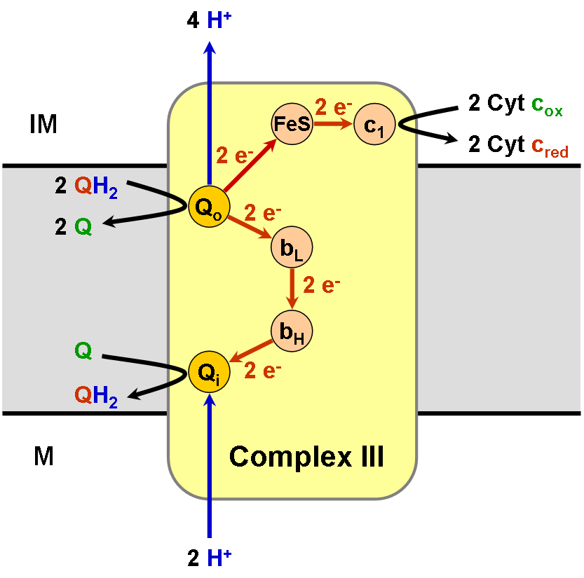
- Transfers electrons from a 2-electron carrier (QH₂) to a 1-electron carrier (cytochrome c)
- Pumps protons across the membrane
- Effectively doubles the number of protons translocated per QH₂
- Qp site (also called Qo site) — on the P-side (IMS side / positive side) — where QH₂ is oxidized
- Qn site (also called Qi site) — on the N-side (matrix side / negative side) — where Q is reduced
First Half of the Q Cycle:
- QH₂ binds at the Qp site
- First electron → Rieske [2Fe-2S] → Cytochrome c₁ → Cytochrome c (which departs as a reduced, mobile carrier)
- 2 H⁺ are released into the IMS (from QH₂ oxidation)
- The remaining electron goes to the low-potential pathway: QH• → heme bL → heme bH → reduces Q to Q•⁻ (semiquinone) at the Qn site
- QH₂ at Qp is now fully oxidized → Q, which leaves
Second Half of the Q Cycle:
- A second QH₂ binds at the Qp site
- Again: first electron → Rieske → cyt c₁ → second cytochrome c (reduced and departs)
- 2 more H⁺ released into IMS
- Second electron → heme bL → heme bH → reduces the semiquinone (Q•⁻) at Qn to QH₂ (picking up 2 H⁺ from the matrix)
- This regenerated QH₂ can re-enter the cycle
Net Q Cycle (per pair of electrons reaching cytochrome c):
QH₂ + 2 cyt c (ox) + 2H⁺(matrix) → Q + 2 cyt c (red) + 4H⁺(IMS)
- 2 QH₂ oxidized at Qp
- 1 Q reduced to QH₂ at Qn (net consumption: 1 QH₂)
- 4 H⁺ released to IMS (2 from each QH₂ oxidation at Qp)
- 2 H⁺ consumed from matrix (for QH₂ formation at Qn)
- Net proton translocation: ~4 H⁺ per 2 electrons reaching O₂ via cyt c

9.3 Inhibitors of Complex III
| Inhibitor | Site of Action | Mechanism |
|---|---|---|
| Antimycin A | Qn (Qi) site | Blocks electron transfer from heme bH to Q at the Qn site; prevents the second half of Q cycle |
| Myxothiazol | Qp (Qo) site | Blocks QH₂ oxidation at the Qp site |
| Stigmatellin | Qp (Qo) site | Blocks electron transfer to Rieske center |
🔴 CLINICAL: Antimycin A and ROS
- When Antimycin A blocks the Qn site, the semiquinone radical (Q•⁻) at the Qp site accumulates
- This semiquinone can donate its electron directly to O₂ → forming superoxide (O₂•⁻)
- Complex III (Qp site) is a major site of mitochondrial ROS production
- Relevant to aging, ischemia-reperfusion injury, and neurodegenerative diseases
🔴 CLINICAL: Complex III Deficiency
- Rare; can be caused by mutations in:
- Cytochrome b (mtDNA — MT-CYB)
- BCS1L (assembly factor for Rieske protein)
- BCS1L mutations cause:
- GRACILE syndrome (Growth Retardation, Aminoaciduria, Cholestasis, Iron overload, Lactic acidosis, Early death) — Finnish heritage
- Björnstad syndrome (sensorineural deafness + pili torti — twisted hair)
- Isolated Complex III deficiency with encephalopathy
- MT-CYB mutations: exercise intolerance, myopathy, cardiomyopathy
10. CYTOCHROME c
10.1 Structure and Function
- Small (~12.4 kDa), highly conserved, water-soluble protein
- Contains one heme c (covalently attached via thioether bonds to two cysteine residues — -CXXCH- motif)
- Located in the intermembrane space
- Peripheral membrane protein — loosely associated with the outer surface of the IMM (bound by electrostatic interactions with cardiolipin)
- Transfers one electron from Complex III (cyt c₁) → Complex IV (CuA)
10.2 Role in Apoptosis
🔴 CLINICAL: Cytochrome c and Apoptosis (Intrinsic Pathway)
- During intrinsic (mitochondrial) apoptosis:
- Pro-apoptotic stimuli (DNA damage, oxidative stress, growth factor withdrawal) activate BH3-only proteins (BID, BIM, BAD, etc.)
- These activate BAX and BAK (pro-apoptotic BCL-2 family members)
- BAX/BAK oligomerize and form pores in the outer mitochondrial membrane → MOMP (Mitochondrial Outer Membrane Permeabilization)
- Cytochrome c is released from the IMS into the cytoplasm
- Cytochrome c binds APAF-1 (Apoptotic Protease-Activating Factor 1) → forms the apoptosome (wheel-of-death)
- The apoptosome activates Caspase-9 (initiator caspase)
- Caspase-9 activates Caspase-3 and Caspase-7 (executioner caspases) → cell death
- Anti-apoptotic proteins (BCL-2, BCL-XL, MCL-1) prevent BAX/BAK activation → prevent cytochrome c release
- Cancer cells often overexpress BCL-2 → resistance to apoptosis
- Venetoclax: BCL-2 inhibitor used in CLL and AML — restores apoptosis by allowing cytochrome c release
- Other proteins released from mitochondria during apoptosis: Smac/DIABLO (inhibits IAPs), AIF (apoptosis-inducing factor — caspase-independent death), endonuclease G, Omi/HtrA2
10.3 Cytochrome c in Evolution
- One of the most conserved proteins in evolution
- Comparing cytochrome c amino acid sequences across species has been used to construct phylogenetic trees
- Human and chimpanzee cytochrome c are identical (100% homology)
- Human and yeast share ~60% identity
11. COMPLEX IV — CYTOCHROME c OXIDASE (EC 1.9.3.1)
11.1 Structure
- ~200 kDa as a functional dimer (each monomer: 13 subunits in mammals)
- 3 mtDNA-encoded subunits: COX I, COX II, COX III (catalytic core)
- 10 nuclear-encoded subunits (regulatory/structural)
- Contains 4 redox-active metal centers:
- CuA center (in COX II): dinuclear copper; first to accept electron from cytochrome c
- Heme a (in COX I): transfers electron to the binuclear center
- Heme a₃ (in COX I): part of the binuclear center
- CuB (in COX I): part of the binuclear center; paired with heme a₃
- Also contains Zn and Mg ions (structural roles)
11.2 Mechanism
- Cytochrome c (reduced) binds to the IMS face of Complex IV and donates 1 electron to CuA
- CuA → heme a → binuclear center (heme a₃–CuB)
- At the binuclear center, O₂ binds (between Fe of heme a₃ and CuB)
- Four electrons are required to fully reduce O₂:
O₂ + 4 e⁻ + 4 H⁺ (matrix) → 2 H₂O
- This occurs in a carefully controlled sequence to avoid release of partially reduced oxygen species (superoxide, peroxide)
11.3 Proton Pumping
- 4 "chemical" protons: consumed in the matrix to make H₂O (scalar protons)
- 4 "pumped" protons: translocated from matrix to IMS (vectorial protons)
- Total: 8 H⁺ removed from the matrix per O₂ (4 used for H₂O + 4 pumped)
- But per 2 electrons (from 1 NADH): 2 H⁺ pumped + 2 H⁺ consumed for 1 H₂O
11.4 Overall Reaction
4 cyt c (red) + O₂ + 8H⁺(matrix) → 4 cyt c (ox) + 2H₂O + 4H⁺(IMS)
2 cyt c (red) + ½O₂ + 4H⁺(matrix) → 2 cyt c (ox) + H₂O + 2H⁺(IMS)
11.5 Inhibitors of Complex IV
| Inhibitor | Mechanism |
|---|---|
| Cyanide (CN⁻) | Binds to Fe³⁺ of heme a₃ → blocks O₂ binding |
| Carbon monoxide (CO) | Binds to Fe²⁺ of heme a₃ → blocks O₂ binding |
| Hydrogen sulfide (H₂S) | Binds to heme a₃ → blocks O₂ binding |
| Azide (N₃⁻) | Binds to Fe³⁺ of heme a₃ |
| Nitric oxide (NO) | Reversible inhibitor; binds to heme a₃ and CuB |
| Phosphine (PH₃) | Insecticide; inhibits Complex IV |
| Formate (from methanol metabolism) | Inhibits Complex IV |
🔴 CLINICAL: Cyanide Poisoning
- Sources: industrial chemicals (electroplating, mining), smoke inhalation (burning of plastics, wool, silk releases HCN), cyanogenic glycosides (bitter almonds, cassava, apple seeds, cherry pits — contain amygdalin), sodium nitroprusside (releases CN⁻ during prolonged infusion)
- Mechanism: CN⁻ binds Fe³⁺ of cytochrome a₃ → complete inhibition of Complex IV → cells cannot use O₂ → histotoxic/cytotoxic hypoxia
- Classic features:
- Bright cherry-red skin (venous blood remains oxygenated because cells can't extract O₂)
- "Bitter almond" breath odor (only 40% of people can detect this — genetically determined)
- High venous PO₂ (venous blood is almost as oxygenated as arterial → narrow A-V O₂ difference)
- Lactic acidosis (severe, from anaerobic glycolysis)
- Rapidly: headache, confusion, seizures, coma, death
- PaO₂ and O₂ saturation may be normal (pulse oximetry and ABG can be misleadingly normal)
- Lab: elevated lactate, elevated venous O₂ saturation, cyanide levels >0.5 mg/L are toxic, >3 mg/L lethal
- Treatment:
- 100% oxygen
- Hydroxocobalamin (Cyanokit®): binds CN⁻ → cyanocobalamin (vitamin B₁₂) → renally excreted. Preferred antidote, especially in smoke inhalation (safe in combined CO/CN poisoning)
- Sodium thiosulfate: provides sulfur donor for rhodanese (thiosulfate sulfurtransferase) → converts CN⁻ → thiocyanate (SCN⁻) → renally excreted. Slow onset.
- Sodium nitrite (or amyl nitrite): induces methemoglobinemia → methemoglobin (Fe³⁺) binds CN⁻ → cyanomethemoglobin → draws CN away from cytochrome oxidase. Caution in smoke inhalation (may already have carboxyhemoglobin → further reduces O₂ carrying capacity)
- Dicobalt edetate (Kelocyanor): used in UK — cobalt directly chelates CN⁻
- Traditional "Cyanide Antidote Kit": amyl nitrite + sodium nitrite + sodium thiosulfate
🔴 CLINICAL: Carbon Monoxide (CO) Poisoning
- CO binds to:
- Hemoglobin (240× greater affinity than O₂) → carboxyhemoglobin (COHb) → leftward shift of O₂-Hb dissociation curve → impaired O₂ delivery
- Cytochrome c oxidase (Complex IV) → Fe²⁺ of heme a₃ → histotoxic hypoxia
- Myoglobin → impaired muscle O₂ utilization
- Sources: incomplete combustion, house fires, car exhaust, charcoal grills, furnaces
- Findings:
- Cherry-red skin (due to COHb, but this is a LATE finding — often patients appear pale/cyanotic)
- Headache (most common early symptom), nausea, confusion, syncope, seizures, coma
- PaO₂ is NORMAL (dissolved O₂ is unaffected)
- O₂ saturation by pulse oximetry is FALSELY NORMAL (pulse ox can't distinguish COHb from OxyHb)
- CO-oximetry is the diagnostic test → directly measures COHb%
- Normal COHb <3% (non-smokers), <10% (smokers); symptomatic >15-20%; lethal >50-60%
- Treatment:
- 100% O₂ via non-rebreather mask (reduces COHb half-life from 5 hours → 90 minutes)
- Hyperbaric oxygen (HBO₂): reduces half-life to ~30 minutes; indicated for COHb >25%, loss of consciousness, neurological symptoms, cardiac ischemia, pregnancy
- Delayed neuropsychiatric sequelae (DNS): cognitive impairment, personality changes weeks later → may be prevented by HBO₂
🔴 CLINICAL: Hydrogen Sulfide (H₂S) Poisoning
- "Knockdown gas" — toxic in sewer gas, volcanic emissions, petroleum industry
- Mechanism: like cyanide, inhibits Complex IV
- Can cause sudden collapse ("sewer gas asphyxia")
- Rotten egg smell at low concentrations; at high concentrations → olfactory nerve paralysis (can no longer smell it — extremely dangerous)
- Treatment: similar to cyanide → nitrites (to form sulfmethemoglobin), 100% O₂, supportive
🔴 CLINICAL: Nitric Oxide (NO) and Complex IV
- NO is a physiological signaling molecule (vasodilator, neurotransmitter)
- At physiological concentrations: reversibly inhibits Complex IV → may regulate mitochondrial respiration and O₂ utilization
- In excess (sepsis, inflammation → iNOS activation): excessive NO → mitochondrial dysfunction → contributes to septic shock pathophysiology
- NO competes with O₂ at the binuclear center → when O₂ is low, NO inhibition is enhanced → may help redistribute O₂ in tissues (metabolic signaling)
🔴 CLINICAL: Methanol and Formate Toxicity
- Methanol → formaldehyde → formic acid/formate (via alcohol dehydrogenase and aldehyde dehydrogenase)
- Formate inhibits cytochrome c oxidase (Complex IV)
- Causes: metabolic acidosis (high anion gap), blindness (retinal/optic nerve toxicity — retinal ganglion cells are especially vulnerable)
- Treatment: fomepizole (4-methylpyrazole — ADH inhibitor) or ethanol (competitive substrate for ADH), dialysis, folate (enhances formate metabolism to CO₂ and H₂O)
12. COMPLEX V — ATP SYNTHASE (F₁F₀-ATP SYNTHASE) (EC 7.1.2.2)
12.1 Overview
- Also called: F₁F₀-ATPase, Complex V
- Not a component of the electron transport chain per se, but is the site of oxidative phosphorylation
- Uses the proton motive force (PMF) generated by Complexes I, III, IV to synthesize ATP from ADP + Pi
- ~600 kDa
- 16 subunits in mammals (2 mtDNA-encoded: ATPase 6 = subunit a, ATPase 8 = subunit A6L)
12.2 Structure
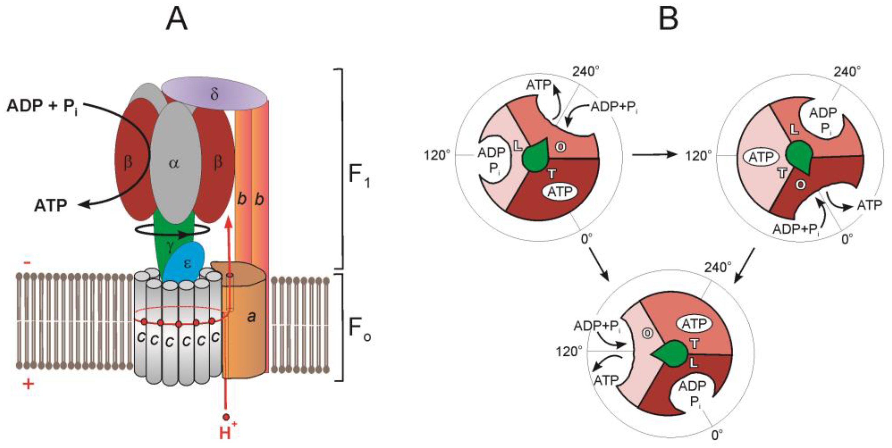
F₁ Component (Soluble, Matrix side — "Factor 1")
- Knob-like structure projecting into the matrix
- Composition: α₃β₃γδε
- 3 α subunits and 3 β subunits: arranged alternately like segments of an orange
- Each β subunit contains a catalytic site for ATP synthesis
- The 3 β subunits exist in 3 different conformations (see binding change mechanism)
- γ subunit: central stalk/axle; rotates within the α₃β₃ ring; asymmetric → drives conformational changes in β subunits
- ε subunit: attached to γ; part of the rotor
- δ subunit: part of the peripheral stalk (in bacterial nomenclature; in mitochondria, the OSCP — Oligomycin Sensitivity Conferring Protein)
F₀ Component (Membrane-embedded — "Factor 0" — "oligomycin-sensitive factor")
- Proton channel
- Composition (in mammals):
- c-ring: ring of 8-15 c subunits (8 in mammals, 10 in yeast); each c subunit has a proton-binding carboxylate (aspartate or glutamate)
- a subunit (subunit 6): has two half-channels for proton translocation; stationary
- b subunit: part of the peripheral/lateral stalk connecting F₀ to F₁ (stator)
- Additional subunits: d, F6, A6L, e, f, g (structural and regulatory)
Peripheral Stalk (Stator)
- Connects F₁ to F₀ without rotating
- Composed of: b, d, F6, OSCP subunits
- Prevents co-rotation of α₃β₃ with γ → allows the conformational changes in β subunits
12.3 The Rotary Catalysis — Boyer's Binding Change Mechanism

Three Conformations of β Subunits:
- O (Open): low affinity for substrates; releases ATP
- L (Loose): binds ADP + Pi loosely
- T (Tight): catalyzes ATP formation; binds ATP very tightly
Catalytic Cycle (per 120° rotation of γ):
- Step 1: ADP + Pi bind to the L-site (Loose)
- Step 2: γ rotates 120° → L-site converts to T-site (Tight) → ATP is synthesized (essentially a spontaneous condensation reaction when substrates are properly positioned)
- Step 3: The previous T-site converts to O-site (Open) → ATP is released
- Step 4: The previous O-site converts to L-site → ready for new ADP + Pi
12.4 Proton Translocation Through F₀
- H⁺ from IMS enters the half-channel in the a subunit (inlet half-channel)
- H⁺ binds to the carboxylate (Asp/Glu) on a c subunit
- This neutralization allows the c subunit to move into the hydrophobic lipid bilayer environment
- As the c-ring rotates (driven by the proton gradient), each c subunit eventually reaches the outlet half-channel on the matrix side
- The H⁺ is released into the matrix
- Depends on the number of c subunits in the ring
- In mammals (8 c subunits): 360°/3 = 120° per ATP; 8 H⁺ per 360° → 8/3 ≈ 2.67 H⁺ per ATP
- Plus 1 H⁺ for the phosphate carrier (Pi/H⁺ symport) → ~3.67 H⁺ per ATP
- Often approximated as ~4 H⁺ per ATP (including phosphate transport)
12.5 ATP/ADP Translocase (Adenine Nucleotide Translocator — ANT)
- Located in IMM
- Antiporter: exchanges ATP⁴⁻ (out to IMS) for ADP³⁻ (in to matrix)
- This exchange is electrogenic — net export of 1 negative charge → driven by the membrane potential (Δψ)
- Consumes energy from the proton gradient
- Accounts for ~25% of the energy of the PMF
| Inhibitor | Effect |
|---|---|
| Atractyloside (from thistle plant) | Binds to cytoplasmic face of ANT → blocks ADP import |
| Bongkrekic acid (from contaminated coconut/corn fermentation — Burkholderia gladioli) | Binds to matrix face → locks ANT in "m-state" |
🔴 CLINICAL: Bongkrekic Acid Poisoning
- From fermented coconut or corn products contaminated with Burkholderia gladioli pathovar cocovenenans
- Outbreaks in Southeast Asia, China
- Inhibits ANT → ATP depletion → multiorgan failure
- Very high mortality rate (40-100%)
12.6 Phosphate Carrier (PiC)
- Symporter: transports Pi (H₂PO₄⁻) with H⁺ into the matrix
- Driven by the ΔpH component of the PMF
- Total cost of ATP synthesis and transport:
- 3-4 H⁺ through ATP synthase
- 1 H⁺ for phosphate import
- Electrical potential used by ANT
12.7 Inhibitors of ATP Synthase (Complex V)
| Inhibitor | Mechanism |
|---|---|
| Oligomycin | Blocks the proton channel in F₀ (binds between a and c subunits) → prevents H⁺ flow → stops both ATP synthesis AND electron transport (because the PMF builds up and back-pressure stops proton pumping by Complexes I, III, IV) |
| DCCD (Dicyclohexylcarbodiimide) | Reacts with the carboxylate (Asp/Glu) on c subunits → blocks proton binding |
| Aurovertin | Binds to β subunit of F₁ → blocks catalytic activity |
| Efrapeptin | Blocks the F₁ catalytic site |
🔴 CLINICAL: Oligomycin Effect
- When oligomycin blocks ATP synthase → protons accumulate in IMS → PMF increases → proton pumping by Complexes I, III, IV stops → electron transport stops → O₂ consumption stops
- This is called "respiratory control" — the tight coupling between electron transport and ATP synthesis
- Shows that under normal conditions, ETC activity is directly coupled to ATP synthesis
🔴 CLINICAL: ATP Synthase Deficiency
- Mutations in MT-ATP6 (subunit a):
- NARP syndrome (Neuropathy, Ataxia, Retinitis Pigmentosa): when mutant mtDNA load is 70-90%
- Maternally Inherited Leigh Syndrome (MILS): when mutant load >90%
- Most common mutation: m.8993T>G or m.8993T>C (both in MT-ATP6)
- MT-ATP8 mutations: rare; cardiomyopathy
- Nuclear-encoded assembly factors: TMEM70 mutations — most common cause of nuclear-encoded ATP synthase deficiency; 3-methylglutaconic aciduria, cardiomyopathy, lactic acidosis
13. THE PROTON MOTIVE FORCE (PMF)
13.1 Definition
- Δψ = membrane potential (voltage gradient) — inside (matrix) is negative; ~150-180 mV in mammals
- ΔpH = pH gradient — matrix is more alkaline (~0.5-1.0 pH units)
- Under physiological conditions: ~80% from Δψ, ~20% from ΔpH
13.2 Total Protons Pumped per NADH
| Complex | H⁺ pumped (per 2 electrons from NADH) |
|---|---|
| Complex I | 4 H⁺ |
| Complex III | 4 H⁺ (via Q cycle) |
| Complex IV | 2 H⁺ (pumped) + 2 H⁺ (consumed for H₂O) |
| Total vectorial H⁺ | 10 H⁺ per NADH |
| Complex | H⁺ pumped (per 2 electrons from FADH₂) |
|---|---|
| Complex III | 4 H⁺ |
| Complex IV | 2 H⁺ (pumped) + 2 H⁺ (consumed) |
| Total vectorial H⁺ | 6 H⁺ per FADH₂ |

14. CHEMIOSMOTIC HYPOTHESIS
Peter Mitchell (1961) — Nobel Prize in Chemistry (1978)
14.1 Key Postulates:
- The ETC complexes are vectorially arranged in the IMM, pumping protons from matrix → IMS
- The IMM is impermeable to protons (and most ions) — the proton gradient is maintained
- The electrochemical proton gradient (PMF) stores energy
- Protons flow back into the matrix only through ATP synthase → this drives ATP synthesis
- The processes of electron transport and ATP synthesis are coupled through the PMF (not by chemical intermediates)
14.2 Evidence Supporting the Hypothesis:
- Intact IMM is required for oxidative phosphorylation (disrupted membranes → no ATP synthesis)
- Uncouplers (which dissipate the proton gradient) abolish ATP synthesis without stopping electron transport
- Artificially created pH gradients across vesicles can drive ATP synthesis
- ATP synthase reconstituted in artificial liposomes produces ATP when a pH gradient is applied
- Ionophores that collapse the gradient inhibit ATP synthesis
15. P/O RATIOS AND ATP YIELD
15.1 Definition
15.2 Revised Values (Current Consensus)
- ~4 H⁺ needed per ATP (including transport costs)
- 10 H⁺ pumped per NADH; 6 H⁺ pumped per FADH₂
| Substrate | H⁺ pumped | ATP per molecule (H⁺/~4) | P/O ratio |
|---|---|---|---|
| NADH | 10 | ~2.5 (10/4) | 2.5 |
| FADH₂ | 6 | ~1.5 (6/4) | 1.5 |
15.3 Total ATP Yield from Complete Oxidation of Glucose
| Reaction | NADH | FADH₂ | GTP/ATP | ATP equivalent |
|---|---|---|---|---|
| Glycolysis: Glucose → 2 Pyruvate | 2 | — | 2 ATP (substrate level) | 2 |
| 2 Pyruvate → 2 Acetyl-CoA (PDH) | 2 | — | — | — |
| 2 turns TCA cycle | 6 | 2 | 2 GTP | 2 |
| Totals | 10 NADH | 2 FADH₂ | 4 ATP/GTP |
- 10 NADH × 2.5 = 25 ATP
- 2 FADH₂ × 1.5 = 3 ATP
- 4 ATP/GTP (substrate level)
- But: glycolytic NADH (2) is in the cytoplasm and must be shuttled into mitochondria
- Malate-Aspartate Shuttle: delivers electrons as NADH → 2.5 ATP each → 5 ATP
- Glycerol-3-Phosphate Shuttle: delivers electrons as FADH₂ → 1.5 ATP each → 3 ATP
| Using Malate-Aspartate Shuttle | Using Glycerol-3-Phosphate Shuttle |
|---|---|
| 25 + 3 + 4 + (2 × 2.5) = 30-32 ATP | 25 + 3 + 4 + (2 × 1.5) = 30 ATP |
16. SHUTTLE SYSTEMS
16.1 Malate-Aspartate Shuttle
- Cytoplasmic oxaloacetate + NADH → malate + NAD⁺ (cytoplasmic malate dehydrogenase)
- Malate crosses IMM via the malate-α-ketoglutarate antiporter
- Matrix malate → oxaloacetate + NADH (mitochondrial malate dehydrogenase)
- Matrix oxaloacetate cannot cross IMM → transaminated to aspartate (by aspartate aminotransferase, using glutamate → α-ketoglutarate)
- Aspartate exits via the glutamate-aspartate antiporter (electrogenic — driven by Δψ)
- Cytoplasmic aspartate → oxaloacetate (by cytoplasmic aspartate aminotransferase)
- α-ketoglutarate returns to matrix via the malate-αKG antiporter
16.2 Glycerol-3-Phosphate Shuttle
- Cytoplasmic DHAP + NADH → glycerol-3-phosphate + NAD⁺ (cytoplasmic glycerol-3-phosphate dehydrogenase — NAD⁺-linked)
- Glycerol-3-phosphate → DHAP + FADH₂ (mitochondrial glycerol-3-phosphate dehydrogenase — FAD-linked, located on outer surface of IMM, has active site facing IMS)
- FADH₂ directly reduces CoQ in the IMM
🔴 CLINICAL: Malate-Aspartate Shuttle and Aralar1/AGC1 Deficiency
- SLC25A12 mutations (encoding mitochondrial aspartate-glutamate carrier isoform 1 — AGC1/Aralar1)
- Causes global cerebral hypomyelination, developmental delay, seizures
- Impaired transfer of reducing equivalents into brain mitochondria
17. INHIBITORS OF THE ETC — COMPREHENSIVE SUMMARY
17.1 Classification by Site
| Complex | Inhibitors |
|---|---|
| Complex I | Rotenone, Piericidin A, Barbiturates (amytal/amobarbital), MPP⁺ |
| Complex II | Malonate, Carboxin, TTFA, 3-Nitropropionic acid |
| Complex III | Antimycin A (Qi/Qn site), Myxothiazol (Qo/Qp site), Stigmatellin (Qo/Qp site) |
| Complex IV | Cyanide (CN⁻), Carbon monoxide (CO), Hydrogen sulfide (H₂S), Azide (N₃⁻), Nitric oxide (NO), Phosphine, Formate |
| Complex V | Oligomycin, DCCD, Aurovertin, Efrapeptin |
| ANT | Atractyloside, Bongkrekic acid |
17.2 Effects of ETC Inhibitors
- Upstream of the block: carriers become more reduced (can't pass electrons forward)
- Downstream of the block: carriers become more oxidized (no electrons coming in)
- O₂ consumption decreases (except with uncouplers)
- ATP synthesis decreases
- NADH/NAD⁺ ratio increases → inhibits TCA cycle and other NAD⁺-dependent reactions
- Lactate production increases → lactic acidosis
18. UNCOUPLERS OF OXIDATIVE PHOSPHORYLATION
18.1 Definition
18.2 Mechanism
- Pick up H⁺ on the IMS side (protonated form is lipid-soluble)
- Cross the IMM in their protonated form
- Release H⁺ on the matrix side
- Return across the membrane in their anionic form
- This short-circuits the proton gradient → collapses Δp
18.3 Effects of Uncoupling
| Parameter | Effect |
|---|---|
| Electron transport | ↑ (accelerated — no back-pressure from PMF) |
| O₂ consumption | ↑↑ |
| ATP synthesis | ↓↓ (abolished) |
| NADH/NAD⁺ ratio | ↓ (more NAD⁺ generated — TCA cycle accelerated) |
| Heat production | ↑↑ (thermogenesis) |
| Proton gradient | ↓↓ (dissipated) |
| Fuel oxidation | ↑↑ |
18.4 Examples of Uncouplers
| Uncoupler | Notes |
|---|---|
| 2,4-dinitrophenol (DNP) | Classical uncoupler; used as a weight-loss drug in 1930s (banned due to toxicity) |
| FCCP (carbonyl cyanide-p-trifluoromethoxyphenylhydrazone) | Research tool |
| CCCP (carbonyl cyanide m-chlorophenylhydrazone) | Research tool |
| Thermogenin (UCP1) | Natural uncoupling protein in brown adipose tissue |
| Valinomycin | K⁺ ionophore; dissipates Δψ component |
| Nigericin | K⁺/H⁺ antiporter ionophore; dissipates ΔpH |
| Salicylates (high dose aspirin) | Weak uncoupling activity |
| Bilirubin (high levels) | Mild uncoupler |
| Calcium ions (high levels) | Uncouple via mitochondrial permeability transition pore (mPTP) |
| Long-chain fatty acids | Can act as mild uncouplers |
🔴 CLINICAL: 2,4-Dinitrophenol (DNP) Toxicity
- Used in the 1930s as a weight-loss agent due to uncoupling → increased metabolic rate → fat burning
- Banned by FDA in 1938 due to severe side effects
- Still available illegally online → causes deaths in body-builders/dieters
- Symptoms: profuse sweating, tachycardia, tachypnea, hyperthermia (potentially fatal malignant hyperthermia-like syndrome)
- Cataracts (with chronic use)
- No specific antidote — treatment is supportive: cooling, dantrolene may help
- Narrow therapeutic index → small dose increase can be lethal
🔴 CLINICAL: Aspirin (Salicylate) Overdose
- High-dose salicylates are:
- Uncouplers of oxidative phosphorylation (weak acid, lipid-soluble)
- Direct stimulants of the respiratory center (medullary)
- Clinical toxicity:
- Respiratory alkalosis (early — direct stimulation of respiratory center)
- Followed by metabolic acidosis (high anion gap — from uncoupling → lactate, ketoacids + salicylate itself)
- Mixed respiratory alkalosis and metabolic acidosis is characteristic
- Hyperthermia, tinnitus, diaphoresis, nausea/vomiting
- Severe: pulmonary edema, cerebral edema, seizures, death
- Treatment: sodium bicarbonate (alkalinize urine and blood → traps salicylate in ionized form in urine), hemodialysis for severe cases, cooling
18.5 Thermogenin (UCP1) and Brown Adipose Tissue (BAT)
🔴 CLINICAL: Non-Shivering Thermogenesis
- Brown adipose tissue (BAT) contains abundant mitochondria (brown color from cytochromes)
- Contains UCP1 (Uncoupling Protein 1 / Thermogenin) — a natural proton channel in the IMM
- UCP1 allows H⁺ to re-enter the matrix without passing through ATP synthase → energy released as HEAT
- Important in:
- Neonates (who cannot shiver) — major source of heat production
- Hibernating animals
- Adults (recently discovered to have metabolically active BAT, especially in supraclavicular, paravertebral, perirenal regions)
- Regulation of UCP1:
- Activated by: free fatty acids, norepinephrine (via β₃-adrenergic receptors → cAMP → PKA → hormone-sensitive lipase → FFA release → FFA activates UCP1)
- Inhibited by: purine nucleotides (GDP, GTP, ADP, ATP) — bind and block the channel
- Cold exposure → sympathetic nervous system → norepinephrine → β₃ receptors on BAT → thermogenesis
- Thyroid hormones (T₃) upregulate UCP1 expression → explains heat intolerance in hyperthyroidism
- BAT and obesity: less active BAT is associated with obesity; activating BAT is a potential anti-obesity strategy
18.6 Other Uncoupling Proteins
| UCP | Location | Proposed Function |
|---|---|---|
| UCP1 | Brown adipose tissue | Thermogenesis |
| UCP2 | Widely expressed (pancreatic β-cells, brain, etc.) | ROS regulation, β-cell function |
| UCP3 | Skeletal muscle, heart | Fatty acid metabolism, ROS protection |
| UCP4 | Brain | Neuroprotection |
| UCP5 (BMCP1) | Brain | Neuroprotection |
🔴 CLINICAL: UCP2 and Type 2 Diabetes
- UCP2 in pancreatic β-cells may reduce ATP production → decreased ATP/ADP ratio → impaired glucose-stimulated insulin secretion
- UCP2 polymorphisms have been associated with type 2 diabetes risk and obesity (though controversial)
- UCP2 knockout mice show improved insulin secretion
19. TIGHT COUPLING, RESPIRATORY CONTROL, AND REGULATION
19.1 Respiratory Control
- Under normal conditions, electron transport is tightly coupled to ATP synthesis
- The rate of O₂ consumption (electron transport) is determined by the availability of ADP
- When ADP levels are low (cell has enough ATP) → no ATP synthesis needed → proton gradient builds up → back-pressure inhibits ETC → O₂ consumption decreases
- When ADP levels are high (ATP is being used) → ADP enters mitochondria → ATP synthase becomes active → proton gradient is consumed → ETC speeds up → O₂ consumption increases
19.2 Respiratory States (Chance & Williams)
| State | Substrate | ADP | O₂ | Rate of respiration |
|---|---|---|---|---|
| State 1 | Low | Low | Adequate | Slow |
| State 2 | Added | Low | Adequate | Slow |
| State 3 (active) | Adequate | Added (high) | Adequate | Fast |
| State 4 (resting/controlled) | Adequate | Depleted (low) | Adequate | Slow |
| State 5 | Adequate | Adequate | Absent | Zero |
- Respiratory Control Ratio (RCR) = State 3 rate / State 4 rate
- Normal RCR = 5-10 (indicates tight coupling)
- Low RCR suggests uncoupling or damaged mitochondria
19.3 Regulation of ETC
| Regulatory Factor | Effect |
|---|---|
| [ADP] | Primary regulator — high ADP → increased respiration |
| [ATP] | High ATP → inhibits Complex IV (allosteric) |
| [NADH]/[NAD⁺] ratio | Controls substrate supply to Complex I |
| [O₂] | Must be adequate (Km of Complex IV for O₂ is very low ~1 μM — efficient even at low O₂) |
| Thyroid hormones | Increase mitochondrial biogenesis, ETC expression, UCP expression → increase BMR |
| Ca²⁺ | Activates PDH, isocitrate DH, α-KG DH → increases NADH supply; also stimulates Complex IV and ATP synthase |
| NO | Reversible inhibition of Complex IV — helps regulate O₂ distribution |
| Allosteric regulation | ATP inhibits cytochrome c oxidase; ADP relieves inhibition |
20. REACTIVE OXYGEN SPECIES (ROS)
20.1 Overview
20.2 Sites of ROS Production
| Site | ROS produced | Mechanism |
|---|---|---|
| Complex I (FMN site and N1a cluster) | O₂•⁻ (superoxide) | Especially during reverse electron transfer (RET) from succinate when Δp is high |
| Complex III (Qp/Qo site) | O₂•⁻ | Semiquinone radical at Qp site donates electron to O₂ |
| Complex II (flavin site) | O₂•⁻ | Minor source |
| Other sources | Monoamine oxidase (OMM), p66^shc, xanthine oxidase, NADPH oxidase |
20.3 Types of ROS
| Species | Formula | Formation |
|---|---|---|
| Superoxide | O₂•⁻ | 1-electron reduction of O₂ |
| Hydrogen peroxide | H₂O₂ | By SOD from superoxide; or 2-electron reduction of O₂ |
| Hydroxyl radical | •OH | Fenton reaction: Fe²⁺ + H₂O₂ → Fe³⁺ + •OH + OH⁻ |
| Peroxynitrite | ONOO⁻ | O₂•⁻ + NO• → ONOO⁻ |
20.4 Antioxidant Defense Systems
| Defense | Mechanism |
|---|---|
| SOD (Superoxide Dismutase) | O₂•⁻ → H₂O₂ + O₂; MnSOD (SOD2) in matrix, Cu/ZnSOD (SOD1) in cytoplasm and IMS |
| Catalase | 2 H₂O₂ → 2 H₂O + O₂ (in peroxisomes) |
| Glutathione peroxidase (GPx) | H₂O₂ + 2 GSH → 2 H₂O + GSSG (requires selenium) |
| Glutathione reductase | GSSG + NADPH → 2 GSH (requires NADPH from pentose phosphate pathway) |
| Thioredoxin / Peroxiredoxin system | Reduces H₂O₂ and organic peroxides |
| Vitamin E (α-tocopherol) | Lipid-soluble radical scavenger; breaks chain of lipid peroxidation |
| Vitamin C (ascorbate) | Water-soluble radical scavenger; regenerates vitamin E |
| CoQ₁₀ (ubiquinol) | Lipid-soluble antioxidant in membranes |
20.5 ROS-Mediated Damage
| Target | Damage |
|---|---|
| DNA | Oxidation of bases (8-oxo-guanine), strand breaks → mutations, cancer |
| Proteins | Carbonylation, disulfide bond formation, loss of function |
| Lipids | Lipid peroxidation → malondialdehyde (MDA), 4-hydroxynonenal (4-HNE) → membrane damage |
| mtDNA | Especially vulnerable (no histones, near ROS source, limited repair) → vicious cycle of mitochondrial dysfunction |
🔴 CLINICAL: ROS and Disease
- Aging: "Mitochondrial free radical theory of aging" — cumulative mtDNA damage → declining ETC function → more ROS → more damage (vicious cycle)
- Ischemia-Reperfusion Injury: during reperfusion, sudden O₂ reintroduction → massive ROS burst from dysfunctional ETC → cell death (relevant in MI, stroke, organ transplantation)
- Neurodegenerative diseases: Parkinson's (Complex I deficiency, ROS in substantia nigra), Alzheimer's, ALS (SOD1 mutations in familial ALS)
- Cancer: ROS cause DNA mutations → oncogenic transformation; paradoxically, many cancer cells have increased ROS which promotes proliferation and genomic instability
- Diabetes: Hyperglycemia → increased ETC substrate → ROS overproduction → diabetic complications (retinopathy, nephropathy, neuropathy)
🔴 CLINICAL: Superoxide Dismutase and ALS
- ~20% of familial ALS (amyotrophic lateral sclerosis) cases involve mutations in SOD1 (Cu/ZnSOD)
- Not a loss-of-function → gain of toxic function (misfolded SOD1 aggregates → motor neuron death)
- Autosomal dominant
🔴 CLINICAL: Friedreich's Ataxia and Iron-Sulfur Clusters
- Autosomal recessive; most common hereditary ataxia
- GAA trinucleotide repeat expansion in FXN gene (frataxin)
- Frataxin: mitochondrial protein involved in iron-sulfur cluster assembly and iron homeostasis
- Frataxin deficiency → impaired Fe-S cluster synthesis → dysfunctional Complexes I, II, III and aconitase
- Mitochondrial iron accumulation → Fenton reaction → ROS damage
- Clinical: progressive gait and limb ataxia (cerebellar and spinocerebellar), dysarthria, hypertrophic cardiomyopathy (major cause of death), diabetes mellitus, absent deep tendon reflexes (peripheral neuropathy), pes cavus, scoliosis, upgoing plantars (corticospinal tract involvement)
- Treatment: idebenone (CoQ analog, antioxidant — limited benefit), omaveloxolone (Nrf2 activator — FDA approved 2023)
21. SUPERCOMPLEXES (RESPIRASOMES)
21.1 Concept
| Supercomplex | Composition | Name |
|---|---|---|
| I + III₂ | Complex I + dimer of Complex III | Supercomplex |
| I + III₂ + IV₁₋₄ | Complex I + Complex III dimer + 1-4 copies of Complex IV | Respirasome |
| III₂ + IV | Complex III dimer + Complex IV | Supercomplex |
21.2 Functional Significance
- Substrate channeling: CoQ and cytochrome c may be channeled between complexes → faster electron transfer → reduced ROS production
- Enhanced efficiency: reduces the distance electrons must travel
- Reduced ROS: shorter transit time → less chance of electron leak to O₂
- Stabilization: individual complexes are stabilized by supercomplex formation (Complex I is unstable without Complex III)
- Cardiolipin is essential for supercomplex assembly
🔴 CLINICAL: Supercomplex Dysfunction
- Heart failure: decreased supercomplex assembly → increased ROS, decreased ATP production
- Barth syndrome: cardiolipin deficiency → impaired supercomplex formation
- Aging: decreased supercomplex levels in aged tissues
22. COMPREHENSIVE CLINICAL CORRELATIONS
22.1 Mitochondrial Diseases — Overview
- Brain: encephalopathy, seizures, stroke-like episodes, cognitive decline, ataxia
- Muscle: myopathy, exercise intolerance, ophthalmoplegia
- Heart: cardiomyopathy, conduction defects
- Eye: optic neuropathy, retinitis pigmentosa, ptosis
- Ear: sensorineural hearing loss
- Endocrine: diabetes mellitus, growth hormone deficiency
- GI: pseudo-obstruction
- Kidney: tubular dysfunction, Fanconi syndrome
- Liver: hepatopathy
- Elevated serum lactate (most consistent finding)
- Elevated lactate:pyruvate ratio (>20:1, indicating NADH/NAD⁺ imbalance)
- Elevated alanine (transamination of pyruvate)
- Ragged red fibers (RRF) on Gomori trichrome stain of muscle biopsy (subsarcolemmal accumulation of abnormal mitochondria)
- COX-negative fibers (cytochrome c oxidase staining absent in affected fibers)
- Strongly SDH-reactive fibers ("ragged blue fibers")
- MRI: stroke-like lesions not conforming to vascular territories, bilateral basal ganglia lesions (Leigh syndrome), cerebellar atrophy
- Genetic testing: mtDNA sequencing, nuclear gene panels, whole exome/genome sequencing
- Enzyme analysis: measurement of individual complex activities in muscle tissue
22.2 Specific Mitochondrial Syndromes
MELAS (Mitochondrial Encephalomyopathy, Lactic Acidosis, Stroke-like episodes)
- Mutation: m.3243A>G in MT-TL1 (tRNA^Leu(UUR)) — ~80% of cases
- Also affects Complex I function predominantly
- Onset: usually childhood-young adulthood (onset before age 40)
- Features:
- Stroke-like episodes (NOT true strokes — do not follow vascular territories; often occipital/parietal)
- Lactic acidosis
- Seizures (often associated with stroke-like episodes)
- Myopathy, exercise intolerance
- Sensorineural hearing loss
- Diabetes mellitus (m.3243A>G is a common cause of maternally inherited diabetes and deafness — MIDD)
- Short stature
- Migraine headaches
- Cognitive decline
- Ragged red fibers on muscle biopsy
- Important: the same m.3243A>G mutation can cause MELAS, MIDD, or progressive external ophthalmoplegia (PEO) — variable phenotype due to heteroplasmy
MERRF (Myoclonic Epilepsy with Ragged Red Fibers)
- Mutation: m.8344A>G in MT-TK (tRNA^Lys) — ~80% of cases
- Features:
- Myoclonus (stimulus-sensitive, progressive)
- Generalized epilepsy
- Myopathy with ragged red fibers
- Cerebellar ataxia
- Lipomas (multiple, symmetric — especially cervical)
- Sensorineural hearing loss
- Short stature, optic atrophy, dementia
- Lactic acidosis
Leigh Syndrome (Subacute Necrotizing Encephalomyelopathy)
- Most common mitochondrial disease in infancy/childhood
- Can be caused by mutations in >75 different genes (both mtDNA and nuclear)
- Complex I (most common), Complex II, Complex III, Complex IV, Complex V, PDH, CoQ₁₀ biosynthesis
- Common mtDNA mutations: MT-ATP6 (m.8993T>G/C), MT-ND genes
- Common nuclear gene mutations: SURF1 (Complex IV assembly), NDUFS4 (Complex I), SDHA
- Pathology: bilateral, symmetric necrotizing lesions in basal ganglia (putamen, caudate), brainstem, thalamus, spinal cord
- Features:
- Developmental regression
- Hypotonia → spasticity
- Respiratory abnormalities (central hypoventilation — brainstem involvement)
- Eye movement abnormalities (nystagmus, ophthalmoplegia)
- Dysphagia
- Lactic acidosis
- Usually fatal in early childhood (but variable)
- MRI: bilateral symmetric T2-hyperintense lesions in basal ganglia and brainstem
🔴 CLINICAL: SURF1 Mutations and Leigh Syndrome
- SURF1 is a Complex IV assembly factor (involved in heme a insertion)
- Autosomal recessive
- Most common nuclear gene cause of COX-deficient Leigh syndrome
- Muscle biopsy: COX-negative fibers with SDH-positive fibers
Kearns-Sayre Syndrome (KSS)
- Caused by large-scale mtDNA deletions (typically 1.3-10 kb; most common: "common deletion" — 4,977 bp deletion removing genes from ATPase8 to ND5)
- Usually sporadic (not maternally inherited — deletions arise de novo in oocyte or early embryogenesis)
- Diagnostic triad (all required):
- Progressive External Ophthalmoplegia (PEO) — ptosis + restricted eye movements
- Pigmentary retinopathy ("salt and pepper" fundus)
- Onset before age 20
- Plus at least one of: cardiac conduction defect, cerebellar ataxia, CSF protein >100 mg/dL
- Other features: short stature, hearing loss, endocrinopathies (diabetes, hypoparathyroidism, growth hormone deficiency), cognitive decline
- Cardiac conduction defects are life-threatening → may require pacemaker (heart block can cause sudden death)
- Ragged red fibers and COX-negative fibers on muscle biopsy
Pearson Syndrome
- Also caused by large-scale mtDNA deletions (same deletions as KSS)
- Presents in infancy (much earlier than KSS)
- Sideroblastic anemia + pancreatic exocrine insufficiency
- Bone marrow: vacuolization of precursors, ringed sideroblasts
- Many die in infancy; survivors may develop KSS in later years
- Same deletion → different phenotype due to tissue distribution of mutant mtDNA
Chronic Progressive External Ophthalmoplegia (CPEO)
- PEO without the full features of KSS
- Can be caused by:
- Single mtDNA deletions (sporadic)
- Nuclear gene mutations affecting mtDNA maintenance → multiple mtDNA deletions:
- POLG (mitochondrial DNA polymerase γ)
- Twinkle/TWNK (mitochondrial helicase)
- ANT1/SLC25A4 (adenine nucleotide translocator)
- RRM2B (ribonucleotide reductase)
- Features: progressive ptosis, ophthalmoplegia, exercise intolerance
🔴 CLINICAL: POLG Mutations
- POLG encodes the catalytic subunit of mitochondrial DNA polymerase γ (responsible for mtDNA replication)
- Autosomal recessive (or dominant)
- Can cause a wide spectrum:
- Alpers-Huttenlocher syndrome: childhood; progressive encephalopathy, intractable seizures, hepatopathy → liver failure (especially with valproate — VALPROATE IS CONTRAINDICATED in POLG mutations as it precipitates fatal hepatotoxicity)
- CPEO (with multiple mtDNA deletions)
- Ataxia-neuropathy spectrum (MIRAS — Mitochondrial Recessive Ataxia Syndrome, SANDO — Sensory Ataxic Neuropathy, Dysarthria, Ophthalmoparesis)
- Myocerebrohepatopathy spectrum
NARP (Neuropathy, Ataxia, Retinitis Pigmentosa)
- Mutation: m.8993T>G (or T>C) in MT-ATP6
- When heteroplasmy level is 70-90% → NARP
- When >90% → Leigh syndrome (maternally inherited Leigh syndrome — MILS)
- Features: peripheral neuropathy (sensorimotor), cerebellar ataxia, retinitis pigmentosa, proximal weakness, developmental delay
Aminoglycoside-Induced Ototoxicity
- Mutation: m.1555A>G in MT-RNR1 (12S rRNA)
- This mutation makes the mitochondrial ribosome more "bacterial-like" → aminoglycosides (which target bacterial ribosomes) also damage mitochondrial translation
- Individuals with this mutation: even a single dose of aminoglycosides can cause permanent bilateral sensorineural hearing loss
- Important genetic counseling point; some hospitals screen for this mutation before aminoglycoside use
22.3 Acquired Conditions Affecting the ETC
Ischemia-Reperfusion Injury
- During ischemia: O₂ deprived → ETC cannot function → NADH/FADH₂ accumulate → succinate accumulates (via reversal of Complex II) → ATP depleted → anaerobic glycolysis → acidosis → Ca²⁺ accumulation → cell damage
- During reperfusion: O₂ reintroduced → accumulated succinate is rapidly oxidized by Complex II → reverse electron transport (RET) through Complex I → massive superoxide production → oxidative damage, cell death, inflammation
- Clinical relevance: myocardial infarction reperfusion, stroke thrombolysis, organ transplantation
- Therapeutic targets: preventing succinate accumulation, inhibiting RET, antioxidants
Sepsis
- NO overproduction (iNOS) → Complex IV inhibition
- ROS-mediated ETC damage
- Mitochondrial dysfunction → bioenergetic failure → organ failure
Cancer (Warburg Effect)
- Many cancer cells rely on aerobic glycolysis ("Warburg effect") despite functional mitochondria
- Some cancers have ETC mutations:
- SDH mutations (paraganglioma, pheochromocytoma — discussed above)
- Fumarate hydratase (FH) mutations → hereditary leiomyomatosis and renal cell cancer (HLRCC)
- Isocitrate dehydrogenase (IDH) mutations → gliomas, AML (oncometabolite 2-hydroxyglutarate)
Drug-Induced Mitochondrial Toxicity
| Drug | Mechanism | Clinical Effect |
|---|---|---|
| NRTIs (zidovudine, stavudine, didanosine) | Inhibit mitochondrial DNA polymerase γ (POLG) | Myopathy, lactic acidosis, lipodystrophy, neuropathy, hepatic steatosis |
| Metformin | Mild Complex I inhibition | Lactic acidosis (rare, mainly in renal failure) |
| Doxorubicin | ROS generation, Complex I inhibition, cardiolipin damage | Cardiomyopathy (dose-dependent, cumulative) |
| Linezolid | Inhibits mitochondrial protein synthesis (targets 16S rRNA — similar to bacterial ribosome) | Lactic acidosis, peripheral neuropathy (with prolonged use >2 weeks) |
| Propofol | Inhibits Complex I and Complex IV, impairs fatty acid oxidation | Propofol infusion syndrome (PRIS): cardiac failure, rhabdomyolysis, lactic acidosis, renal failure, lipemia; associated with high doses (>4 mg/kg/hr) for >48 hours, especially in critically ill children |
| Valproate | Depletes CoA, carnitine; inhibits β-oxidation; inhibits Complex IV; toxic in POLG mutations | Hepatotoxicity, hyperammonemia, Reye-like syndrome |
| Chloramphenicol | Inhibits mitochondrial ribosomes | Aplastic anemia |
| Amiodarone | Inhibits Complexes I and II | Phospholipidosis, liver damage, thyroid dysfunction |
| Statins | Decrease CoQ₁₀ synthesis | Myalgia/myopathy (mechanism debated) |
22.4 Malignant Hyperthermia
🔴 CLINICAL: Malignant Hyperthermia
- Not a primary ETC disorder but involves catastrophic thermogenesis
- Autosomal dominant; mutations in RYR1 (Ryanodine Receptor 1) — ~70% of cases — or CACNA1S (dihydropyridine receptor)
- Triggered by volatile anesthetics (halothane, sevoflurane, desflurane, isoflurane) and/or succinylcholine
- Mechanism: uncontrolled Ca²⁺ release from sarcoplasmic reticulum → sustained muscle contraction → massively increased ATP consumption → accelerated ETC activity → excessive heat production, CO₂ production
- Features: rapidly rising temperature, muscle rigidity (especially masseter/jaw rigidity), tachycardia, hypercarbia, metabolic acidosis, rhabdomyolysis, hyperkalemia
- Treatment: dantrolene (blocks RyR1 Ca²⁺ release), active cooling, treat hyperkalemia and acidosis
- Not the same mechanism as DNP uncoupling but shares the feature of excessive heat production
22.5 Thyroid Hormones and ETC
- Hyperthyroidism: increased expression of ETC components + increased UCP expression → increased O₂ consumption, heat production → heat intolerance, weight loss, sweating, tachycardia, increased BMR
- Hypothyroidism: decreased ETC activity → cold intolerance, weight gain, fatigue, decreased BMR
23. SUMMARY TABLES
Table 1: Complete Summary of ETC Complexes
| Feature | Complex I | Complex II | Complex III | Complex IV | Complex V |
|---|---|---|---|---|---|
| Name | NADH:CoQ oxidoreductase | Succinate:CoQ oxidoreductase | CoQH₂:cyt c oxidoreductase | Cytochrome c oxidase | ATP synthase |
| Subunits | 45 | 4 | 11 (per monomer) | 13 | 16 |
| mtDNA-encoded | 7 (ND1-6, ND4L) | 0 | 1 (cyt b) | 3 (COX I-III) | 2 (ATP6, ATP8) |
| Prosthetic groups | FMN, 8 Fe-S clusters | FAD, 3 Fe-S clusters, heme b | Heme bL, heme bH, [2Fe-2S] Rieske, heme c₁ | Heme a, heme a₃, CuA, CuB | None (catalytic) |
| Electron donor | NADH | Succinate (FADH₂) | CoQH₂ | Cytochrome c | — |
| Electron acceptor | CoQ | CoQ | Cytochrome c | O₂ | — |
| H⁺ pumped | 4 | 0 | 4 (via Q cycle) | 2 (+ 2 for H₂O) | ~4 H⁺ consumed per ATP |
| Inhibitors | Rotenone, piericidin A, barbiturates, MPP⁺ | Malonate, carboxin, TTFA, 3-NPA | Antimycin A, myxothiazol, stigmatellin | CN⁻, CO, H₂S, N₃⁻, NO | Oligomycin, DCCD |
| Key diseases | LHON, Leigh syndrome, Complex I deficiency | Paraganglioma-pheochromocytoma | GRACILE, Björnstad | Leigh (SURF1) | NARP, MILS |
Table 2: Proton Accounting
| Per NADH | Complex I | Complex III | Complex IV (pumped) | Total H⁺ pumped |
|---|---|---|---|---|
| 4 H⁺ | 4 H⁺ | 2 H⁺ | 10 H⁺ |
| Per FADH₂ | Complex I | Complex III | Complex IV (pumped) | Total H⁺ pumped |
|---|---|---|---|---|
| 0 (bypassed) | 4 H⁺ | 2 H⁺ | 6 H⁺ |
Table 3: ATP Yield
| H⁺ per molecule | ATP per molecule | |
|---|---|---|
| NADH | 10 | ~2.5 |
| FADH₂ | 6 | ~1.5 |
Table 4: Vitamins and Minerals in ETC
| Vitamin/Mineral | Role in ETC |
|---|---|
| Riboflavin (B₂) | Precursor of FMN (Complex I) and FAD (Complex II) |
| Niacin (B₃) | Precursor of NAD⁺ |
| Pantothenic acid (B₅) | Part of CoA (needed for acetyl-CoA — substrate for TCA) |
| Iron | Fe-S clusters (Complexes I-III), heme groups (cytochromes) |
| Copper | CuA and CuB in Complex IV |
| Sulfur | Fe-S clusters |
| Coenzyme Q₁₀ | Mobile electron carrier (synthesized endogenously) |
| Ubiquinone biosynthesis | Requires tyrosine, mevalonate pathway intermediates |
Table 5: Inhibitors vs. Uncouplers
| Parameter | Inhibitor | Uncoupler |
|---|---|---|
| Electron transport | ↓ Blocked | ↑ Accelerated |
| O₂ consumption | ↓ | ↑↑ |
| ATP synthesis | ↓ | ↓ |
| Heat production | ↓ | ↑↑ |
| Proton gradient | ↑ (builds up, if at Complex V) or normal | ↓↓ (dissipated) |
| NADH/NAD⁺ | ↑ (upstream of block) | ↓ |
FINAL INTEGRATIVE SUMMARY
- Inhibition (at any complex — leading to electron transport cessation, ATP depletion, and lactic acidosis)
- Uncoupling (dissipation of the proton gradient — electron transport continues but ATP is not made; energy is released as heat)
- Genetic disorders (mutations in either mtDNA or nuclear genes encoding ETC subunits, assembly factors, or mtDNA maintenance proteins — producing a vast spectrum of multisystem diseases preferentially affecting high-energy tissues)
- Normal energy metabolism
- The pathophysiology of mitochondrial diseases
- Mechanisms of toxicity (cyanide, CO, drug-induced mitochondrial toxicity)
- The basis of thermogenesis
- The role of ROS in aging, neurodegeneration, and ischemia-reperfusion injury
- Emerging therapeutic targets in cancer, obesity, and metabolic disease
-
Section 1 — Introduction/Overview
- Full ETC schematic showing all 5 complexes in the inner mitochondrial membrane, electron flow from NADH/FADH₂ to O₂, and proton pumping
-
Section 2 — Mitochondrial Anatomy
- Diagram of the inner mitochondrial membrane and the five subunits of the ETC embedded within it, alongside cristae structure
-
Section 4 — Thermodynamic Basis
- Free energy diagram showing the energetically downhill flow of electrons — large drops at Complexes I, III, and IV (the three proton-pumping sites)
-
Section 6 — Complex I
- L-shaped Complex I schematic with hydrophilic peripheral arm (FMN + Fe-S clusters) projecting into the matrix and hydrophobic membrane arm containing ND2/ND4/ND5 antiporter-like subunits
-
Section 9 — Complex III / Q Cycle
- Two images here: the Complex III/cytochrome bc₁ structure showing the Qo and Qi sites, and the modified Q cycle schematic showing how semiquinone accumulation at the Qp site generates superoxide (directly relevant to ROS pathophysiology and Antimycin A toxicity)
-
Section 12 — ATP Synthase
- Two images: the F₁F₀ structure showing the c-ring rotor, central γ stalk, peripheral stator, and α₃β₃ hexamer; plus the E. coli ATP synthase annotated structural diagram for clarity
-
Section 13 — Proton Motive Force
- Integrated ETC + ATP synthase overview showing the complete proton accounting (10 H⁺ per NADH, 6 H⁺ per FADH₂) and the chemiosmotic coupling
-
Section 21 — Supercomplexes / Final Summary
- Respirasome schematic showing Complexes I, III₂, IV₁ with cofactor positions (FMN in blue, Fe-S in purple, quinols in green, hemes in red, copper in orange)
Rewrite
What would you like me to do with the rewrite?
No
What should the rewrite focus on?
Rewrite everything because I'm unable to find the previous context
electron transport chain complexes I II III IV V inner mitochondrial membrane schematic diagram
ATP synthase F1F0 rotary mechanism binding change Boyer structure diagram
Q cycle complex III cytochrome bc1 ubiquinol mechanism proton pumping diagram
THE ELECTRON TRANSPORT CHAIN (ETC) — A COMPREHENSIVE TREATISE
TABLE OF CONTENTS
- Introduction & Overview
- Location and Structural Organization
- Historical Background
- Thermodynamic Basis
- Components of the ETC
- Complex I (NADH: Ubiquinone Oxidoreductase)
- Complex II (Succinate: Ubiquinone Oxidoreductase)
- Ubiquinone (Coenzyme Q)
- Complex III (Ubiquinol: Cytochrome c Oxidoreductase)
- Cytochrome c
- Complex IV (Cytochrome c Oxidase)
- Complex V (ATP Synthase) — Oxidative Phosphorylation
- The Proton Motive Force
- Chemiosmotic Hypothesis
- P/O Ratios and ATP Yield
- Shuttle Systems
- Inhibitors of ETC
- Uncouplers of ETC
- Regulation of ETC
- Reactive Oxygen Species (ROS)
- Supercomplex Organization (Respirasomes)
- Clinical Correlations (Comprehensive)
- Summary Tables
1. INTRODUCTION & OVERVIEW
Key Concepts:
- Oxidation = loss of electrons
- Reduction = gain of electrons
- Electrons flow from carriers with more negative (lower) reduction potential to carriers with more positive (higher) reduction potential
- The overall reactions:
NADH + H⁺ + ½O₂ → NAD⁺ + H₂O (ΔG°' = −220 kJ/mol = −52.6 kcal/mol)
FADH₂ + ½O₂ → FAD + H₂O (ΔG°' = −152 kJ/mol = −36.4 kcal/mol)
2. LOCATION AND STRUCTURAL ORGANIZATION
2.1 Mitochondrial Anatomy
| Structure | Features |
|---|---|
| Outer Mitochondrial Membrane (OMM) | Permeable to molecules <5 kDa via porins (VDAC — Voltage-Dependent Anion Channel); contains monoamine oxidase (MAO) |
| Intermembrane Space (IMS) | ~pH 7.0 (slightly acidic relative to matrix); contains cytochrome c, adenylate kinase |
| Inner Mitochondrial Membrane (IMM) | Highly folded into cristae; IMPERMEABLE to most ions and molecules; contains ETC complexes, ATP synthase, cardiolipin; protein:lipid ratio ≈ 75:25 |
| Mitochondrial Matrix | ~pH 7.8 (slightly alkaline); contains TCA cycle enzymes, β-oxidation enzymes, mtDNA, mitoribosomes, PDH complex |

2.2 Cardiolipin
- Diphosphatidylglycerol — a unique phospholipid found almost exclusively in the IMM
- Contains 4 fatty acid chains (usually linoleic acid)
- Essential for the function of Complex III, Complex IV, and ATP synthase
- Provides structural support and maintains the impermeability of the IMM to protons
🔴 CLINICAL: Barth Syndrome
- X-linked recessive disorder
- Mutation in the tafazzin gene (TAZ) on Xq28
- Tafazzin is a transacylase required for cardiolipin remodeling
- Results in abnormal cardiolipin → impaired ETC function
- Features: dilated cardiomyopathy, skeletal myopathy, neutropenia, 3-methylglutaconic aciduria, growth retardation
- Predominantly affects males
2.3 Mitochondrial DNA (mtDNA)
- Circular, double-stranded DNA — 16,569 base pairs
- Encodes 37 genes: 13 polypeptides (all ETC/ATP synthase subunits), 22 tRNAs, 2 rRNAs
- Maternal inheritance (mitochondria come from the ovum)
- No histones, limited repair mechanisms → high mutation rate (~10x nuclear DNA)
- Heteroplasmy: a cell can contain a mixture of normal and mutant mtDNA
- Threshold effect: disease manifests when the proportion of mutant mtDNA exceeds a critical threshold
| Complex | mtDNA-encoded subunits |
|---|---|
| Complex I | ND1, ND2, ND3, ND4, ND4L, ND5, ND6 (7 subunits) |
| Complex II | None (all 4 subunits nuclear-encoded) |
| Complex III | Cytochrome b (1 subunit) |
| Complex IV | COX I, COX II, COX III (3 subunits) |
| Complex V | ATPase 6, ATPase 8 (2 subunits) |
| Total | 13 subunits |
🔴 CLINICAL: Mitochondrial Inheritance Patterns
- All children of an affected mother may be affected (but variable expressivity due to heteroplasmy)
- An affected father CANNOT transmit the disease
- Tissues with high energy demands (brain, heart, skeletal muscle, retina, kidney) are most affected
3. HISTORICAL BACKGROUND
| Year | Contribution |
|---|---|
| 1897 | Buchner — cell-free fermentation |
| 1900 | Warburg — identified "Atmungsferment" (respiratory enzyme), later identified as cytochrome oxidase |
| 1925 | Keilin — rediscovered cytochromes (a, b, c) using spectroscopy |
| 1937 | Kalckar — linked oxidation to phosphorylation |
| 1948 | Kennedy & Lehninger — localized oxidative phosphorylation to mitochondria |
| 1961 | Peter Mitchell — proposed the Chemiosmotic Hypothesis (Nobel Prize 1978) |
| 1964 | Hatefi — isolated the four respiratory complexes |
| 1979 | Anderson — sequenced human mtDNA |
| 1994 | John Walker — determined the crystal structure of ATP synthase (Nobel Prize 1997 with Paul Boyer) |
4. THERMODYNAMIC BASIS
4.1 Standard Reduction Potential (E°')
| Redox Pair | E°' (Volts) |
|---|---|
| NAD⁺/NADH | −0.32 V |
| FAD/FADH₂ (free) | −0.22 V |
| FAD/FADH₂ (in Complex II) | +0.03 V |
| CoQ/CoQH₂ | +0.04 V |
| Cytochrome b (Fe³⁺/Fe²⁺) | +0.07 V |
| Cytochrome c₁ (Fe³⁺/Fe²⁺) | +0.22 V |
| Cytochrome c (Fe³⁺/Fe²⁺) | +0.25 V |
| Cytochrome a (Fe³⁺/Fe²⁺) | +0.29 V |
| Cytochrome a₃ (Fe³⁺/Fe²⁺) | +0.55 V |
| ½O₂/H₂O | +0.82 V |
4.2 Free Energy Relationship
- n = number of electrons transferred
- F = Faraday constant (96,485 J/V·mol = 23.06 kcal/V·mol)
- ΔE°' = E°'(acceptor) − E°'(donor)
4.3 Sites of Sufficient Free Energy for ATP Synthesis
- Complex I: NADH → CoQ (ΔE = 0.36 V → ΔG = −69.5 kJ/mol)
- Complex III: CoQH₂ → Cytochrome c (ΔE = 0.21 V → ΔG = −40.5 kJ/mol)
- Complex IV: Cytochrome c → O₂ (ΔE = 0.57 V → ΔG = −110 kJ/mol)
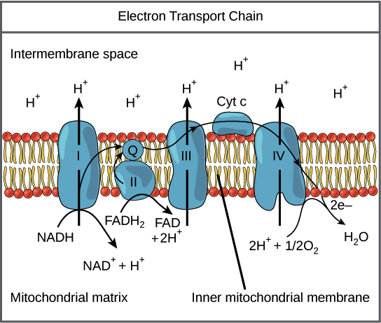
5. COMPONENTS OF THE ETC — ELECTRON CARRIERS
5.1 Types of Electron Carriers
| Carrier Type | Carries | Examples |
|---|---|---|
| NAD⁺/NADH | 2 electrons + 1 H⁺ (hydride ion) | Soluble in matrix |
| FAD/FADH₂ | 2 electrons + 2 H⁺ | Prosthetic group (tightly bound) |
| FMN/FMNH₂ | 2 electrons + 2 H⁺ | Prosthetic group in Complex I |
| Coenzyme Q (Ubiquinone) | 2 electrons + 2 H⁺ | Mobile carrier in lipid bilayer |
| Iron-Sulfur Centers (Fe-S) | 1 electron | Multiple types: [2Fe-2S], [3Fe-4S], [4Fe-4S] |
| Cytochromes (heme iron) | 1 electron (Fe²⁺ ↔ Fe³⁺) | Cyt b, c₁, c, a, a₃ |
| Copper Centers | 1 electron (Cu⁺ ↔ Cu²⁺) | CuA, CuB in Complex IV |
5.2 Flavin Nucleotides (FMN and FAD)
- Derived from Riboflavin (Vitamin B₂)
- Can accept 1 or 2 electrons (can form a semiquinone radical intermediate)
- FMN is the first electron acceptor in Complex I
- FAD is the prosthetic group in Complex II (succinate dehydrogenase)
5.3 Iron-Sulfur Clusters
- Also called non-heme iron proteins
- Contain iron atoms coordinated with inorganic sulfide (S²⁻) and/or cysteine sulfur from the protein
- Types:
- [2Fe-2S] — Rieske center in Complex III (unique: one Fe coordinated by 2 histidines instead of cysteines)
- [4Fe-4S] — most common, found in Complexes I, II, III
- [3Fe-4S] — found in Complex II
- Transfer only 1 electron at a time despite having multiple iron atoms
- Complex I contains 8 Fe-S clusters forming an electron "wire" ~95 Å long
5.4 Heme Groups and Cytochromes
| Cytochrome | Heme Type | Location | Unique Features |
|---|---|---|---|
| Cyt b (bL and bH) | Heme B (protoporphyrin IX) | Complex III | Two b-type cytochromes in Q cycle |
| Cyt c₁ | Heme C (covalently attached) | Complex III | Part of the complex |
| Cyt c | Heme C | IMS (mobile carrier) | Small, water-soluble; also involved in apoptosis |
| Cyt a | Heme A | Complex IV | Contains formyl group and hydroxyethylfarnesyl side chain |
| Cyt a₃ | Heme A | Complex IV | Contains the binuclear center (with CuB) where O₂ binds |
5.5 Copper Centers
- CuA in Complex IV: dinuclear copper center; first electron acceptor from cytochrome c
- CuB in Complex IV: paired with heme a₃ in the binuclear center; directly involved in O₂ binding and reduction
6. COMPLEX I — NADH:UBIQUINONE OXIDOREDUCTASE (EC 1.6.5.3)
6.1 Structure
- Largest respiratory complex
- ~1,000 kDa (1 MDa) in mammals
- 45 subunits in mammals (7 encoded by mtDNA: ND1–ND6, ND4L; 38 nuclear-encoded)
- Shaped like an "L" or a boot:
- Hydrophilic arm (peripheral arm): projects into the matrix; contains FMN and all Fe-S clusters
- Hydrophobic arm (membrane arm): embedded in the IMM; contains the proton-pumping machinery
- Contains:
- 1 FMN (flavin mononucleotide)
- 8 iron-sulfur clusters (N1a, N1b, N2, N3, N4, N5, N6a, N6b)
- The Fe-S clusters form a ~95 Å electron transfer wire
6.2 Mechanism
- NADH binds to the hydrophilic arm and donates 2 electrons as a hydride ion (H⁻) to FMN → FMNH₂
- FMNH₂ passes electrons one at a time through the chain of 8 Fe-S clusters
- The terminal Fe-S cluster N2 (highest potential in the chain) donates electrons to ubiquinone (CoQ)
- CoQ is reduced: Q → QH• (semiquinone) → QH₂ (ubiquinol)
- The energy released drives conformational changes in the membrane arm that translocate 4 H⁺ from the matrix to the IMS
6.3 Proton Pumping Mechanism
- The membrane arm contains three antiporter-like subunits (ND2, ND4, ND5) each pumping 1 H⁺
- A fourth proton is pumped at the junction near the ubiquinone-binding site (ND1/ND6 region)
- Total: 4 H⁺ pumped per NADH (per 2 electrons)
- Mechanism: long-range conformational coupling — a "piston-like" mechanism transmitted through a long amphipathic helix
6.4 Overall Reaction
NADH + H⁺ + Q + 4H⁺(matrix) → NAD⁺ + QH₂ + 4H⁺(IMS)
6.5 Inhibitors of Complex I
| Inhibitor | Source/Type | Mechanism |
|---|---|---|
| Rotenone | Plant insecticide (from Derris plant roots) | Blocks electron transfer from Fe-S cluster N2 to ubiquinone |
| Piericidin A | Streptomyces antibiotic | CoQ analog; competes at Q-binding site |
| Barbiturates (Amobarbital/Amytal) | Pharmaceutical | Block at the same site as rotenone |
| MPP⁺ | Active metabolite of MPTP | Inhibits Complex I |
🔴 CLINICAL: Rotenone and Parkinson's Disease
- Chronic rotenone exposure in animal models produces Parkinsonism including selective dopaminergic neuron death and Lewy body-like inclusions
- Dopaminergic neurons in the substantia nigra pars compacta are especially vulnerable due to high energy demands and high oxidative stress
- Epidemiological studies link pesticide exposure (rotenone, paraquat) to increased Parkinson's disease risk
🔴 CLINICAL: MPTP Toxicity (Drug-induced Parkinsonism)
- MPTP (1-methyl-4-phenyl-1,2,3,6-tetrahydropyridine) — contaminant of synthetic heroin ("designer drug")
- MPTP crosses the blood-brain barrier → taken up by astrocytes → converted to MPP⁺ by MAO-B
- MPP⁺ is selectively taken up by dopaminergic neurons via the dopamine transporter (DAT)
- MPP⁺ inhibits Complex I → ATP depletion, ROS generation → selective death of dopaminergic neurons
- Produces acute, irreversible Parkinsonism
- Prevention: MAO-B inhibitors (selegiline/rasagiline) block MPTP → MPP⁺ conversion
🔴 CLINICAL: Leber's Hereditary Optic Neuropathy (LHON)
- Mitochondrial inheritance (maternal)
- Point mutations in mtDNA genes encoding Complex I subunits
- Most common mutations: m.11778G>A (ND4, ~70% of cases), m.3460G>A (ND1), m.14484T>C (ND6)
- Presents in young adults (15-35 years), more commonly in males (male:female = 4:1)
- Acute or subacute painless bilateral central vision loss (sequential, one eye then the other within weeks to months)
- Loss of central vision with centrocecal scotoma (central blind spot)
- Optic disc pseudoedema, peripapillary telangiectatic microangiopathy (does NOT leak on fluorescein angiography)
- Eventually → optic atrophy
- No consistently effective treatment; idebenone (CoQ analog) may have some benefit
- Smoking and alcohol use may trigger/worsen the disease
🔴 CLINICAL: Complex I Deficiency
- Most common cause of mitochondrial disease in children
- Clinical spectrum: Leigh syndrome, MELAS-like syndrome, neonatal lactic acidosis, hypertrophic cardiomyopathy, leukodystrophy
- Lab findings: elevated lactate, elevated lactate:pyruvate ratio (>20:1), elevated alanine
7. COMPLEX II — SUCCINATE:UBIQUINONE OXIDOREDUCTASE (EC 1.3.5.1)
7.1 Structure
- Smallest respiratory complex: ~124 kDa
- 4 subunits — ALL nuclear-encoded (the ONLY ETC complex entirely nuclear-encoded)
- SDHA: flavoprotein subunit; contains covalently bound FAD and the succinate-binding site
- SDHB: iron-sulfur protein; contains three Fe-S clusters: [2Fe-2S], [4Fe-4S], [3Fe-4S]
- SDHC and SDHD: membrane-anchor subunits; contain heme b and the ubiquinone-binding site
- Part of both the TCA cycle (as succinate dehydrogenase, enzyme #6) and the ETC
7.2 Mechanism
- Succinate is oxidized to fumarate at the SDHA subunit
- FAD is reduced to FADH₂ (covalently bound — does NOT dissociate)
- Electrons pass through the three Fe-S clusters in SDHB: [2Fe-2S] → [4Fe-4S] → [3Fe-4S]
- Electrons are donated to ubiquinone (CoQ) at the Q-binding site → ubiquinol (QH₂)
7.3 Key Points
- Does NOT pump protons — insufficient free energy change (ΔG ≈ −6 kJ/mol for FAD → CoQ transfer)
- FADH₂ generated here enters the chain at the level of CoQ, bypassing Complex I
- Therefore, FADH₂ from Complex II generates fewer ATP than NADH (1.5 vs. 2.5 ATP)
7.4 Overall Reaction
Succinate + Q → Fumarate + QH₂ (No proton pumping)
7.5 Inhibitors of Complex II
| Inhibitor | Mechanism |
|---|---|
| Malonate | Competitive inhibitor of succinate (structural analog — dicarboxylic acid) |
| Carboxin | Blocks Q-binding site |
| TTFA (thenoyltrifluoroacetone) | Blocks Q-binding site |
| 3-Nitropropionic acid (3-NPA) | Irreversible inhibitor; causes striatal necrosis |
🔴 CLINICAL: Hereditary Paraganglioma-Pheochromocytoma Syndrome
- Germline mutations in SDHB, SDHC, SDHD (and also SDHA, SDHAF2)
- These are tumor suppressor genes (loss-of-function mutations)
- SDH deficiency → accumulation of succinate → inhibits prolyl hydroxylases (PHDs) → stabilization of HIF-1α → pseudohypoxic signaling → angiogenesis, proliferation (oncometabolite mechanism)
- Clinical manifestations: Paragangliomas, Pheochromocytomas, GISTs, Renal cell carcinoma
- SDHB mutations: highest malignancy risk (~30-40% metastatic)
- SDHD mutations: predominantly paternally inherited expression (genomic imprinting)
- Screening: plasma/urine metanephrines and catecholamines
- IHC: loss of SDHB staining in tumor tissue is a marker
🔴 CLINICAL: 3-Nitropropionic Acid (3-NPA) Toxicity
- Found in moldy sugarcane (Arthrinium saccharicola fungus)
- Irreversibly inhibits Complex II (SDH)
- Causes acute encephalopathy with selective bilateral striatal (basal ganglia) necrosis
- Mimics Huntington's disease pathology; used as an experimental model for HD
7.6 Other FADH₂-Linked Dehydrogenases
| Enzyme | Pathway | Location |
|---|---|---|
| ETF dehydrogenase (ETF:QO) | β-oxidation of fatty acids | IMM |
| Glycerol-3-phosphate dehydrogenase (mitochondrial) | Glycerol-3-phosphate shuttle | Outer surface of IMM |
| Dihydroorotate dehydrogenase | Pyrimidine biosynthesis | IMM |
| Choline dehydrogenase | Choline metabolism | IMM |
| Proline dehydrogenase | Proline metabolism | IMM |
🔴 CLINICAL: Multiple Acyl-CoA Dehydrogenase Deficiency (MADD) / Glutaric Aciduria Type II
- Deficiency of ETF or ETF:ubiquinone oxidoreductase (ETF-QO)
- Biochemical: elevated multiple acylcarnitines, organic acids (glutaric, ethylmalonic, adipic, suberic acids)
- Clinical: severe neonatal form or milder late-onset form (lipid storage myopathy)
- Treatment: riboflavin (B₂) — many cases are riboflavin-responsive
8. UBIQUINONE (COENZYME Q / CoQ₁₀)
8.1 Structure
- 2,3-dimethoxy-5-methyl-1,4-benzoquinone with an isoprenoid side chain
- In humans: 10 isoprenoid units (CoQ₁₀ or ubiquinone-50)
- Long hydrophobic tail → freely dissolves in the lipid bilayer of the IMM
- Small, lipid-soluble, mobile electron carrier — NOT a protein
8.2 Redox States
- Ubiquinone (Q) — fully oxidized
- Semiquinone radical (QH• or Q•⁻) — partially reduced (one electron)
- Ubiquinol (QH₂) — fully reduced (two electrons)
8.3 Function
- Collects electrons from Complexes I and II (and other FADH₂-linked dehydrogenases)
- Shuttles electrons to Complex III
- The CoQ pool contains ~10× more CoQ than Complex III → functions as a mobile electron buffer
8.4 Biosynthesis
- Synthesized endogenously via the mevalonate pathway (same pathway as cholesterol synthesis)
- The benzoquinone ring is derived from tyrosine (or phenylalanine)
- The isoprenoid tail is from the mevalonate pathway (farnesyl pyrophosphate → decaprenyl-PP)
🔴 CLINICAL: CoQ₁₀ Deficiency
- Primary CoQ₁₀ deficiency: autosomal recessive mutations in CoQ₁₀ biosynthetic pathway genes (COQ2, COQ4, COQ6, COQ7, COQ8A, COQ8B, COQ9, PDSS1, PDSS2)
- Clinical phenotypes: Cerebellar ataxia (most common), Steroid-resistant nephrotic syndrome (COQ2, COQ6, COQ8B), Encephalomyopathy, Isolated myopathy, Multisystem infantile form
- Secondary CoQ₁₀ deficiency: seen in mitochondrial diseases, aging, statin therapy
- Treatment: oral CoQ₁₀ supplementation (one of the few treatable mitochondrial disorders)
🔴 CLINICAL: Statins and CoQ₁₀
- Statins (HMG-CoA reductase inhibitors) inhibit the mevalonate pathway
- Reduces not only cholesterol but also CoQ₁₀ and dolichol synthesis
- May contribute to statin-associated myopathy/myalgia (biologically plausible; evidence mixed)
8.5 CoQ₁₀ as an Antioxidant
- Ubiquinol (QH₂) is a potent lipid-soluble antioxidant
- Protects membrane lipids from peroxidation
- Regenerates vitamin E (α-tocopherol) from its radical form
9. COMPLEX III — UBIQUINOL:CYTOCHROME c OXIDOREDUCTASE (EC 1.10.2.2)
9.1 Structure
- ~240 kDa as a functional homodimer (each monomer ~11 subunits in mammals)
- 11 subunits per monomer: 1 mtDNA-encoded (cytochrome b) + 10 nuclear-encoded
- Key catalytic subunits:
- Cytochrome b: contains two heme b groups — heme bL (low potential) and heme bH (high potential)
- Rieske iron-sulfur protein: contains [2Fe-2S] cluster — one Fe coordinated by 2 histidines (unique; gives higher reduction potential: +0.28 V)
- Cytochrome c₁: contains heme c₁
9.2 The Q Cycle (Mitchell's Q Cycle)
- Transfers electrons from a 2-electron carrier (QH₂) to a 1-electron carrier (cytochrome c)
- Pumps protons across the membrane
- Effectively doubles the number of protons translocated per QH₂
- Qp site (Qo site) — P-side (IMS side / positive side) — where QH₂ is oxidized
- Qn site (Qi site) — N-side (matrix side / negative side) — where Q is reduced
First Half of the Q Cycle:
- QH₂ binds at the Qp site
- First electron → Rieske [2Fe-2S] → Cytochrome c₁ → Cytochrome c (departs reduced)
- 2 H⁺ released into the IMS
- Second electron → low-potential pathway: QH• → heme bL → heme bH → reduces Q to Q•⁻ (semiquinone) at the Qn site
Second Half of the Q Cycle:
- A second QH₂ binds at the Qp site
- First electron → Rieske → cyt c₁ → second cytochrome c (reduced and departs)
- 2 more H⁺ released into IMS
- Second electron → heme bL → heme bH → reduces the semiquinone (Q•⁻) at Qn to QH₂ (picking up 2 H⁺ from the matrix)
Net Q Cycle (per pair of electrons reaching cytochrome c):
QH₂ + 2 cyt c (ox) + 2H⁺(matrix) → Q + 2 cyt c (red) + 4H⁺(IMS)
9.3 Inhibitors of Complex III
| Inhibitor | Site of Action | Mechanism |
|---|---|---|
| Antimycin A | Qn (Qi) site | Blocks electron transfer from heme bH to Q at the Qn site; prevents second half of Q cycle |
| Myxothiazol | Qp (Qo) site | Blocks QH₂ oxidation at the Qp site |
| Stigmatellin | Qp (Qo) site | Blocks electron transfer to Rieske center |
🔴 CLINICAL: Antimycin A and ROS
- When Antimycin A blocks the Qn site, the semiquinone radical (Q•⁻) at the Qp site accumulates
- This semiquinone can donate its electron directly to O₂ → forming superoxide (O₂•⁻)
- Complex III (Qp site) is a major site of mitochondrial ROS production
🔴 CLINICAL: Complex III Deficiency
- Rare; caused by mutations in:
- Cytochrome b (mtDNA — MT-CYB)
- BCS1L (assembly factor for Rieske protein)
- BCS1L mutations cause:
- GRACILE syndrome (Growth Retardation, Aminoaciduria, Cholestasis, Iron overload, Lactic acidosis, Early death) — Finnish heritage
- Björnstad syndrome (sensorineural deafness + pili torti — twisted hair)
- MT-CYB mutations: exercise intolerance, myopathy, cardiomyopathy
10. CYTOCHROME c
10.1 Structure and Function
- Small (~12.4 kDa), highly conserved, water-soluble protein
- Contains one heme c (covalently attached via thioether bonds to two cysteine residues — -CXXCH- motif)
- Located in the intermembrane space
- Peripheral membrane protein — loosely associated with the outer surface of the IMM via cardiolipin
- Transfers one electron from Complex III (cyt c₁) → Complex IV (CuA)
10.2 Role in Apoptosis
🔴 CLINICAL: Cytochrome c and Apoptosis (Intrinsic Pathway)
- During intrinsic (mitochondrial) apoptosis:
- Pro-apoptotic stimuli activate BH3-only proteins (BID, BIM, BAD, etc.)
- These activate BAX and BAK → oligomerize → pores in OMM → MOMP
- Cytochrome c is released from the IMS into the cytoplasm
- Cytochrome c + APAF-1 → forms the apoptosome
- Apoptosome activates Caspase-9 (initiator) → Caspase-3 and -7 (executioners) → cell death
- Anti-apoptotic proteins (BCL-2, BCL-XL, MCL-1) prevent cytochrome c release
- Venetoclax: BCL-2 inhibitor used in CLL and AML
- Other mitochondrial proteins released: Smac/DIABLO (inhibits IAPs), AIF (caspase-independent death), endonuclease G, Omi/HtrA2
10.3 Cytochrome c in Evolution
- One of the most conserved proteins in evolution — used to construct phylogenetic trees
- Human and chimpanzee cytochrome c are identical (100% homology)
- Human and yeast share ~60% identity
11. COMPLEX IV — CYTOCHROME c OXIDASE (EC 1.9.3.1)
11.1 Structure
- ~200 kDa as a functional dimer (each monomer: 13 subunits in mammals)
- 3 mtDNA-encoded subunits: COX I, COX II, COX III (catalytic core)
- 10 nuclear-encoded subunits (regulatory/structural)
- Contains 4 redox-active metal centers:
- CuA center (in COX II): dinuclear copper; first to accept electron from cytochrome c
- Heme a (in COX I): transfers electron to the binuclear center
- Heme a₃ (in COX I): part of the binuclear center
- CuB (in COX I): part of the binuclear center; paired with heme a₃
11.2 Mechanism
- Cytochrome c (reduced) binds to the IMS face → donates 1 electron to CuA
- CuA → heme a → binuclear center (heme a₃–CuB)
- At the binuclear center, O₂ binds between Fe of heme a₃ and CuB
- Four electrons required to fully reduce O₂:
O₂ + 4 e⁻ + 4 H⁺ (matrix) → 2 H₂O
- Carefully controlled sequence avoids release of partially reduced oxygen species
11.3 Proton Pumping
- 4 "chemical" protons: consumed in the matrix to make H₂O (scalar protons)
- 4 "pumped" protons: translocated from matrix to IMS (vectorial protons)
- Total: 8 H⁺ removed from the matrix per O₂
11.4 Overall Reaction
4 cyt c (red) + O₂ + 8H⁺(matrix) → 4 cyt c (ox) + 2H₂O + 4H⁺(IMS)
11.5 Inhibitors of Complex IV
| Inhibitor | Mechanism |
|---|---|
| Cyanide (CN⁻) | Binds to Fe³⁺ of heme a₃ → blocks O₂ binding |
| Carbon monoxide (CO) | Binds to Fe²⁺ of heme a₃ → blocks O₂ binding |
| Hydrogen sulfide (H₂S) | Binds to heme a₃ → blocks O₂ binding |
| Azide (N₃⁻) | Binds to Fe³⁺ of heme a₃ |
| Nitric oxide (NO) | Reversible inhibitor; binds to heme a₃ and CuB |
| Phosphine (PH₃) | Insecticide; inhibits Complex IV |
| Formate (from methanol metabolism) | Inhibits Complex IV |
🔴 CLINICAL: Cyanide Poisoning
- Sources: industrial chemicals (electroplating, mining), smoke inhalation (burning of plastics/wool/silk releases HCN), cyanogenic glycosides (bitter almonds, cassava, apple seeds — contain amygdalin), sodium nitroprusside (prolonged infusion)
- Mechanism: CN⁻ binds Fe³⁺ of cytochrome a₃ → complete inhibition of Complex IV → histotoxic/cytotoxic hypoxia
- Classic features:
- Bright cherry-red skin (venous blood remains oxygenated — cells can't extract O₂)
- "Bitter almond" breath odor (only 40% of people can detect this — genetically determined)
- High venous PO₂ (narrow A-V O₂ difference)
- Severe lactic acidosis
- PaO₂ and O₂ saturation may be normal (pulse oximetry and ABG can be misleadingly normal)
- Treatment:
- 100% oxygen
- Hydroxocobalamin (Cyanokit®): binds CN⁻ → cyanocobalamin (B₁₂) → renal excretion. Preferred antidote (safe in combined CO/CN poisoning)
- Sodium thiosulfate: sulfur donor for rhodanese → converts CN⁻ → thiocyanate (SCN⁻) → renal excretion. Slow onset.
- Sodium nitrite / amyl nitrite: induces methemoglobinemia → MetHb (Fe³⁺) binds CN⁻. Caution in smoke inhalation (already has COHb)
- Dicobalt edetate (Kelocyanor): used in UK — cobalt chelates CN⁻
🔴 CLINICAL: Carbon Monoxide (CO) Poisoning
- CO binds to:
- Hemoglobin (240× greater affinity than O₂) → carboxyhemoglobin (COHb) → leftward shift of O₂-Hb dissociation curve
- Cytochrome c oxidase (Complex IV) → Fe²⁺ of heme a₃ → histotoxic hypoxia
- Myoglobin → impaired muscle O₂ utilization
- Sources: incomplete combustion, house fires, car exhaust, charcoal grills
- Findings:
- Headache (most common early symptom), nausea, confusion, syncope, seizures, coma
- PaO₂ is NORMAL (dissolved O₂ unaffected)
- O₂ saturation by pulse oximetry is FALSELY NORMAL (can't distinguish COHb from OxyHb)
- CO-oximetry is the diagnostic test
- Normal COHb <3% (non-smokers); symptomatic >15-20%; lethal >50-60%
- Treatment:
- 100% O₂ via non-rebreather mask (reduces COHb half-life from 5 hours → 90 minutes)
- Hyperbaric oxygen (HBO₂): reduces half-life to ~30 minutes; indicated for COHb >25%, loss of consciousness, neurological symptoms, cardiac ischemia, pregnancy
🔴 CLINICAL: Hydrogen Sulfide (H₂S) Poisoning
- "Knockdown gas" — toxic in sewer gas, volcanic emissions, petroleum industry
- Mechanism: like cyanide, inhibits Complex IV
- Rotten egg smell at low concentrations; at high concentrations → olfactory nerve paralysis (extremely dangerous — can no longer smell it)
- Treatment: nitrites (to form sulfmethemoglobin), 100% O₂, supportive
🔴 CLINICAL: Methanol and Formate Toxicity
- Methanol → formaldehyde → formic acid/formate (via alcohol dehydrogenase and aldehyde dehydrogenase)
- Formate inhibits cytochrome c oxidase (Complex IV)
- Causes: metabolic acidosis (high anion gap), blindness (retinal ganglion cell toxicity)
- Treatment: fomepizole (ADH inhibitor) or ethanol (competitive substrate), dialysis, folate
12. COMPLEX V — ATP SYNTHASE (F₁F₀-ATP SYNTHASE) (EC 7.1.2.2)
12.1 Overview
- Also called: F₁F₀-ATPase, Complex V
- Uses the proton motive force (PMF) generated by Complexes I, III, IV to synthesize ATP from ADP + Pi
- ~600 kDa
- 16 subunits in mammals (2 mtDNA-encoded: ATPase 6 = subunit a, ATPase 8 = subunit A6L)
12.2 Structure
F₁ Component (Soluble, Matrix side)
- Knob-like structure projecting into the matrix
- Composition: α₃β₃γδε
- 3 α subunits and 3 β subunits: arranged alternately like segments of an orange
- Each β subunit contains a catalytic site for ATP synthesis
- The 3 β subunits exist in 3 different conformations simultaneously (see binding change mechanism)
- γ subunit: central stalk/axle; rotates within the α₃β₃ ring; asymmetric → drives conformational changes in β subunits
- ε subunit: attached to γ; part of the rotor
- δ subunit (OSCP — Oligomycin Sensitivity Conferring Protein): part of the peripheral stalk
F₀ Component (Membrane-embedded)
- Proton channel driving rotation
- Composition (in mammals):
- c-ring: ring of 8 c subunits in mammals (10 in yeast); each c subunit has a proton-binding carboxylate (Asp or Glu)
- a subunit (subunit 6): has two half-channels for proton translocation; stationary
- b subunit: part of the peripheral/lateral stalk (stator)
- Additional subunits: d, F6, A6L, e, f, g (structural and regulatory)
Peripheral Stalk (Stator)
- Connects F₁ to F₀ without rotating
- Composed of: b, d, F6, OSCP subunits
- Prevents co-rotation of α₃β₃ with γ
12.3 Boyer's Binding Change Mechanism

Three Conformations of β Subunits:
- O (Open): low affinity for substrates; releases ATP
- L (Loose): binds ADP + Pi loosely
- T (Tight): catalyzes ATP formation; binds ATP very tightly
Catalytic Cycle (per 120° rotation of γ):
- ADP + Pi bind to the L-site → γ rotates 120° → L-site converts to T-site → ATP is synthesized
- Previous T-site converts to O-site → ATP is released
- Previous O-site converts to L-site → ready for new ADP + Pi
12.4 Proton Translocation Through F₀
- H⁺ from IMS enters the inlet half-channel in the a subunit
- H⁺ binds to the carboxylate (Asp/Glu) on a c subunit
- Neutralization allows the c subunit to move into the hydrophobic lipid bilayer
- As the c-ring rotates (driven by the proton gradient), each c subunit eventually reaches the outlet half-channel on the matrix side
- The H⁺ is released into the matrix
- In mammals (8 c subunits): 8 H⁺ per 360° → 8/3 ≈ 2.67 H⁺ per ATP
- Plus 1 H⁺ for the phosphate carrier (Pi/H⁺ symport) → ~3.67 H⁺ per ATP
- Often approximated as ~4 H⁺ per ATP
12.5 ATP/ADP Translocase (Adenine Nucleotide Translocator — ANT)
- Located in IMM
- Antiporter: exchanges ATP⁴⁻ (out to IMS) for ADP³⁻ (in to matrix)
- Electrogenic — driven by the membrane potential (Δψ)
| Inhibitor | Effect |
|---|---|
| Atractyloside (from thistle plant) | Binds to cytoplasmic face of ANT → blocks ADP import |
| Bongkrekic acid (contaminated coconut/corn — Burkholderia gladioli) | Binds to matrix face → locks ANT in "m-state" |
🔴 CLINICAL: Bongkrekic Acid Poisoning
- From fermented coconut or corn products contaminated with Burkholderia gladioli pathovar cocovenenans
- Outbreaks in Southeast Asia, China
- Inhibits ANT → ATP depletion → multiorgan failure; very high mortality (40-100%)
12.6 Inhibitors of ATP Synthase (Complex V)
| Inhibitor | Mechanism |
|---|---|
| Oligomycin | Blocks the proton channel in F₀ (between a and c subunits) → prevents H⁺ flow → stops ATP synthesis AND electron transport (PMF back-pressure) |
| DCCD (Dicyclohexylcarbodiimide) | Reacts with the carboxylate (Asp/Glu) on c subunits → blocks proton binding |
| Aurovertin | Binds to β subunit of F₁ → blocks catalytic activity |
| Efrapeptin | Blocks the F₁ catalytic site |
🔴 CLINICAL: ATP Synthase Deficiency
- Mutations in MT-ATP6 (subunit a):
- NARP syndrome (Neuropathy, Ataxia, Retinitis Pigmentosa): mutant mtDNA load 70-90%
- Maternally Inherited Leigh Syndrome (MILS): mutant load >90%
- Most common mutation: m.8993T>G or m.8993T>C
- TMEM70 mutations: most common nuclear-encoded ATP synthase deficiency; 3-methylglutaconic aciduria, cardiomyopathy, lactic acidosis
13. THE PROTON MOTIVE FORCE (PMF)
13.1 Definition
- Δψ = membrane potential (voltage gradient) — inside (matrix) is negative; ~150-180 mV in mammals
- ΔpH = pH gradient — matrix is more alkaline (~0.5-1.0 pH units)
- Under physiological conditions: ~80% from Δψ, ~20% from ΔpH
13.2 Total Protons Pumped
| Complex | H⁺ pumped per NADH | H⁺ pumped per FADH₂ |
|---|---|---|
| Complex I | 4 H⁺ | 0 (bypassed) |
| Complex III | 4 H⁺ | 4 H⁺ |
| Complex IV (pumped) | 2 H⁺ | 2 H⁺ |
| Total vectorial H⁺ | 10 H⁺ | 6 H⁺ |

14. CHEMIOSMOTIC HYPOTHESIS
Peter Mitchell (1961) — Nobel Prize in Chemistry (1978)
14.1 Key Postulates:
- The ETC complexes are vectorially arranged in the IMM, pumping protons from matrix → IMS
- The IMM is impermeable to protons — the proton gradient is maintained
- The electrochemical proton gradient (PMF) stores energy
- Protons flow back into the matrix only through ATP synthase → drives ATP synthesis
- Electron transport and ATP synthesis are coupled through the PMF — not by chemical intermediates
14.2 Evidence Supporting the Hypothesis:
- Intact IMM is required for oxidative phosphorylation
- Uncouplers (which dissipate the proton gradient) abolish ATP synthesis without stopping electron transport
- Artificially created pH gradients across vesicles can drive ATP synthesis
- ATP synthase reconstituted in artificial liposomes produces ATP when a pH gradient is applied
- Ionophores that collapse the gradient inhibit ATP synthesis
15. P/O RATIOS AND ATP YIELD
15.1 Definition
15.2 Revised Values (Current Consensus)
| Substrate | H⁺ pumped | ATP per molecule | P/O ratio |
|---|---|---|---|
| NADH | 10 | ~2.5 (10/4) | 2.5 |
| FADH₂ | 6 | ~1.5 (6/4) | 1.5 |
15.3 Total ATP Yield from Complete Oxidation of Glucose
| Reaction | NADH | FADH₂ | GTP/ATP |
|---|---|---|---|
| Glycolysis: Glucose → 2 Pyruvate | 2 (cytoplasmic) | — | 2 ATP |
| 2 Pyruvate → 2 Acetyl-CoA (PDH) | 2 | — | — |
| 2 turns TCA cycle | 6 | 2 | 2 GTP |
| Totals | 10 NADH | 2 FADH₂ | 4 ATP/GTP |
- 8 mitochondrial NADH × 2.5 = 20 ATP
- 2 cytoplasmic NADH — yield depends on shuttle used (see Section 16)
- 2 FADH₂ × 1.5 = 3 ATP
- 4 ATP/GTP (substrate level)
| Using Malate-Aspartate Shuttle | Using Glycerol-3-Phosphate Shuttle |
|---|---|
| 20 + (2 × 2.5) + 3 + 4 = ~30-32 ATP | 20 + (2 × 1.5) + 3 + 4 = ~30 ATP |
16. SHUTTLE SYSTEMS
16.1 Malate-Aspartate Shuttle
- Cytoplasmic OAA + NADH → malate + NAD⁺ (cytoplasmic malate dehydrogenase)
- Malate crosses IMM via the malate-α-ketoglutarate antiporter
- Matrix malate → OAA + NADH (mitochondrial malate dehydrogenase)
- Matrix OAA → transaminated to aspartate (by aspartate aminotransferase, glutamate → α-KG)
- Aspartate exits via the glutamate-aspartate antiporter (electrogenic — driven by Δψ)
- Cytoplasmic aspartate → OAA (by cytoplasmic aspartate aminotransferase)
- α-KG returns to matrix via malate-αKG antiporter
16.2 Glycerol-3-Phosphate Shuttle
- Cytoplasmic DHAP + NADH → glycerol-3-phosphate + NAD⁺ (cytoplasmic G3P dehydrogenase — NAD⁺-linked)
- G3P → DHAP + FADH₂ (mitochondrial G3P dehydrogenase — FAD-linked, on outer surface of IMM)
- FADH₂ directly reduces CoQ in the IMM
🔴 CLINICAL: Malate-Aspartate Shuttle and Aralar1/AGC1 Deficiency
- SLC25A12 mutations (AGC1/Aralar1 — mitochondrial aspartate-glutamate carrier)
- Causes global cerebral hypomyelination, developmental delay, seizures
17. INHIBITORS OF THE ETC — COMPREHENSIVE SUMMARY
17.1 Classification by Site
| Complex | Inhibitors |
|---|---|
| Complex I | Rotenone, Piericidin A, Barbiturates (amytal), MPP⁺ |
| Complex II | Malonate, Carboxin, TTFA, 3-Nitropropionic acid |
| Complex III | Antimycin A (Qi/Qn site), Myxothiazol (Qo/Qp site), Stigmatellin (Qo/Qp site) |
| Complex IV | Cyanide (CN⁻), Carbon monoxide (CO), H₂S, Azide (N₃⁻), NO, Phosphine, Formate |
| Complex V | Oligomycin, DCCD, Aurovertin, Efrapeptin |
| ANT | Atractyloside, Bongkrekic acid |
17.2 Effects of ETC Inhibitors
- Upstream of the block: carriers become more reduced
- Downstream of the block: carriers become more oxidized
- O₂ consumption decreases
- ATP synthesis decreases
- NADH/NAD⁺ ratio increases → inhibits TCA cycle
- Lactic acidosis
18. UNCOUPLERS OF OXIDATIVE PHOSPHORYLATION
18.1 Definition
18.2 Mechanism
- Pick up H⁺ on the IMS side (protonated form is lipid-soluble)
- Cross the IMM in their protonated form
- Release H⁺ on the matrix side
- Return in their anionic form
- Short-circuits the proton gradient → collapses Δp
18.3 Effects of Uncoupling
| Parameter | Effect |
|---|---|
| Electron transport | ↑ (accelerated — no back-pressure from PMF) |
| O₂ consumption | ↑↑ |
| ATP synthesis | ↓↓ (abolished) |
| NADH/NAD⁺ ratio | ↓ (more NAD⁺ — TCA cycle accelerated) |
| Heat production | ↑↑ (thermogenesis) |
| Proton gradient | ↓↓ (dissipated) |
| Fuel oxidation | ↑↑ |
18.4 Examples of Uncouplers
| Uncoupler | Notes |
|---|---|
| 2,4-dinitrophenol (DNP) | Classical uncoupler; used as weight-loss drug in 1930s (banned due to toxicity) |
| FCCP | Research tool |
| CCCP | Research tool |
| Thermogenin (UCP1) | Natural uncoupling protein in brown adipose tissue |
| Valinomycin | K⁺ ionophore; dissipates Δψ component |
| Nigericin | K⁺/H⁺ antiporter; dissipates ΔpH |
| Salicylates (high dose aspirin) | Weak uncoupling activity |
| Bilirubin (high levels) | Mild uncoupler |
| Long-chain fatty acids | Can act as mild uncouplers |
🔴 CLINICAL: 2,4-Dinitrophenol (DNP) Toxicity
- Used in the 1930s as a weight-loss agent — uncoupling → increased metabolic rate → fat burning
- Banned by FDA in 1938; still available illegally online
- Symptoms: profuse sweating, tachycardia, tachypnea, hyperthermia (potentially fatal)
- Cataracts with chronic use
- No specific antidote — supportive: cooling, dantrolene may help
- Narrow therapeutic index → small dose increase can be lethal
🔴 CLINICAL: Aspirin (Salicylate) Overdose
- High-dose salicylates:
- Uncouple oxidative phosphorylation (weak acid, lipid-soluble)
- Direct stimulant of the respiratory center (medullary)
- Clinical toxicity:
- Respiratory alkalosis (early — direct stimulation of respiratory center)
- Metabolic acidosis (high anion gap — from uncoupling → lactate, ketoacids + salicylate itself)
- Mixed respiratory alkalosis and metabolic acidosis is characteristic
- Hyperthermia, tinnitus, diaphoresis, nausea/vomiting
- Treatment: sodium bicarbonate (alkalinize urine → traps salicylate in ionized form), hemodialysis for severe cases, cooling
18.5 Thermogenin (UCP1) and Brown Adipose Tissue (BAT)
🔴 CLINICAL: Non-Shivering Thermogenesis
- Brown adipose tissue (BAT) — abundant mitochondria (brown color from cytochromes)
- Contains UCP1 (Uncoupling Protein 1 / Thermogenin) — a natural proton channel in the IMM
- UCP1 allows H⁺ to re-enter the matrix without passing through ATP synthase → energy released as HEAT
- Important in:
- Neonates (who cannot shiver) — major source of heat production
- Hibernating animals
- Adults (metabolically active BAT in supraclavicular, paravertebral, perirenal regions)
- Regulation of UCP1:
- Activated by: free fatty acids, norepinephrine (via β₃-adrenergic receptors → cAMP → PKA → hormone-sensitive lipase → FFA release → FFA activates UCP1)
- Inhibited by: purine nucleotides (GDP, GTP, ADP, ATP) — bind and block the channel
- Thyroid hormones (T₃) upregulate UCP1 → explains heat intolerance in hyperthyroidism
18.6 Other Uncoupling Proteins
| UCP | Location | Proposed Function |
|---|---|---|
| UCP1 | Brown adipose tissue | Thermogenesis |
| UCP2 | Widely expressed (pancreatic β-cells, brain) | ROS regulation, β-cell function |
| UCP3 | Skeletal muscle, heart | Fatty acid metabolism, ROS protection |
| UCP4 | Brain | Neuroprotection |
| UCP5 (BMCP1) | Brain | Neuroprotection |
19. TIGHT COUPLING, RESPIRATORY CONTROL, AND REGULATION
19.1 Respiratory Control
- The rate of O₂ consumption (electron transport) is determined by the availability of ADP
- Low ADP → no ATP synthesis needed → proton gradient builds up → back-pressure inhibits ETC → O₂ consumption decreases (State 4)
- High ADP → ADP enters mitochondria → ATP synthase active → proton gradient consumed → ETC speeds up → O₂ consumption increases (State 3)
19.2 Respiratory States (Chance & Williams)
| State | Substrate | ADP | O₂ | Rate of respiration |
|---|---|---|---|---|
| State 1 | Low | Low | Adequate | Slow |
| State 2 | Added | Low | Adequate | Slow |
| State 3 (active) | Adequate | Added (high) | Adequate | Fast |
| State 4 (resting) | Adequate | Depleted (low) | Adequate | Slow |
| State 5 | Adequate | Adequate | Absent | Zero |
- Respiratory Control Ratio (RCR) = State 3 rate / State 4 rate
- Normal RCR = 5-10 (tight coupling); low RCR = uncoupling or damaged mitochondria
19.3 Regulation of ETC
| Regulatory Factor | Effect |
|---|---|
| [ADP] | Primary regulator — high ADP → increased respiration |
| [ATP] | High ATP → inhibits Complex IV (allosteric) |
| [NADH]/[NAD⁺] ratio | Controls substrate supply to Complex I |
| [O₂] | Must be adequate (Km of Complex IV for O₂ ≈ 1 μM — efficient even at low O₂) |
| Thyroid hormones | Increase mitochondrial biogenesis, ETC expression, UCP expression → increase BMR |
| Ca²⁺ | Activates PDH, isocitrate DH, α-KG DH → increases NADH supply; stimulates Complex IV and ATP synthase |
| NO | Reversible inhibition of Complex IV — regulates O₂ distribution |
20. REACTIVE OXYGEN SPECIES (ROS)
20.1 Overview
20.2 Sites of ROS Production
| Site | ROS produced | Mechanism |
|---|---|---|
| Complex I (FMN site and N1a cluster) | O₂•⁻ (superoxide) | Especially during reverse electron transfer (RET) from succinate when Δp is high |
| Complex III (Qp/Qo site) | O₂•⁻ | Semiquinone radical at Qp site donates electron to O₂ |
| Complex II (flavin site) | O₂•⁻ | Minor source |
| Other sources | Monoamine oxidase (OMM), p66^shc, xanthine oxidase, NADPH oxidase |
20.3 Types of ROS
| Species | Formula | Formation |
|---|---|---|
| Superoxide | O₂•⁻ | 1-electron reduction of O₂ |
| Hydrogen peroxide | H₂O₂ | By SOD from superoxide; or 2-electron reduction of O₂ |
| Hydroxyl radical | •OH | Fenton reaction: Fe²⁺ + H₂O₂ → Fe³⁺ + •OH + OH⁻ |
| Peroxynitrite | ONOO⁻ | O₂•⁻ + NO• → ONOO⁻ |
20.4 Antioxidant Defense Systems
| Defense | Mechanism |
|---|---|
| SOD (Superoxide Dismutase) | O₂•⁻ → H₂O₂ + O₂; MnSOD (SOD2) in matrix, Cu/ZnSOD (SOD1) in cytoplasm/IMS |
| Catalase | 2 H₂O₂ → 2 H₂O + O₂ (in peroxisomes) |
| Glutathione peroxidase (GPx) | H₂O₂ + 2 GSH → 2 H₂O + GSSG (requires selenium) |
| Glutathione reductase | GSSG + NADPH → 2 GSH (requires NADPH from pentose phosphate pathway) |
| Thioredoxin / Peroxiredoxin system | Reduces H₂O₂ and organic peroxides |
| Vitamin E (α-tocopherol) | Lipid-soluble radical scavenger |
| Vitamin C (ascorbate) | Water-soluble radical scavenger; regenerates vitamin E |
| CoQ₁₀ (ubiquinol) | Lipid-soluble antioxidant in membranes |
20.5 ROS-Mediated Damage
| Target | Damage |
|---|---|
| DNA | Oxidation of bases (8-oxo-guanine), strand breaks → mutations, cancer |
| Proteins | Carbonylation, disulfide bond formation, loss of function |
| Lipids | Lipid peroxidation → MDA, 4-HNE → membrane damage |
| mtDNA | Especially vulnerable (no histones, near ROS source, limited repair) → vicious cycle |
🔴 CLINICAL: ROS and Disease
- Aging: cumulative mtDNA damage → declining ETC function → more ROS → more damage (vicious cycle) — "Mitochondrial free radical theory of aging"
- Ischemia-Reperfusion Injury: reperfusion → massive ROS burst from dysfunctional ETC (relevant in MI, stroke, organ transplantation)
- Neurodegenerative diseases: Parkinson's (Complex I deficiency, ROS in substantia nigra), Alzheimer's, ALS
- Diabetes: hyperglycemia → increased ETC substrate → ROS overproduction → diabetic complications
🔴 CLINICAL: Superoxide Dismutase and ALS
- ~20% of familial ALS cases involve mutations in SOD1 (Cu/ZnSOD)
- Gain of toxic function (misfolded SOD1 aggregates → motor neuron death) — NOT loss-of-function
- Autosomal dominant
🔴 CLINICAL: Friedreich's Ataxia and Iron-Sulfur Clusters
- Autosomal recessive; most common hereditary ataxia
- GAA trinucleotide repeat expansion in FXN gene (frataxin)
- Frataxin → involved in iron-sulfur cluster assembly and iron homeostasis
- Frataxin deficiency → impaired Fe-S cluster synthesis → dysfunctional Complexes I, II, III and aconitase
- Mitochondrial iron accumulation → Fenton reaction → ROS damage
- Clinical: progressive gait and limb ataxia, dysarthria, hypertrophic cardiomyopathy (major cause of death), diabetes mellitus, absent deep tendon reflexes, pes cavus, scoliosis, upgoing plantars
- Treatment: omaveloxolone (Nrf2 activator — FDA approved 2023), idebenone (limited benefit)
21. SUPERCOMPLEXES (RESPIRASOMES)
21.1 Concept
| Supercomplex | Composition | Name |
|---|---|---|
| I + III₂ | Complex I + dimer of Complex III | Supercomplex |
| I + III₂ + IV₁₋₄ | Complex I + Complex III dimer + 1-4 copies of Complex IV | Respirasome |
| III₂ + IV | Complex III dimer + Complex IV | Supercomplex |
21.2 Functional Significance
- Substrate channeling: CoQ and cytochrome c channeled between complexes → faster electron transfer
- Reduced ROS: shorter transit time → less chance of electron leak to O₂
- Stabilization: Complex I is unstable without Complex III
- Cardiolipin is essential for supercomplex assembly
🔴 CLINICAL: Supercomplex Dysfunction
- Heart failure: decreased supercomplex assembly → increased ROS, decreased ATP production
- Barth syndrome: cardiolipin deficiency → impaired supercomplex formation
- Aging: decreased supercomplex levels in aged tissues
22. COMPREHENSIVE CLINICAL CORRELATIONS
22.1 Mitochondrial Diseases — Overview
- Brain: encephalopathy, seizures, stroke-like episodes, ataxia
- Muscle: myopathy, exercise intolerance, ophthalmoplegia
- Heart: cardiomyopathy, conduction defects
- Eye: optic neuropathy, retinitis pigmentosa, ptosis
- Ear: sensorineural hearing loss
- Endocrine: diabetes mellitus, growth hormone deficiency
- GI: pseudo-obstruction
- Kidney: tubular dysfunction, Fanconi syndrome
- Elevated serum lactate (most consistent finding)
- Elevated lactate:pyruvate ratio (>20:1)
- Elevated alanine
- Ragged red fibers (RRF) on Gomori trichrome stain of muscle biopsy
- COX-negative fibers on cytochrome c oxidase staining
- MRI: stroke-like lesions not conforming to vascular territories, bilateral basal ganglia lesions (Leigh syndrome), cerebellar atrophy
22.2 Specific Mitochondrial Syndromes
MELAS (Mitochondrial Encephalomyopathy, Lactic Acidosis, Stroke-like episodes)
- Mutation: m.3243A>G in MT-TL1 (tRNA^Leu(UUR)) — ~80% of cases
- Features: stroke-like episodes (NOT true strokes — don't follow vascular territories; often occipital/parietal), lactic acidosis, seizures, myopathy, sensorineural hearing loss, diabetes mellitus (MIDD — maternally inherited diabetes and deafness), short stature, migraine, cognitive decline, ragged red fibers
MERRF (Myoclonic Epilepsy with Ragged Red Fibers)
- Mutation: m.8344A>G in MT-TK (tRNA^Lys) — ~80% of cases
- Features: myoclonus (stimulus-sensitive), generalized epilepsy, myopathy with RRF, cerebellar ataxia, multiple symmetric lipomas (especially cervical), sensorineural hearing loss, optic atrophy, dementia, lactic acidosis
Leigh Syndrome (Subacute Necrotizing Encephalomyelopathy)
- Most common mitochondrial disease in infancy/childhood
- Caused by mutations in >75 different genes (both mtDNA and nuclear)
- Complex I (most common), Complex II, Complex III, Complex IV, Complex V, PDH, CoQ₁₀ biosynthesis
- Common: MT-ATP6 (m.8993T>G/C), SURF1 (Complex IV assembly), NDUFS4 (Complex I)
- Pathology: bilateral, symmetric necrotizing lesions in basal ganglia, brainstem, thalamus, spinal cord
- Features: developmental regression, hypotonia → spasticity, respiratory abnormalities (central hypoventilation), eye movement abnormalities, lactic acidosis
- MRI: bilateral symmetric T2-hyperintense lesions in basal ganglia and brainstem
Kearns-Sayre Syndrome (KSS)
- Large-scale mtDNA deletions (most common: "common deletion" — 4,977 bp); usually sporadic
- Diagnostic triad (ALL required):
- Progressive External Ophthalmoplegia (PEO) — ptosis + restricted eye movements
- Pigmentary retinopathy ("salt and pepper" fundus)
- Onset before age 20
- Plus at least one of: cardiac conduction defect, cerebellar ataxia, CSF protein >100 mg/dL
- Other features: short stature, hearing loss, endocrinopathies, cognitive decline
- Cardiac conduction defects are life-threatening → may require pacemaker
- Ragged red fibers and COX-negative fibers on muscle biopsy
Pearson Syndrome
- Same large-scale mtDNA deletions as KSS; presents in infancy
- Sideroblastic anemia + pancreatic exocrine insufficiency
- Bone marrow: vacuolization of precursors, ringed sideroblasts
- Survivors may develop KSS in later years
NARP (Neuropathy, Ataxia, Retinitis Pigmentosa)
- Mutation: m.8993T>G (or T>C) in MT-ATP6
- Heteroplasmy level 70-90% → NARP; >90% → Leigh syndrome (MILS)
- Features: peripheral neuropathy, cerebellar ataxia, retinitis pigmentosa, proximal weakness, developmental delay
Aminoglycoside-Induced Ototoxicity
- Mutation: m.1555A>G in MT-RNR1 (12S rRNA)
- Makes mitochondrial ribosome more "bacterial-like" → aminoglycosides damage mitochondrial translation
- Even a single dose of aminoglycosides → permanent bilateral sensorineural hearing loss
🔴 CLINICAL: POLG Mutations
- POLG encodes mitochondrial DNA polymerase γ (responsible for mtDNA replication)
- Wide spectrum:
- Alpers-Huttenlocher syndrome: progressive encephalopathy, intractable seizures, hepatopathy → liver failure (VALPROATE IS CONTRAINDICATED in POLG mutations — precipitates fatal hepatotoxicity)
- CPEO (with multiple mtDNA deletions)
- Ataxia-neuropathy spectrum (MIRAS, SANDO)
22.3 Acquired Conditions Affecting the ETC
Ischemia-Reperfusion Injury
- Ischemia: O₂ deprivation → ETC stops → succinate accumulates → ATP depleted → anaerobic glycolysis → acidosis → Ca²⁺ accumulation
- Reperfusion: O₂ reintroduced → accumulated succinate rapidly oxidized by Complex II → reverse electron transport (RET) through Complex I → massive superoxide production → oxidative damage
- Clinical: MI reperfusion, stroke thrombolysis, organ transplantation
Drug-Induced Mitochondrial Toxicity
| Drug | Mechanism | Clinical Effect |
|---|---|---|
| NRTIs (zidovudine, stavudine, didanosine) | Inhibit mitochondrial DNA polymerase γ (POLG) | Myopathy, lactic acidosis, lipodystrophy, neuropathy, hepatic steatosis |
| Metformin | Mild Complex I inhibition | Lactic acidosis (rare — mainly in renal failure) |
| Doxorubicin | ROS generation, Complex I inhibition, cardiolipin damage | Cardiomyopathy (dose-dependent, cumulative) |
| Linezolid | Inhibits mitochondrial protein synthesis | Lactic acidosis, peripheral neuropathy (prolonged use >2 weeks) |
| Propofol | Inhibits Complex I and Complex IV, impairs fatty acid oxidation | Propofol infusion syndrome (PRIS): cardiac failure, rhabdomyolysis, lactic acidosis, renal failure; high doses >4 mg/kg/hr for >48 hours |
| Valproate | Depletes CoA, carnitine; inhibits β-oxidation; inhibits Complex IV | Hepatotoxicity, hyperammonemia, fatal in POLG mutations |
| Chloramphenicol | Inhibits mitochondrial ribosomes | Aplastic anemia |
| Statins | Decrease CoQ₁₀ synthesis | Myalgia/myopathy |
Malignant Hyperthermia
🔴 CLINICAL: Malignant Hyperthermia
- Autosomal dominant; mutations in RYR1 (Ryanodine Receptor 1) — ~70% — or CACNA1S
- Triggered by volatile anesthetics (halothane, sevoflurane, desflurane, isoflurane) and/or succinylcholine
- Mechanism: uncontrolled Ca²⁺ release from SR → sustained muscle contraction → massively increased ATP consumption → accelerated ETC → excessive heat production
- Features: rapidly rising temperature, muscle rigidity (masseter/jaw rigidity is early sign), tachycardia, hypercarbia, metabolic acidosis, rhabdomyolysis, hyperkalemia
- Treatment: dantrolene (blocks RyR1 Ca²⁺ release), active cooling, treat hyperkalemia and acidosis
22.4 Thyroid Hormones and ETC
- Hyperthyroidism: increased ETC expression + increased UCP expression → increased O₂ consumption, heat production → heat intolerance, weight loss, sweating, tachycardia, increased BMR
- Hypothyroidism: decreased ETC activity → cold intolerance, weight gain, fatigue, decreased BMR
23. SUMMARY TABLES
Table 1: Complete Summary of ETC Complexes
| Feature | Complex I | Complex II | Complex III | Complex IV | Complex V |
|---|---|---|---|---|---|
| Name | NADH:CoQ oxidoreductase | Succinate:CoQ oxidoreductase | CoQH₂:cyt c oxidoreductase | Cytochrome c oxidase | ATP synthase |
| Subunits | 45 | 4 | 11 (per monomer) | 13 | 16 |
| mtDNA-encoded | 7 (ND1-6, ND4L) | 0 | 1 (cyt b) | 3 (COX I-III) | 2 (ATP6, ATP8) |
| Prosthetic groups | FMN, 8 Fe-S clusters | FAD, 3 Fe-S clusters, heme b | Heme bL, heme bH, [2Fe-2S] Rieske, heme c₁ | Heme a, heme a₃, CuA, CuB | None (catalytic) |
| Electron donor | NADH | Succinate (FADH₂) | CoQH₂ | Cytochrome c | — |
| Electron acceptor | CoQ | CoQ | Cytochrome c | O₂ | — |
| H⁺ pumped | 4 | 0 | 4 (via Q cycle) | 2 (+ 2 for H₂O) | ~4 H⁺ consumed per ATP |
| Key inhibitors | Rotenone, piericidin A, amytal, MPP⁺ | Malonate, carboxin, TTFA, 3-NPA | Antimycin A, myxothiazol, stigmatellin | CN⁻, CO, H₂S, N₃⁻, NO | Oligomycin, DCCD |
| Key diseases | LHON, Leigh syndrome, Complex I deficiency | Paraganglioma-pheochromocytoma | GRACILE, Björnstad | Leigh (SURF1) | NARP, MILS |
Table 2: Proton Accounting
| Per NADH | Complex I | Complex III | Complex IV (pumped) | Total |
|---|---|---|---|---|
| 4 H⁺ | 4 H⁺ | 2 H⁺ | 10 H⁺ |
| Per FADH₂ | Complex I | Complex III | Complex IV (pumped) | Total |
|---|---|---|---|---|
| 0 (bypassed) | 4 H⁺ | 2 H⁺ | 6 H⁺ |
Table 3: ATP Yield
| H⁺ per molecule | ATP per molecule | |
|---|---|---|
| NADH | 10 | ~2.5 |
| FADH₂ | 6 | ~1.5 |
Table 4: Vitamins and Minerals in ETC
| Vitamin/Mineral | Role in ETC |
|---|---|
| Riboflavin (B₂) | Precursor of FMN (Complex I) and FAD (Complex II) |
| Niacin (B₃) | Precursor of NAD⁺ |
| Pantothenic acid (B₅) | Part of CoA (needed for acetyl-CoA — substrate for TCA) |
| Iron | Fe-S clusters (Complexes I-III), heme groups (cytochromes) |
| Copper | CuA and CuB in Complex IV |
| Sulfur | Fe-S clusters |
| CoQ₁₀ | Mobile electron carrier (synthesized via mevalonate pathway) |
Table 5: Inhibitors vs. Uncouplers
| Parameter | Inhibitor | Uncoupler |
|---|---|---|
| Electron transport | ↓ Blocked | ↑ Accelerated |
| O₂ consumption | ↓ | ↑↑ |
| ATP synthesis | ↓ | ↓ |
| Heat production | ↓ | ↑↑ |
| Proton gradient | ↑ (if at Complex V) or normal | ↓↓ (dissipated) |
| NADH/NAD⁺ | ↑ (upstream of block) | ↓ |
Table 6: Mitochondrial Syndromes Quick Reference
| Syndrome | Mutation | Key Features |
|---|---|---|
| MELAS | m.3243A>G (MT-TL1) | Stroke-like episodes, lactic acidosis, MIDD |
| MERRF | m.8344A>G (MT-TK) | Myoclonus, epilepsy, RRF, lipomas |
| LHON | m.11778G>A (ND4) most common | Painless bilateral central vision loss, young males |
| KSS | Large mtDNA deletion (sporadic) | PEO + pigmentary retinopathy + onset <20 + cardiac conduction defect |
| Pearson | Large mtDNA deletion | Sideroblastic anemia + pancreatic exocrine insufficiency (infancy) |
| NARP | m.8993T>G (MT-ATP6), 70-90% | Neuropathy, ataxia, retinitis pigmentosa |
| MILS | m.8993T>G (MT-ATP6), >90% | Leigh syndrome (maternally inherited) |
| Leigh | >75 genes (SURF1, MT-ATP6, NDUFS4…) | Bilateral basal ganglia + brainstem necrosis |
| Friedreich's ataxia | GAA repeat in FXN | Ataxia, hypertrophic cardiomyopathy, absent DTRs |
| Barth syndrome | TAZ (tafazzin) | Dilated cardiomyopathy, neutropenia, 3-MGA aciduria |
FINAL INTEGRATIVE SUMMARY
- Inhibition (at any complex — leading to electron transport cessation, ATP depletion, and lactic acidosis)
- Uncoupling (dissipation of the proton gradient — electron transport continues but ATP is not made; energy is released as heat)
- Genetic disorders (mutations in either mtDNA or nuclear genes encoding ETC subunits, assembly factors, or mtDNA maintenance proteins — producing a vast spectrum of multisystem diseases preferentially affecting high-energy tissues)
- Section 1 - Full ETC schematic with all 5 complexes in the inner mitochondrial membrane
- Section 2 - Detailed diagram showing all five complexes with NADH/FADH₂ entry points, CoQ and cytochrome c as mobile carriers, and electron flow to O₂
- Section 4 - Simplified ETC diagram showing the three energy-drop sites (Complexes I, III, IV) and proton pumping
- Section 9 - Wikipedia Q Cycle diagram showing Complex III, the Qo/Qp and Qi/Qn sites, electron bifurcation, and proton release
- Section 12 - F₁F₀ ATP synthase structure with the rotary mechanism clearly labeled
- Section 12 (also) - Boyer's binding change mechanism showing the three β-subunit conformations (O, L, T)
- Section 13 - Complete ETC + ATP synthase overview showing proton accounting
- Final Summary - Full ETC diagram as an integrative closing image
The whole content is again gone
ls /home/daytona/workspace/ 2>/dev/null && mkdir -p /home/daytona/workspace/etc-treatise