
Sub conjuntivl hea morhage
Reading File
Subconjunctival hemorrhage (SCH) is bleeding between the conjunctiva and sclera, typically appearing as a bright red patch on the white of the eye. It is usually benign and self-limiting, resolving in 1-2 weeks. Common causes include Valsalva maneuver, trauma, hypertension, or it may be spontaneous.
Searching sources...
Finding Sources
Searching PubMed
subconjunctival hemorrhage
Good - I have multiple textbook sources. Let me read from the most comprehensive ones in parallel.
Reading File
Reading File
Reading File
Let me also get the treatment section from the Wills Eye Manual and fetch the clinical image.
Reading File
Loading Image
I now have comprehensive information from multiple authoritative sources. Here is the full answer:
Subconjunctival Hemorrhage
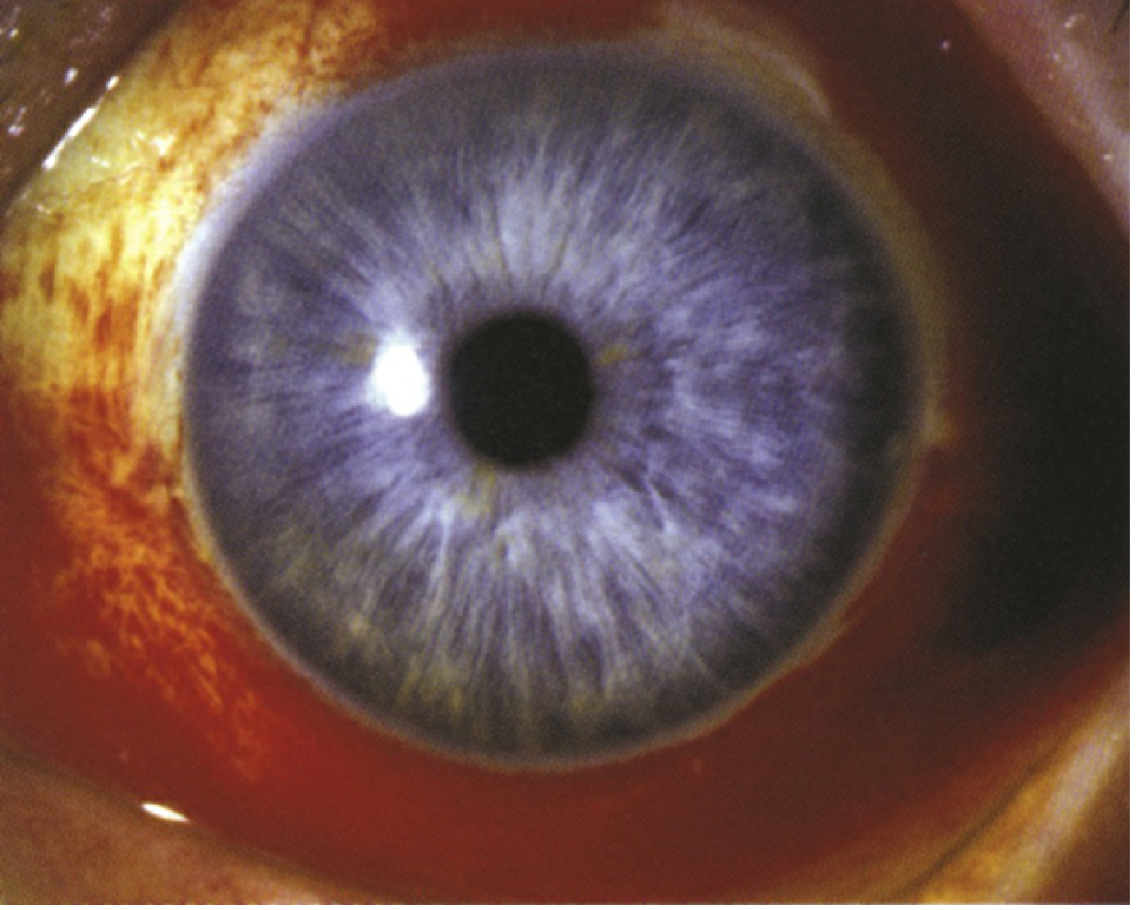
Subconjunctival hemorrhage - Wills Eye Manual
Subconjunctival hemorrhage (SCH) is bleeding between the conjunctiva and the underlying sclera. It appears as a sharply demarcated, bright red patch on the white of the eye where no individual vessels are visible through the blood.
Definition / Pathophysiology
Subconjunctival blood vessels rupture either spontaneously or after a sudden acute rise in venous pressure. The blood spreads smoothly beneath the conjunctiva, gently elevating it. No individual vessels are visible behind the hemorrhage (distinguishing it from conjunctival injection). - Rosen's Emergency Medicine, p. 879
Symptoms & Signs
-
Usually asymptomatic - the patient often first notices it in the mirror
-
Mild foreign body sensation possible, but no change in visual acuity
-
Blood is visible under the conjunctiva, often sectoral but can be circumferential (360 degrees)
-
Associated chemosis (conjunctival edema) may be present in larger hemorrhages
-
Wills Eye Manual, p. 335
Etiology / Causes
| Category | Examples |
|---|---|
| Valsalva maneuver | Coughing, sneezing, vomiting, heavy lifting, straining with constipation |
| Traumatic | Blunt/penetrating injury; may mask retrobulbar hemorrhage or ruptured globe |
| Systemic disease | Hypertension, diabetes mellitus |
| Medications | Aspirin, clopidogrel, warfarin, ticagrelor, dabigatran, rivaroxaban, apixaban, edoxaban |
| Coagulopathy | Bleeding disorders, thrombocytopenia |
| Other | Topical steroid therapy, orbital mass (rare), idiopathic |
Warning Signs Requiring Urgent Evaluation
A 360-degree hemorrhage with any of the following mandates workup for globe perforation:
- Pain or chemosis
- Decreased visual acuity
- Photophobia
- High-impact mechanism of injury
Also check for retrobulbar hemorrhage if there is proptosis, restricted extraocular motility, or elevated IOP. - Rosen's Emergency Medicine, p. 879
Workup
- History - bleeding/clotting problems, medications (anticoagulants), eye rubbing, trauma, heavy lifting, Valsalva, recurrent SCH, cough history
- Check blood pressure - hypertension is a common underlying cause
- Ocular exam - slit lamp + fluorescein dye to check for corneal abrasion, globe perforation; check IOP and extraocular motility in traumatic cases
- Labs if recurrent: PT, aPTT, CBC with differential (rule out thrombocytopenia or leukemia), LFTs, Protein C and S
- CT/MRI orbits if proptosis or restricted motility is present (rule out orbital mass)
- Wills Eye Manual, pp. 335-336
Differential Diagnosis
- Kaposi sarcoma - red/purple elevated conjunctival lesion; check HIV status
- Conjunctival lymphoma or amyloid with secondary hemorrhage
- Episcleritis - sectoral redness with visible dilated vessels (vessels can be moved with a cotton-tip applicator, unlike SCH)
- Conjunctival laceration (more painful, requires evaluation for globe perforation)
- Scleritis (painful, deep injection)
Treatment
-
No treatment required in the vast majority of cases
-
Artificial tears (q.i.d.) for mild irritation
-
Discourage elective use of aspirin/NSAIDs where possible
-
Do NOT stop blood thinners without clearance from the patient's primary physician
-
Manage underlying hypertension
-
Wills Eye Manual, p. 336
Follow Up / Prognosis
- Resolves spontaneously within 2 to 4 weeks (Tintinalli: usually within 2 weeks)
- Reassurance is the mainstay of management for uncomplicated cases
- Return if blood does not fully resolve or recurrence occurs
- Refer to internist if hypertension or bleeding diathesis is identified
Key Takeaways
- Benign, self-limiting condition in most cases
- The alarming appearance often causes significant patient anxiety - reassurance is key
- Always rule out serious underlying trauma (ruptured globe, retrobulbar hemorrhage) in the correct context
- Recurrent episodes warrant coagulation studies and systemic workup
Sources: Wills Eye Manual 6e, p. 335-336 | Tintinalli's Emergency Medicine | Rosen's Emergency Medicine, p. 879
This is a shared conversation. Sign in to Orris to start your own chat.