
SURGERY TOPIC FOR FINAL YEAR MBBS • Ischemic colitis – aetiology, pathogenesis, clinical features, complications, management
Reading File
Now let me search the medical library for ischemic colitis content in parallel.
Finding Sources
Finding Sources
Searching PubMed
"ischemic colitis" AND management
Excellent findings. Now let me read the most comprehensive sections in parallel - Sabiston, Current Surgical Therapy, and Schwartz's (the surgical textbooks most relevant for final year MBBS surgery).
Reading File
Reading File
Reading File
Excellent content found. Now let me read more of these rich sections - the risk factors, clinical features, diagnosis and management from Sabiston and Current Surgical Therapy.
Reading File
Reading File
Excellent content. Let me continue reading the management and complications sections.
Reading File
Reading File
Let me get the remaining surgical management and complications sections.
Reading File
Reading File
I now have comprehensive content from Sabiston and Current Surgical Therapy. Let me also fetch the key diagram image and check the Schwartz's section for any additional information.
Loading Image
Reading File
I now have rich, comprehensive content from three major surgical textbooks (Sabiston, Schwartz's, Current Surgical Therapy 14e). I have everything needed to write a thorough final-year MBBS surgery answer. Let me compile it now.
Ischemic Colitis - A Comprehensive Surgery Guide (Final Year MBBS)
Sources: Sabiston Textbook of Surgery 21e, Schwartz's Principles of Surgery 11e, Current Surgical Therapy 14e
Definition & Epidemiology
Ischemic colitis (IC) is a condition in which blood supply to colonocytes fails to meet metabolic demands. It is the most common cause of GI ischemia, with an incidence of 7.1 to 44 cases per 100,000 persons per year. Patients generally present after age 60, with a higher prevalence in women. About 20% of patients ultimately require operative intervention, usually in an emergent setting. - Current Surgical Therapy 14e
Aetiology
Vascular Anatomy - Key to Understanding
The colon is supplied by the SMA (ileocolic, right colic, middle colic arteries) and the IMA (left colic, sigmoid arteries, superior rectal/hemorrhoidal artery). Two collateral networks protect against ischemia:
- Marginal artery of Drummond - runs parallel to the mesenteric margin of the entire colon; absent at the splenic flexure in up to 18-50% of people
- Arc of Riolan (meandering mesenteric artery) - connects SMA to IMA near the mesenteric root; its presence indicates major mesenteric artery obstruction
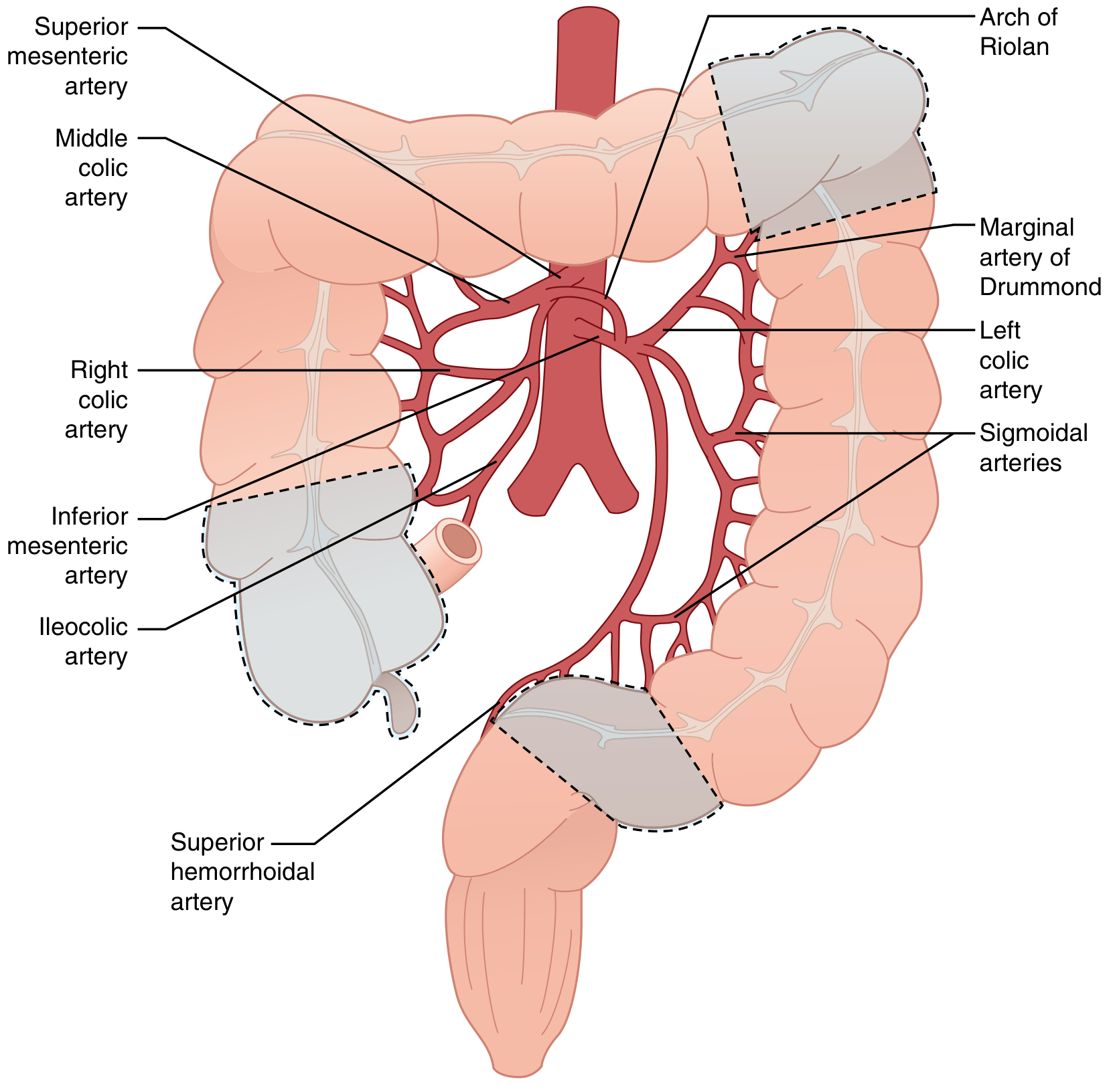
Watershed Areas (Most Vulnerable Sites)
These are zones between two major arterial territories, classically vulnerable to ischemia - responsible for ~80% of IC cases:
| Watershed Area | Location | Between |
|---|---|---|
| Griffith's point | Splenic flexure | SMA and IMA territories |
| Sudeck's point | Rectosigmoid junction | IMA and superior rectal artery |
The rectum is relatively spared due to its rich dual blood supply.
Classification by Mechanism
| Type | Examples |
|---|---|
| Nonocclusive (most common) | Shock, heart failure, sepsis, drugs, colon obstruction, hypotension |
| Occlusive (arterial) | Thrombosis, embolism (e.g., atrial fibrillation) |
| Occlusive (venous) | Venous thrombosis |
| Postoperative (special entity) | After aortic/AAA surgery (IMA sacrifice), cardiac surgery with prolonged cross-clamp time, post-colonoscopy |
- Sabiston Textbook of Surgery 21e
Pathogenesis
- Initial ischemic phase: During hypotension, blood is shunted away from splanchnic circulation toward the brain and heart. Mucosal injury begins within 20 minutes to 1 hour of decreased blood flow.
- Transmural progression: If ischemia continues, injury spreads from mucosa → submucosa → muscularis → serosa. Transmural infarction occurs within 8-16 hours.
- Reperfusion injury: When blood flow is restored, reactive oxygen species (ROS) are released, causing lipid peroxidation and cell necrosis - causing additional injury beyond the initial ischemic insult.
- Histological sequence: Initial changes on the antimesenteric side of the mucosa. Progresses to loss of surface epithelium, crypt necrosis, mucin depletion, hyalinosis of the lamina propria, and eventually ghost crypts.
Depth of Injury Determines Outcome
| Type | Depth | Outcome |
|---|---|---|
| Mucosal (mild) | Mucosa only | Reversible; heals with conservative management |
| Transmural (severe) | Full thickness | Life-threatening; leads to perforation, peritonitis, sepsis, death |
Risk Factors
Patient factors:
- Advanced age, female sex
- Atherosclerotic disease (ischemic heart disease, cerebrovascular disease, peripheral vascular disease)
- Diabetes mellitus, hypertension, COPD, CKD/renal disease
- Heart failure, atrial fibrillation, septic shock, hemorrhagic shock
- IBD, IBS, constipation
- COVID-19 (recently identified association)
Drug associations:
- Diuretics, digoxin (cardiovascular drugs)
- Psychotropic medications
- Immunosuppressants, NSAIDs
- Cocaine, amphetamines (vasoconstrictors)
- Oral contraceptives (thrombogenic)
Procedural:
-
Aortic surgery (AAA repair - most common postoperative cause)
-
Cardiac surgery with prolonged cross-clamping
-
Endovascular abdominal procedures (including chemoembolization)
-
Post-colonoscopy
-
Sabiston 21e, Box 95.6
Isolated Right-Sided Colon Ischemia (IRCI) - Special Entity
IRCI carries worse prognosis:
- 30-day mortality: 20.3% vs 9.0% in non-IRCI
- Surgical intervention required in 40.9% vs 10.3% in non-IRCI
- More associated with coronary artery disease and CKD on hemodialysis
- Must always prompt CT angiography to exclude acute mesenteric ischemia
Clinical Features
Symptoms
- Classic triad: Sudden-onset crampy abdominal pain (usually left lower quadrant) + urgent desire to defecate + hematochezia within 24 hours of pain
- Tenesmus
- Rectal bleeding: more common in non-IRCI (69.9%) than IRCI (39.4%)
Signs by Severity
| Severity | Features |
|---|---|
| Mild | Bloody diarrhea, minimal tenderness; no systemic signs |
| Moderate | Abdominal pain, tenderness, fever, leukocytosis |
| Severe (gangrenous/transmural) | Increasing abdominal tenderness, guarding, rebound, fever, paralytic ileus; peritonitis; haemodynamic instability |
| Fulminant colitis (rare variant) | Sudden onset toxic colitis with peritonitis; rapidly progressive |
Key exam point: Pain out of proportion to examination, especially WITHOUT rectal bleeding, should raise suspicion for IRCI or acute mesenteric ischemia.
Investigations
Laboratory
- Leukocytosis (frequent)
- Elevated lactate (>2.5 mmol/L - poor prognostic sign)
- Elevated LDH, urea, creatinine
- Low hemoglobin, low albumin
- Metabolic acidosis with base deficit in severe cases
- Stool: C. difficile toxin, culture/sensitivity, ova and parasites (to exclude infectious cause)
Imaging
Plain X-ray (AXR):
- "Thumbprinting" - rounded densities along sides of gas-filled colon; represents mucosal edema and submucosal hemorrhage (nonspecific)
- Free air under diaphragm = perforation → immediate surgery
CT Abdomen (with IV and oral contrast) - PRIMARY MODALITY:
- Segmental bowel wall thickening
- Pericolonic fat stranding
- Thumbprinting
- Pneumatosis intestinalis (gas in colonic wall) - severe transmural disease
- Portal/mesenteric venous gas - transmural infarction; indicates need for emergency surgery
- Free air = perforation
CT angiography (multiphasic) indicated when IRCI is suspected or pain is severe without bleeding, to exclude major mesenteric arterial occlusion.
Contrast enema: Largely obsolete; may be used for chronic ischemic strictures only. Contraindicated in acute phase.
Endoscopy - Gold Standard
Early colonoscopy within 48 hours (except in acute peritonitis or suspected severe transmural ischemia):
- Segmental erythema, edema, friable mucosa
- Petechial hemorrhage
- Mucosal ulceration
- Submucosal hemorrhagic nodules
- Involvement of watershed areas
- "Single stripe sign": single linear ulcer along the antimesenteric wall - rare but specific for IC
- Abrupt transition between injured and normal mucosa; rectal sparing (supports ischemia over IBD)
- In severe ischemia: gray-green or black mucosa
- After 48 hrs: ulcerations develop as submucosal hemorrhages resolve
Endoscopy cannot distinguish partial-thickness from full-thickness ischemia. Avoid overinsufflation.
Histology findings: Mucosal necrosis, loss of surface epithelium, ghost crypts, mucin depletion, hyalinosis of lamina propria, paucity of acute inflammatory cells.
Complications
| Complication | Notes |
|---|---|
| Bowel gangrene and perforation | Life-threatening; causes peritonitis, sepsis |
| Chronic ischemic colitis | Ongoing/recurrent pain, bloody diarrhea, sepsis; higher complication rate; requires surgical resection |
| Colonic stricture | Occurs in 10-15%; presents with constipation, narrow stools, abdominal pain; diagnosed with contrast enema/CT/endoscopy; symptomatic strictures require elective resection |
| Sepsis / Multiorgan failure | Especially in transmural cases |
| Death | In-hospital mortality for severe IC requiring surgery: 37%; up to 47% in acute surgical intervention series |
- Schwartz's Principles of Surgery 11e; Sabiston 21e
Management
Conservative (Non-Operative) - 80% of patients
Indications: Mild to moderate IC without peritonitis or perforation.
- Bowel rest (NPO) + IV fluids (aggressive resuscitation)
- Broad-spectrum antibiotics (to prevent bacterial translocation)
- Nasogastric tube if ileus is present
- Optimize hemodynamic status - treat heart failure, sepsis, hypotension
- Discontinue causative drugs (vasoconstrictors, NSAIDs, etc.)
- Adjuncts: phosphodiesterase-5 inhibitors, pentoxifylline, prostaglandin E1 (improve mucosal blood flow)
- Serial clinical monitoring: if no improvement or worsening within 2-3 days, escalate
Indications for Emergency Surgery (Operative)
| Indication |
|---|
| Peritonitis on examination |
| Hemodynamic instability not responding to resuscitation |
| Free intraperitoneal air (perforation) |
| Pneumatosis intestinalis + portal venous gas on CT with clinical IC |
| Massive lower GI bleeding |
| Fulminant colitis |
| Failure to improve / worsening on medical management |
Surgical Approach
- Incision: Midline laparotomy (diagnostic laparoscopy in selected cases)
- Principle: Resect all grossly ischemic colon; assess entire small and large intestine
- Extent of resection: Anatomic resection ensuring adequate blood supply to remaining colon; intraoperative indocyanine green (ICG) infrared angiography increasingly used to assess bowel viability
- Anastomosis vs stoma:
- Right-sided IC: Primary anastomosis safe if bowel well-perfused
- Left-sided IC: Hartmann's procedure (end colostomy + rectal stump) is standard; reversal after 6-8 weeks; primary anastomosis +/- protective diverting ileostomy in selected stable cases
- Pancolitis: Total colectomy with end ileostomy
- Second-look laparotomy: If viability uncertain, leave abdomen open (temporary closure), return in 12-48 hours to assess further necrosis
- No revascularization: Unlike small bowel ischemia, large vessel occlusion is rarely the cause; vascular reconstruction is not typically indicated
Post-Operative Follow-Up
- Colonoscopy after recovery to:
- Evaluate for strictures
- Rule out IBD, malignancy (mimics on endoscopy)
Mortality Risk Factors (Post-colectomy)
- Elderly age, poor functional status, multiple comorbidities
- Preoperative septic shock, blood transfusions, acute renal failure
- Delay from admission to surgery
- Peak preoperative lactate >2.5 mmol/L
- ASA class 4, subtotal/total colectomy, need for dialysis
Summary Algorithm
Suspected IC
     ↓
History + Exam + Labs (CBC, metabolic panel, lactate, stool cultures)
     ↓
CT Abdomen (with contrast)
     ↓
┌──────────────────────────────────────────┐
│ Signs of perforation/transmural ischemia │
│ (free air, portal gas, pneumatosis +     │
│  peritonitis, hemodynamic instability)   │
└──────────────────────────────────────────┘
        ↓ YES                   ↓ NO
   Emergency Surgery       Early Colonoscopy (within 48h)
   (Midline lap,                ↓
   resect ischemic colon,  Conservative Management
   Hartmann's for left-    (Bowel rest, IVF, ABx)
   sided disease)               ↓
                         Reassess in 2-3 days
                         ↓ No improvement or worsening
                         → Repeat imaging/endoscopy → Surgery
High-Yield Points for Exam
- Most common GI ischemia and most common form is nonocclusive (due to low-flow states)
- Watershed areas: Griffith's point (splenic flexure) and Sudeck's point (rectosigmoid) - both watershed, ~80% of cases
- Classic presentation: Crampy LLQ pain + urgency + hematochezia within 24 hours
- Thumbprinting on AXR/CT = mucosal edema/submucosal hemorrhage
- Gold standard diagnosis: Colonoscopy within 48 hours; single stripe sign is specific
- 80% managed conservatively with bowel rest, IVF, broad-spectrum antibiotics
- Peritonitis + portal venous gas + pneumatosis intestinalis = emergency surgery
- Hartmann's procedure for left-sided IC (NOT primary anastomosis)
- IRCI (isolated right colon ischemia) = worst prognosis; suspect acute mesenteric ischemia
- Long-term complications: Stricture (10-15%), chronic segmental ischemia (15-20%)
This is a shared conversation. Sign in to Orris to start your own chat.