
Examination of knee joint in detail
Reading File
Now let me search the medical library for knee joint examination content in parallel.
Finding Sources
Finding Sources
Excellent sources found. Let me read the key sections in parallel - Bailey & Love (clinical exam), S Das (surgical exam), Rosen's (special tests), and Gray's Anatomy.
Reading File
Reading File
Reading File
Reading File
Good content retrieved. Let me now read more from Bailey & Love (the most comprehensive surgical source) and the Rosen's special tests section, in parallel.
Reading File
Reading File
Reading File
Excellent content gathered. Let me get the remaining Bailey & Love special tests section plus S Das meniscal tests, and fetch the key diagnostic images.
Reading File
Reading File
Reading File
Now let me fetch the key clinical images and continue reading Bailey & Love special tests.
Loading Image
Loading Image
Reading File
I now have comprehensive content from multiple authoritative sources. Let me compile the full, detailed response.
Examination of the Knee Joint
The knee is the largest synovial joint in the body. It has three compartments: medial tibiofemoral, lateral tibiofemoral, and patellofemoral. Examination follows the standard "Look - Feel - Move - Special Tests" sequence and must always be performed on both limbs for comparison.
Anatomy Relevant to Examination
The structural components you are assessing during examination:
- Extensor mechanism: Quadriceps muscle - quadriceps tendon - patella - patellar tendon - tibial tuberosity
- ACL: Primary restraint to anterior tibial displacement
- PCL: Primary restraint to posterior tibial displacement
- MCL: Resists valgus and external rotation forces
- LCL: Resists varus forces
- Medial and lateral menisci: Load-bearing fibrocartilage; medial is injured more commonly than lateral
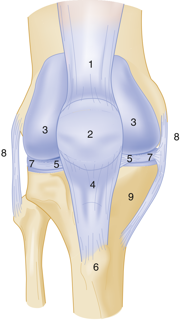
Palpation sites: 1 - Quadriceps tendon; 2 - Prepatellar bursitis/patella; 3 - Retinacular pain; 4 - Patellar tendon; 5 - Fat pad; 6 - Tibial tubercle (Osgood-Schlatter); 7 - Meniscus/joint line; 8 - Collateral ligaments; 9 - Pes anserine
History
Before any examination, take a focused history:
- Mechanism of injury: Direction and force of the blow determines likely structure injured
- Blow to lateral side of knee with weight bearing → medial collateral ligament injury
- Backward thrust on the anterior tibia with knee flexed → PCL injury (classic "dashboard injury")
- Forced hyperextension → ACL injury (isolated ACL tears are uncommon)
- Rotational injury with knee flexed and weight bearing → meniscal tear (medial meniscus injured by internal femoral rotation on tibia; lateral meniscus by external rotation)
- Haemarthrosis vs. effusion: Rapid swelling within 2 hours = haemarthrosis (suggests ACL tear, tibial plateau fracture, patellar dislocation); delayed swelling = serous effusion (meniscal tear, ligament sprain)
- Locking: Cannot extend the knee past a certain degree (not locking in flexion). Caused by a bucket-handle meniscal tear or loose body. The joint can flex freely but cannot fully extend.
- Giving way: Feeling of instability, especially on stairs or uneven ground
- Lifestyle: Activity level guides expectations and management
1. LOOK (Inspection)
Expose both lower limbs fully. Inspect from front, sides, and back, including the popliteal fossa.
Standing inspection
- Alignment (front view): Look for varus (bow-leg - measure interknee distance) or valgus (knock-knee - measure intermalleolar distance)
- Side view: Look for fixed flexion deformity or recurvatum (hyperextension)
- Skin: Scars (prior surgery/trauma), erythema (infection/inflammatory arthritis), sinuses
- Soft tissues: Quadriceps wasting (a hallmark of chronic knee pathology - measure thigh circumference 15 cm above the patella and compare sides)
- Swelling: Effusion produces a characteristic horseshoe-shaped fullness around the suprapatellar region and on both sides of the patella. Look for prepatellar bursitis (housemaid's knee - swelling directly over the patella) vs. infrapatellar bursitis (clergyman's knee - over patellar tendon)
- Patella height: Patella alta (high-riding - patellar tendon rupture) or patella baja (low-riding)
Gait inspection
- Antalgic gait: Short stance phase on the affected side (pain-related)
- Varus thrust: Sudden collapse into greater varus on weight bearing = lateral compartment disease
- Quadriceps avoidance gait: Walking with the knee in extension to avoid quadriceps activation = ACL deficiency
2. FEEL (Palpation)
The patient lies supine with both legs fully exposed.
Temperature
Run the dorsum of your hand along the front of the joint. Warmth indicates inflammation (inflammatory arthritis, infection, haemarthrosis).
Soft tissues
- Palpate the quadriceps and patellar tendons for a palpable gap (tendon rupture)
- Palpate popliteal fossa for a Baker's cyst (posterior fullness)
Tests for effusion
Fluid displacement / Stroke test (small effusion):
- Stroke fluid out of the medial gutter upward into the suprapatellar pouch
- Place your hand over the suprapatellar pouch and stroke downward
- Watch the medial gutter - a positive result is a ripple of fluid appearing on the medial side
Patellar tap test (large effusion):
- Place one hand on the suprapatellar pouch compressing fluid down; with two fingers of the other hand, briskly press the patella downward - a "tap" or ballottement of the patella against the femoral condyles confirms a tense effusion
Bony palpation
Palpate systematically:
- Tibial tuberosity: Tenderness = Osgood-Schlatter disease in adolescents; tibial plateau fracture in trauma
- Patella borders: Gap = transverse fracture; bony irregularity = comminuted fracture
- Inferior pole of patella: Tenderness = jumper's knee (patellar tendinopathy)
- Joint line (medial and lateral): Best felt with knee at 90°. Medial joint line tenderness is the most sensitive test for a medial meniscal tear
- Collateral ligament origins/insertions: Tenderness at the femoral attachment of the MCL = MCL injury (sprain or tear)
- Femoral condyles: Tenderness = fracture or osteochondritis dissecans
- Fibular head: Tenderness = LCL avulsion or proximal fibula fracture; test by "springing the fibula" (compress tibia and fibula distally - pain at the fracture site)
Point of tenderness - key diagnostic guide (S Das)
| Location of Tenderness | Likely Diagnosis |
|---|---|
| Femoral attachment of MCL | MCL injury |
| Joint line (between ligamentum patellae and MCL) | Medial meniscal tear (mid-substance) |
| Anterior to MCL at joint level | Medial meniscal injury (anterior horn) |
| Posterior to MCL at joint line | Posterior horn of medial meniscus |
| Both sides of patellar tendon | Infrapatellar fat pad injury |
| Lateral joint line (often with a palpable cyst) | Lateral meniscal cyst |
Neurovascular assessment
Always check the dorsalis pedis and posterior tibial pulses. The popliteal artery runs close to the posterior capsule and may be injured in knee dislocations or supracondylar femoral fractures.
3. MOVE (Range of Motion)
Normal range: Flexion 0-135°, Extension 0° (or -5 to -10° hyperextension).
- Active then passive movements: Ask the patient to flex and extend first; then examine passively
- Hyperextension test: Place one hand on the anterior distal femur, lift the distal tibia with the other. Note the heel height off the couch or measure the angle - hyperextension >10° is abnormal
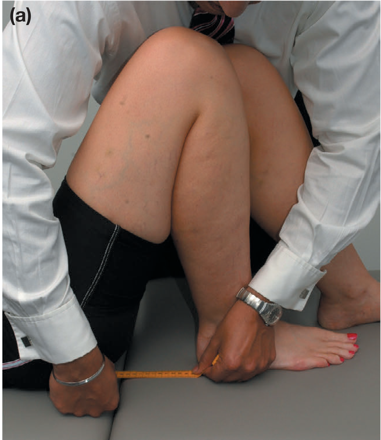
- Fixed flexion deformity: If apparent, sit the patient with knees hanging over the couch edge (eliminates hip flexion artifact). Attempt passive full extension - if not achievable, this is a true flexion deformity (common in OA)
- Extensor lag test: Ask the patient to straight-leg raise to 10°, then flex the knee and attempt to re-extend it in mid-air. Inability to maintain full extension = positive lag = quadriceps mechanism disruption (quadriceps tendon, patellar fracture, patellar tendon, or severe muscle weakness)
4. SPECIAL TESTS
A. Tests for Anterior Instability (ACL)
Lachman's Test (most sensitive for ACL - sensitivity ~85%, specificity ~94%)
- Patient supine, knee at 20-30° flexion (heel rests on couch)
- One hand around the distal femur (thumb on lateral femoral condyle), the other around proximal tibia (thumb on tibial tuberosity)
- Apply a brisk anteriorly directed force to the tibia
- Positive: Excessive anterior tibial translation (>5 mm) with a soft end-point (no firm stop) = ACL tear
- Grading: 1+ (0-5 mm), 2+ (5-10 mm), 3+ (>10 mm)
Anterior Drawer Test (less sensitive than Lachman, especially acutely)
- Knee flexed at 90°, hip at 45°; examiner sits gently on the patient's foot (ensure hamstrings are relaxed)
- Both hands encircle the proximal tibia and pull it anteriorly
- Positive: Tibia slides forward >5 mm on the femur = ACL tear (note: requires medial meniscus or meniscotibial ligament co-injury to be positive)
- A false-positive can occur if the posterior sag sign (PCL injury) is not first excluded
Pivot Shift Test (confirms ACL-deficient rotational instability)
- Patient supine; examiner holds the patient's foot in slight internal rotation and applies a valgus force with the knee in extension
- Slowly flex the knee: at 20-30° of flexion, the lateral tibial plateau suddenly reduces with a visible/palpable clunk
- Positive: A "pivot shift" clunk = combined ACL + posterolateral corner injury
- This test is often easier to perform under anaesthesia
B. Tests for Posterior Instability (PCL)
Posterior Drawer Test
- Knee at 90° flexion, foot stabilised
- Both thumbs on tibial tuberosity; push the tibia posteriorly
- Positive: Posterior tibial displacement >5 mm or soft end-point = PCL tear
- Positive in ~85% of operative PCL tears
Posterior Sag Sign (sensitivity 79% in the acute phase)
- Patient supine, pillow under distal thigh, knee at 45-90°, heel resting on the stretcher
- Observe the tibial tuberosity from the side
- Positive: Tibia sags backward due to gravity = PCL insufficiency
- Important: A missed posterior sag can cause a false-positive anterior drawer test (tibia starts posterior, then moves forward to neutral when pulled)
C. Collateral Ligament Tests
Valgus Stress Test (MCL)
- Knee at 30° flexion (relaxes the posterior capsule; at 0° the cruciates provide stability, masking a collateral tear)
- Apply a valgus force; palpate the medial joint line with the index finger
- Positive: Excessive medial joint line opening with a soft end-point = MCL tear
- Laxity also in full extension (0°) = injury to the posterior capsule and/or PCL as well
Varus Stress Test (LCL)
- Knee at 30° flexion; apply a varus force; palpate the lateral joint line
- Positive: Excessive lateral joint line opening = LCL tear
- Laxity in full extension = posterolateral corner (PLC) injury
Grading of collateral laxity:
- Grade I: Firm end-point, <5 mm opening (partial tear)
- Grade II: Soft end-point, 5-10 mm opening (partial/near-complete)
- Grade III: No end-point, >10 mm opening (complete rupture)
D. Meniscal Tests
McMurray's Test (best known meniscal test)
- Patient supine, knee fully flexed, hip at 45°
- For medial meniscus: Palpate medial joint line; apply a valgus stress and externally rotate the tibia; slowly extend the knee
- For lateral meniscus: Apply varus stress and internally rotate the tibia; slowly extend
- Positive: A palpable or audible click at the joint line, or reproduction of pain = meniscal tear
- The click occurs as the torn meniscus flips over the femoral condyle during extension
Joint Line Tenderness
- Simply palpate the medial and lateral joint lines with the knee at 90°
- The most sensitive clinical test for meniscal tears (though not very specific)
Apley's Grinding Test
- Patient prone, knee flexed to 90°
- Apply downward compression and rotate the tibia (grinding the meniscus between the femur and tibia)
- Positive: Pain on compression + rotation = meniscal tear
- Compare with distraction: if pain is worse with compression than distraction, the meniscus (not the ligament) is implicated
E. Patellofemoral Tests
Patellar Apprehension Test (Fairbank's Test) - for patellar instability
- Patient supine, quadriceps relaxed, knee at 30° flexion
- Gently push the patella laterally
- Positive: Patient shows apprehension (contracts quadriceps or grabs your hand to prevent lateral subluxation) = history of patella dislocation or patellar instability
J-Sign / Patellar Tracking
- Patient sits with legs hanging over the edge of the couch, knees at 90°
- Ask the patient to slowly extend the knee
- Positive: Near full extension, the patella jumps laterally (tracing a "J" shape instead of a smooth straight path) = patellofemoral maltracking
Patellar Grind Test
- With the knee extended, press the patella inferiorly and ask the patient to contract the quadriceps
- Positive: Pain and/or crepitus = patellofemoral chondromalacia or OA
5. ASSESSMENT FOR EFFUSION CONTENT
When aspiration is considered, the character of fluid guides diagnosis:
- Clear/straw-coloured: Inflammatory arthritis, meniscal tear
- Haemarthrosis (blood): ACL tear, tibial plateau fracture, patellar dislocation
- Lipohemarthrosis (fat globules in blood): Intra-articular fracture (fat from medullary cavity enters the joint - visible on lateral X-ray as a fluid-fat level)
- Pus: Septic arthritis (emergency)
6. OTTAWA KNEE RULES (Deciding on X-Ray)
Knee X-ray is indicated if any ONE of the following is present:
| Ottawa Knee Rules | Pittsburgh Knee Rules |
|---|---|
| Age >55 | Age <12 or >50 |
| Isolated patellar tenderness | Inability to walk 4 steps |
| Isolated fibular head tenderness | |
| Knee flexion <90° | |
| Inability to weight bear 4 steps immediately after injury OR in the ED | |
| Sensitivity 98.5%, specificity 49% | Sensitivity 100%, specificity 79% |
Summary Checklist
| Step | What to Assess |
|---|---|
| Look (standing) | Alignment, varus/valgus, muscle wasting, gait |
| Look (supine) | Skin/scars, swelling pattern, limb posture, patellar height |
| Feel | Temperature, effusion (stroke test/patellar tap), joint line tenderness, bony palpation, pulses |
| Move | Flexion/extension ROM, hyperextension, extensor lag test |
| ACL tests | Lachman (most sensitive), anterior drawer, pivot shift |
| PCL tests | Posterior drawer, posterior sag sign |
| Collateral tests | Valgus stress (MCL), varus stress (LCL) at 30° flexion |
| Meniscal tests | Joint line tenderness, McMurray's, Apley's grinding |
| Patellofemoral | Patellar apprehension, J-sign tracking, grind test |
| Neurovascular | Dorsalis pedis and posterior tibial pulses |
Sources:
- Bailey and Love's Short Practice of Surgery, 28th Edition, pp. 512-515 - Clinical Examination of the Knee
- S Das: A Manual on Clinical Surgery, 13th Edition, pp. 207-210 - Examination of Injuries Around the Knee Joint
- Rosen's Emergency Medicine: Concepts and Clinical Practice - Knee Injuries Examination Maneuvers and Ottawa/Pittsburgh Rules
- Gray's Anatomy for Students - Examination of the Knee Joint, Special Tests
This is a shared conversation. Sign in to Orris to start your own chat.