
ECG change in MI, arrhythmias, heart blocks, axis deviation
Reading File
This covers a full ECG revision set. The main areas are:
- Myocardial infarction ECG evolution: hyperacute T waves, ST elevation/depression, Q waves, T-wave inversion, reciprocal changes, infarct localisation.
- Arrhythmias: sinus tachycardia/bradycardia, atrial fibrillation/flutter, SVT, ventricular tachycardia/fibrillation, ectopics.
- Heart blocks: first-degree AV block, Mobitz I and II, complete heart block, bundle branch blocks.
- Axis deviation: normal axis, left/right axis deviation, extreme axis, rapid determination using leads I and aVF.
A practical approach is to learn the ECG reading sequence first, then recognize life-threatening patterns: STEMI, VT/VF, high-grade AV block, and marked bradycardia.
Asking for Preferences
Which topic should I explain in detail first?
This covers a full ECG revision set. The main areas are:
- Myocardial infarction ECG evolution: hyperacute T waves, ST elevation/depression, Q waves, T-wave inversion, reciprocal changes, infarct localisation.
- Arrhythmias: sinus tachycardia/bradycardia, atrial fibrillation/flutter, SVT, ventricular tachycardia/fibrillation, ectopics.
- Heart blocks: first-degree AV block, Mobitz I and II, complete heart block, bundle branch blocks.
- Axis deviation: normal axis, left/right axis deviation, extreme axis, rapid determination using leads I and aVF.
A practical approach is to learn the ECG reading sequence first, then recognize life-threatening patterns: STEMI, VT/VF, high-grade AV block, and marked bradycardia.
Reading File
Choose one topic to start in detail:
- MI ECG changes
- Arrhythmias
- Heart blocks
- Axis deviation
Arrhythmia
Reading File
Finding Sources
Finding Sources
Finding Sources
Searching PubMed
"Cardiac Arrhythmias"[MeSH Terms]
Searching the Web
American Heart Association adult tachyarrhythmia bradycardia ACLS algorithm current
Searching Images
ECG examples atrial fibrillation atrial flutter supraventricular tachycardia ventricular tachycardia ventricular fibrillation
Finding Sources
Finding Sources
Finding Sources
Finding Sources
Reading File
Reading File
Reading File
Reading File
Arrhythmia: ECG-focused revision
Arrhythmia (dysrhythmia) means an abnormal heart rhythm due to abnormal impulse formation, conduction, or both.
Quick ECG approach
Ask these four questions:
- Rate: slow (<60/min), normal, or fast (>100/min)?
- Regularity: regular or irregular?
- P waves: present, absent, abnormal, or unrelated to QRS?
- QRS width: narrow (<120 ms) or wide (≥120 ms)?
A practical rhythm classification:
| Rhythm | Likely diagnoses |
|---|---|
| Regular, narrow-complex tachycardia | Sinus tachycardia, AVNRT/AVRT (SVT), atrial tachycardia, flutter with fixed block |
| Irregular, narrow-complex tachycardia | Atrial fibrillation, multifocal atrial tachycardia, frequent PACs |
| Regular, wide-complex tachycardia | VT unless proven otherwise, SVT with bundle-branch block, antidromic AVRT |
| Irregular, wide-complex tachycardia | Polymorphic VT, AF with WPW, AF with aberrancy, VF |
Most supraventricular tachycardias arise in the atria or AV junction and are usually narrow-complex, though a bundle-branch block or accessory pathway can produce a wide QRS. Harrison’s Principles of Internal Medicine, 22E (2025), Ch. 253.
1. Sinus arrhythmias
Sinus bradycardia
- Rate <60/min
- Normal P wave before every QRS, regular rhythm
- Causes: athletes, sleep, vagal stimulation, inferior MI, hypothyroidism, drugs such as beta-blockers, verapamil/diltiazem, digoxin.
Sinus tachycardia
- Rate >100/min, often 100 to 160/min
- Normal P before each QRS, regular rhythm
- Usually a response to another condition: fever, pain, dehydration, anemia, hypoxia, anxiety, sepsis, thyrotoxicosis.
- Treat the cause, not merely the ECG rate.
Sinus arrhythmia
- Normal P-QRS relationship but cyclic variation in R-R interval, commonly with respiration.
- Usually benign, especially in young people.
2. Premature beats
Premature atrial contraction (PAC)
- Early abnormal P wave, often followed by a normal narrow QRS.
- May reset the sinus node, causing a non-compensatory pause.
- Common and often benign.
Premature ventricular contraction (PVC)
- Early, broad and bizarre QRS complex.
- No preceding P wave.
- Usually followed by a full compensatory pause.
- Can be unifocal or multifocal. Frequent PVCs, couplets, syncope, structural heart disease, or ischemia require assessment.
3. Supraventricular tachyarrhythmias
Atrial fibrillation (AF)
ECG
- Irregularly irregular R-R intervals.
- No distinct P waves.
- Fine/coarse fibrillatory baseline.
- QRS usually narrow unless aberrancy or pre-excitation exists.
Clinical point
- Main risks: rapid ventricular rate, heart failure exacerbation, and atrial thromboembolism/stroke.
- Management principles: assess stability, control ventricular rate or restore rhythm when appropriate, and assess need for anticoagulation.
Atrial flutter
ECG
- Regular atrial activity with saw-tooth flutter waves, best seen in II, III, aVF, often V1.
- Atrial rate often about 300/min.
- With 2:1 AV conduction, ventricular rate is often close to 150/min.
- May be regular or variable if the AV block varies.
Paroxysmal SVT: AVNRT or AVRT
ECG
- Sudden-onset, regular, narrow-complex tachycardia, often 150 to 250/min.
- P waves are absent, hidden within QRS, or retrograde just after the QRS.
- AVNRT is due to a re-entry circuit around the AV node.
- AVRT uses an accessory pathway, such as in WPW syndrome.
Key contrast
- Sinus tachycardia generally has gradual onset/offset and identifiable sinus P waves.
- SVT typically starts and stops abruptly.
Atrial tachycardia
- Regular narrow-complex tachycardia.
- Abnormal P-wave morphology before QRS.
- May occur with digoxin toxicity, pulmonary disease, or structural heart disease.
Multifocal atrial tachycardia (MAT)
ECG
- Irregular rhythm.
- At least 3 different P-wave morphologies.
- Variable PR intervals.
- Rate >100/min.
- Classically associated with COPD/hypoxia.
4. Ventricular arrhythmias
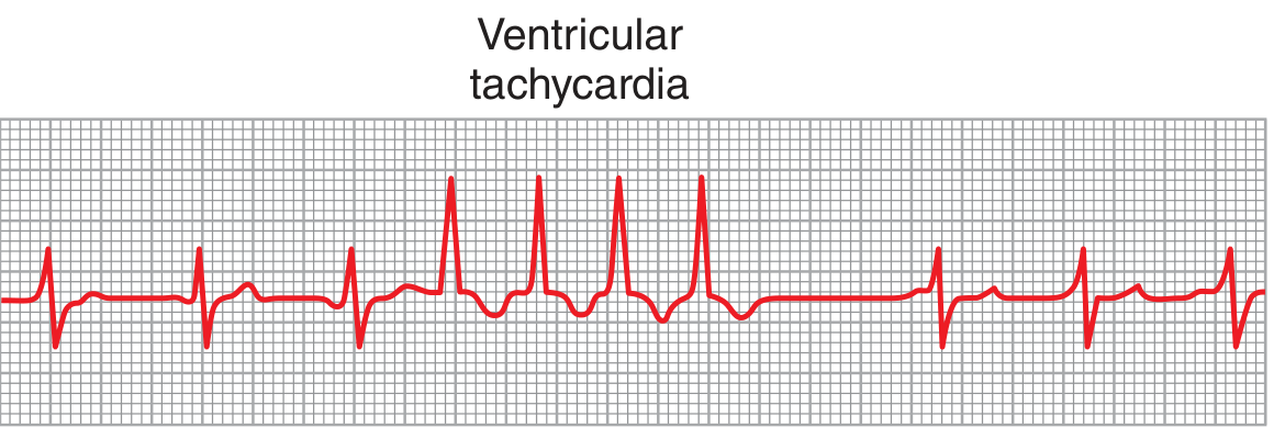
Ventricular tachycardia (VT)
ECG
- Three or more consecutive ventricular beats at >100/min.
- Usually broad QRS tachycardia.
- May be monomorphic or polymorphic.
- AV dissociation, capture beats, or fusion beats strongly support VT.
Exam rule: In a patient with structural heart disease or prior MI, a wide-complex tachycardia should be considered VT until proven otherwise.
- Monomorphic VT: same QRS shape beat-to-beat, often due to scar-related re-entry after MI.
- Polymorphic VT: changing QRS morphology/amplitude. Consider acute ischemia or prolonged QT.
- Torsades de pointes: a polymorphic VT with apparent “twisting” QRS complexes around the baseline, associated with QT prolongation.
VT may impair cardiac output and can deteriorate into VF. Guyton and Hall Textbook of Medical Physiology, 14e, Figure 13.14.
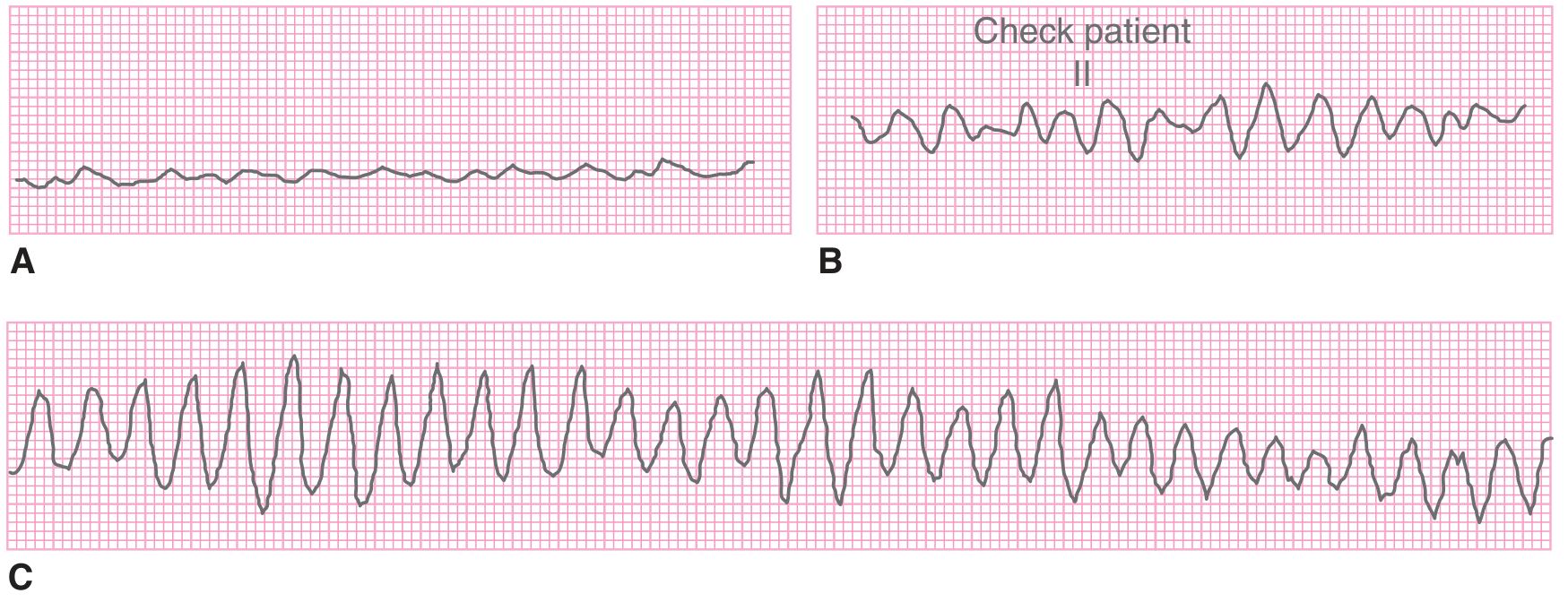
Ventricular fibrillation (VF)
ECG
- Chaotic, irregular waveform.
- No recognizable P waves, QRS complexes, or T waves.
- No effective cardiac output.
This is a cardiac-arrest rhythm: start CPR and defibrillate urgently.
Asystole
- Near-flat line with no organized electrical activity.
- Confirm in more than one lead and exclude loose leads.
- Non-shockable arrest rhythm: CPR, adrenaline/epinephrine per resuscitation protocol, and treatment of reversible causes.
5. Dangerous rhythm recognition
| Rhythm | ECG clue | Immediate principle |
|---|---|---|
| Unstable tachyarrhythmia with pulse | Hypotension, shock, ischemic chest pain, acute heart failure, altered consciousness | Synchronized cardioversion |
| Regular narrow SVT, stable | Regular narrow tachycardia | Vagal manoeuvres, then adenosine when appropriate |
| VT with pulse, unstable | Broad tachycardia plus instability | Synchronized cardioversion |
| Pulseless VT/VF | No pulse with VT/VF ECG | CPR + defibrillation |
| Torsades | Polymorphic VT with long QT | IV magnesium, correct electrolytes, stop QT-prolonging drugs |
| AF with WPW | Irregular broad-complex tachycardia, very rapid | Urgent expert management; avoid isolated AV-nodal blockers |
The current AHA tachyarrhythmia algorithm recommends immediate synchronized cardioversion for tachyarrhythmia causing hypotension, altered mental status, signs of shock, ischemic chest discomfort, or acute heart failure.
High-yield memory points
- Irregularly irregular with absent P waves = AF.
- Saw-tooth waves = atrial flutter.
- Regular narrow fast rhythm with hidden P waves = SVT.
- Three P-wave shapes + irregular rhythm = MAT.
- Wide-complex tachycardia = VT until proven otherwise.
- Twisting polymorphic VT + long QT = torsades de pointes.
- Chaotic line with no QRS = VF.
- Pulseless VT/VF are shockable. Asystole/PEA are not shockable.
If this is for a real patient with chest pain, syncope, severe breathlessness, low blood pressure, or palpitations with collapse, seek emergency care immediately.
This is a shared conversation. Sign in to Orris to start your own chat.