
Tltypes of abcess
types of abscess classification medical
types of abscess diagram classification
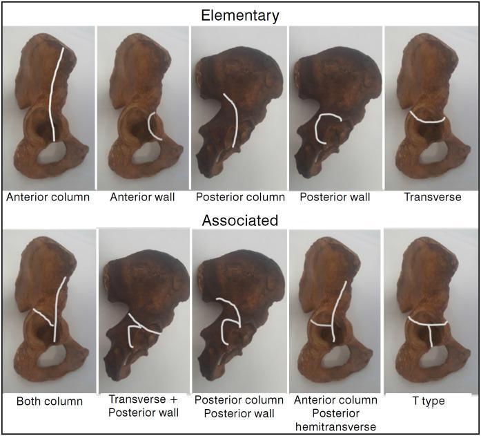
Educational diagram illustrating the Judet-Letournel classification of acetabular fractures using anatomical specimens of the hemipelvis. The visual is divided into two rows: 'Elementary' and 'Associated' fracture types. White lines superimposed on the specimens delineate specific fracture patterns. The 'Elementary' row features five simple patterns: Anterior column (vertical line through the pelvic brim), Anterior wall (localized articular segment), Posterior column (ischiadic notch through obturator foramen), Posterior wall (localized posterior rim), and Transverse (horizontal line bisecting the acetabulum). The 'Associated' row demonstrates complex configurations: Both column, Transverse plus posterior wall, Posterior column plus posterior wall, Anterior column with posterior hemitransverse (a vertical anterior column line joined by a horizontal posterior component), and T-type (a transverse fracture with an additional vertical stem extending through the obturator foramen). This orthopedic diagram serves as a clinical reference for orthopedic surgeons and medical students to understand pelvic trauma morphology and surgical planning.
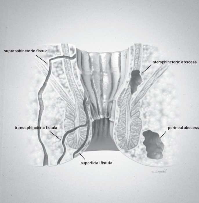
**Imaging Modality:** Medical illustration/Anatomical diagram. **Anatomical Region:** Coronal cross-section of the anorectal region, including the anal canal, rectum, internal and external anal sphincters, and surrounding perianal soft tissues. **Observed Pathology:** The illustration classifies various types of anorectal fistulas and abscesses based on their anatomical trajectory and location. **Characteristic Visual Features:** * **Fistula Tracts:** Three distinct epithelial-lined tracks are labeled. A **superficial fistula** is shown confined to the perianal skin and distal anal canal. A **transsphincteric fistula** is depicted crossing both the internal and external sphincter muscles into the ischioanal fossa. A **suprasphincteric fistula** is illustrated looping above the puborectalis muscle before descending to the skin surface. * **Abscess Formations:** Two localized collections of fluid/pus are identified. An **intersphincteric abscess** is positioned within the space between the internal and external sphincter muscles. A **perineal (perianal) abscess** is shown as a larger pocket located in the subcutaneous tissue adjacent to the anal verge. **Key Diagnostic Features:** The diagram emphasizes the relationship between inflammatory tracts and the sphincter complex, following the Parks classification system for fistula-in-ano. Suitable for clinical education on perianal sepsis and surgical planning.
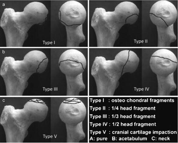
This orthopaedic anatomical diagram illustrates the Chiron classification (2004) for femoral head fractures, shown across five categorized types (Type I to Type V). Each type is presented using a dry bone model of the proximal femur from two perspectives: a lateral/oblique view and a frontal view of the femoral head. Fracture lines are demarcated by solid black lines to indicate the morphology and size of the fracture fragments. Type I displays small osteochondral fragments at the superior-lateral aspect. Type II shows a 1/4 head fragment on the anterior-superior surface. Type III demonstrates a 1/3 head fragment extending from superior to posterior. Type IV depicts a vertical fracture dividing approximately 1/2 of the femoral head. Type V illustrates cranial cartilage impaction with multiple intersecting lines across the superior articular surface. The classification is clinically relevant for orthopedic surgeons and residents for determining the severity of femoral head trauma and informing surgical decisions, such as fragment fixation versus excision based on size and location.
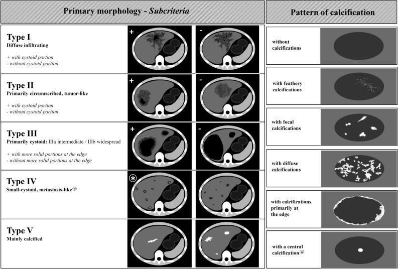
This educational infographic details the Echinococcus multilocularis Ulm Classification for Computed Tomography (EMUC-CT), a standardized system for evaluating hepatic Alveolar Echinococcosis (AE). The diagram is divided into two sections: Primary Morphology and Patterns of Calcification. The left section illustrates five morphological types (I-V) using axial CT cross-sections of the liver. Type I (diffuse infiltrating) and Type II (primarily circumscribed, tumor-like) are sub-categorized by the presence or absence of a cystoid portion. Type III (primarily cystoid) distinguishes between intermediate and widespread involvement, further categorized by solid peripheral portions. Type IV displays small-cystoid, metastasis-like lesions, while Type V represents mainly calcified, inactive lesions. The right panel illustrates six distinct calcification patterns within hepatic lesions: feathery (branching), focal (isolated spots), diffuse (widespread), edge-weighted (peripheral), and central. The classification system is designed to improve diagnostic accuracy and clinical assessment of disease activity in patients with Alveolar Echinococcosis.
perianal abscess anorectal abscess anatomy
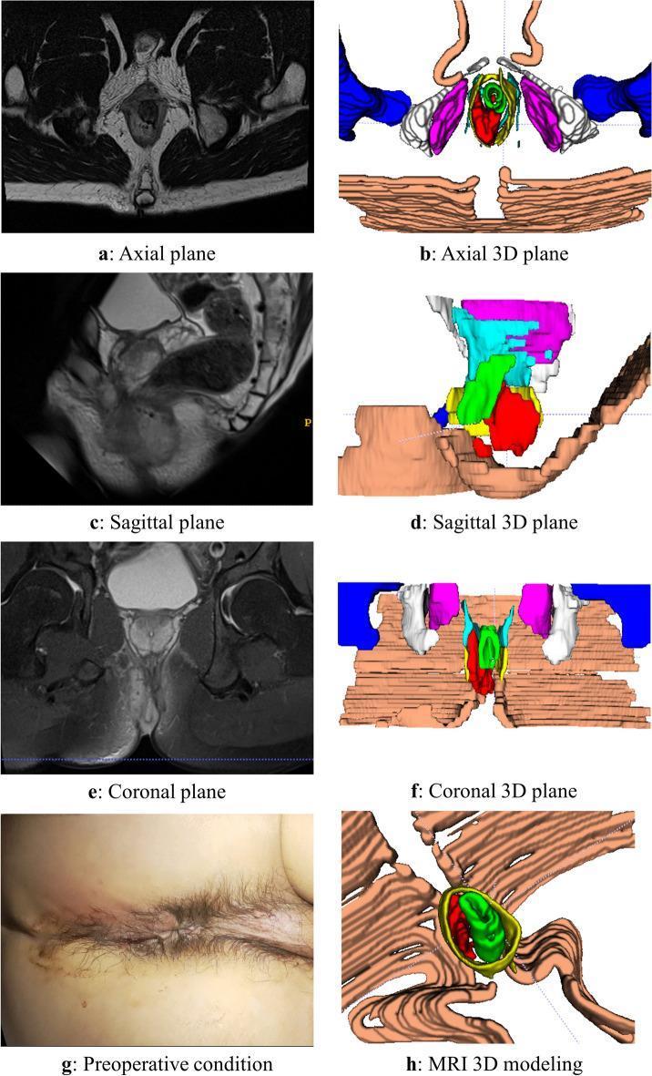
This composite educational image illustrates a case of an intersphincteric anorectal abscess through multi-modal diagnostic views. The panel includes original MRI scans and corresponding color-coded 3D reconstructions across axial (a, b), sagittal (c, d), and coronal (e, f) planes. The MRI images demonstrate the complex anatomy of the perianal region with the abscess appearing as a localized, high-signal intensity lesion on T2-weighted imaging. In the 3D models, the red structure represents the abscess, highlighting its spatial relationship to the anal canal, internal and external sphincters (represented by concentric green and yellow layers), and the levator ani muscle. Panel (g) provides a clinical photograph of the preoperative perianal region, and panel (h) displays a detailed 3D modeling view. This visual aid is designed to teach colorectal surgeons and radiologists the volumetric assessment and anatomical mapping of perianal fistulas and abscesses, emphasizing the value of 3D reconstruction in clarifying tissue demarcations that may be obscured in standard 2D grayscale radiology.
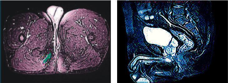
This medical image displays two false-colored pelvic CT scans used for anorectal evaluation. Image (a) on the left is an axial cross-section of the upper thighs and perineum, presented in a pinkish hue. It shows the gluteal and adductor musculature, with a specific focus on the perianal region where a suspected anal fistula or abscess is highlighted with a green digital overlay. Image (b) on the right is a sagittal reconstruction in a blue hue, visualizing the lumbosacral spine, the rectum, and a contrast-filled urinary bladder appearing as a bright white hyperdense structure. The images are used to compare standard diagnostic quality in a control group versus an optimized experimental group, specifically for mapping fistula tracks, identifying internal openings, and assessing involvement of the perianal sphincters. These scans assist in surgical planning for anorectal conditions by delineating the relationship between pathology and surrounding pelvic anatomy.
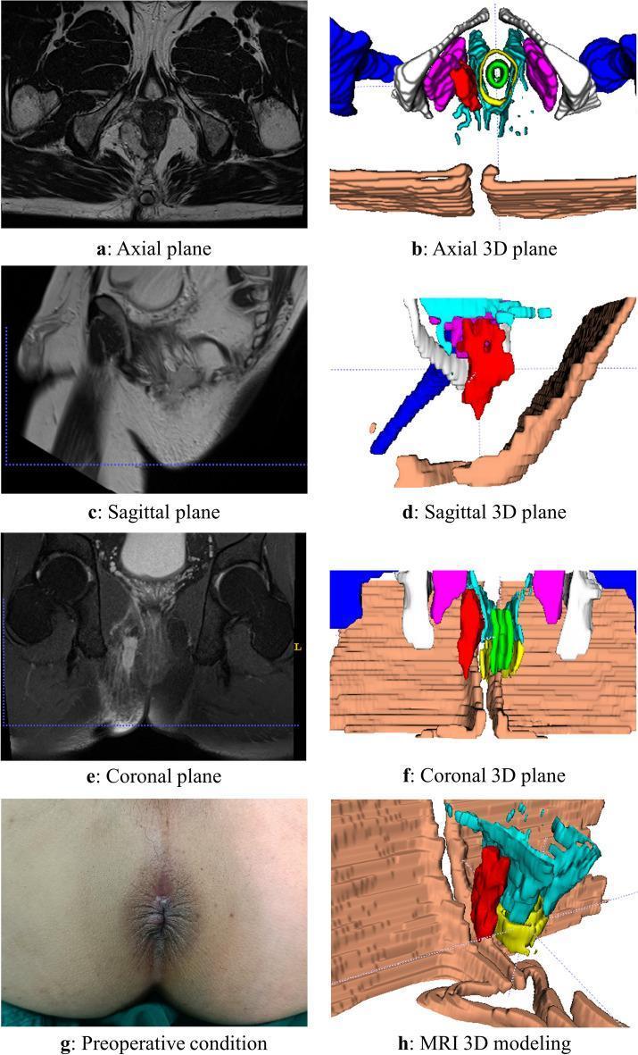
A multi-panel medical graphic illustrating an ischiorectal abscess through imaging, 3D reconstruction, and clinical photography. The panel includes original MRI scans in axial (a), sagittal (c), and coronal (e) planes showing a complex fluid collection in the ischiorectal fossa. Adjacent to these are color-coded 3D models (b, d, f, h) that map the abscess (highlighted in red) relative to pelvic anatomy: the levator ani and pelvic floor muscles (blue and brown), the anal canal/sphincters (green and yellow), and pelvic bones (white/gray). The 3D model allows for the assessment of the abscess's spatial extent, demonstrating its position inferior to the levator ani and lateral to the anal canal, which is diagnostic of an ischiorectal location. Panel (g) provides clinical context with a preoperative photograph of the perianal region. This resource is designed for advanced surgical planning and educational visualization of anorectal infections and their complex anatomical relationships within the pelvic floor.
Types of Abscess
I. Superficial / Skin (Cutaneous) Abscesses
| Type | Description |
|---|---|
| Furuncle (Boil) | Staphylococcal infection of a single hair follicle and surrounding tissue; presents as a fluctuant, tender nodule |
| Carbuncle | Larger, deeper abscess involving multiple hair follicles; common on nape of neck, back, thighs; may cause fever and septicemia |
| Hidradenitis suppurativa | Recurrent abscess formation in apocrine gland areas (axillae, groin); chronic, often resistant to therapy |
| Pilonidal abscess | In the natal cleft/sacrococcygeal area; driven by a nidus of hair; high recurrence risk after drainage |
| Subcutaneous abscess | General soft tissue pus collection beneath skin |
| Incisional/Wound abscess | Forms 7-10 days after surgery along the surgical wound line; up to 75% present after hospital discharge |
- ROSEN's Emergency Medicine, p. 1250-1253
II. Head and Neck Abscesses
| Type | Description |
|---|---|
| Dental / Periapical abscess | Around the apex of a tooth root; caused by carious teeth or failed root canals |
| Periodontal abscess | In the periodontal (gum) tissue surrounding a tooth |
| Peritonsillar abscess (Quinsy) | Collection between the tonsil capsule and the pharyngeal constrictor; the most common deep head/neck abscess |
| Retropharyngeal abscess | In the retropharyngeal space; more common in children; airway emergency |
| Parapharyngeal abscess | Lateral to pharynx; can extend to mediastinum |
| Submandibular abscess (Ludwig's angina) | Bilateral submandibular space infection; life-threatening airway compromise |
| Brain abscess | Focal suppuration within brain parenchyma; sources include otitis media, sinusitis, hematogenous spread |
| Otogenic abscess | Extension from otitis media/mastoiditis |
III. Thoracic Abscesses
| Type | Description |
|---|---|
| Lung abscess | Cavitating necrotic pneumonia; often from aspiration or hematogenous seeding |
| Empyema (pleural abscess) | Pus in the pleural space; usually follows pneumonia, surgery, or trauma |
| Mediastinal abscess | Complication of esophageal perforation, cardiac surgery, or descending neck infections |
| Breast abscess | Lactational (most common, from mastitis) or non-lactational (periductal/subareolar); can be managed with needle aspiration + antibiotics in selected cases |
IV. Abdominal and Pelvic Abscesses
| Type | Description |
|---|---|
| Subphrenic abscess | Below the diaphragm; follows abdominal surgery or perforated viscus |
| Liver (pyogenic hepatic) abscess | Bacterial (often from portal pyemia via appendicitis, diverticulitis) or amoebic; right lobe most common |
| Perinephric abscess | Around the kidney; from ascending UTI or hematogenous spread |
| Psoas abscess | Primary (hematogenous, TB) or secondary (Crohn's disease, vertebral osteomyelitis, appendicitis) |
| Appendiceal abscess | Walled-off perforation of the appendix; may be managed conservatively |
| Diverticular / Pericolic abscess | From perforated diverticulum; staged management (Hartmann's vs. drainage) |
| Pelvic abscess | Post-operative or from pelvic inflammatory disease (PID) / tubo-ovarian abscess |
| Tubo-ovarian abscess | Complication of PID; Neisseria gonorrhoeae, Chlamydia, anaerobes |
V. Anorectal Abscesses
| Type | Location |
|---|---|
| Perianal (superficial/subcutaneous) | Subcutaneous tissue adjacent to anal verge - most common (~60%) |
| Ischiorectal (ischioanal) | In the ischiorectal fossa, lateral to external sphincter |
| Intersphincteric | Between internal and external sphincters |
| Supralevator | Above the levator ani; rarest and most complex |
VI. Special / Named Abscesses
| Type | Notes |
|---|---|
| Brodie's abscess | Chronic bone abscess; subacute osteomyelitis, usually distal tibia metaphysis in children |
| Cold abscess | Lacking heat/redness; classically tuberculous (psoas or spinal - Pott's disease); also seen in actinomycosis |
| Collar-stud abscess | Dumbbell-shaped, crosses deep fascia through a small defect; classically seen in TB lymphadenitis |
| Metastatic/Pyaemic abscess | Multiple small abscesses in various organs from bacteraemia (e.g., S. aureus endocarditis) |
| Bartholin's abscess | In the Bartholin's gland of the vulva; polymicrobial or STI-related |
| Bezold's abscess | Extension of mastoiditis through the tip of the mastoid |
Key Principles
-
Treatment: Incision and drainage (I&D) is the mainstay. Antibiotics alone are insufficient for established abscesses; they are added when the cavity is closed after drainage or when systemic infection is present.
-
Pus composition: Dead/dying neutrophils + bacterial toxins + liquefied tissue debris.
-
Chronicity: Inadequately treated abscesses may form a sinus (one opening) or fistula (two openings). Organisms associated with chronic abscesses include Mycobacterium and Actinomyces.
-
MRSA: Now the most common pathogen in community-acquired skin and soft tissue abscesses; treat with TMP-SMX or clindamycin in addition to I&D.
-
Bailey and Love's Short Practice of Surgery, p. 4244-4256
-
ROSEN's Emergency Medicine, p. 1250-1264
Classification of abcess
collar stud abscess cold abscess tuberculosis lymph node
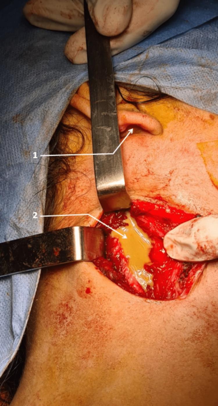
This clinical photograph captures an intra-operative view of a surgical site in the cervical region, positioned near the earlobe (marked as '1'). The image demonstrates the drainage of a 'cold abscess' secondary to lymph node tuberculosis. Two metallic hand-held retractors are used to provide surgical exposure, revealing a deep tissue cavity. Within this cavity, there is a prominent collection of yellowish, viscous, purulent material (pus, marked as '2'), which is characteristic of the caseous necrosis and liquefaction seen in tuberculous lymphadenitis. The surrounding soft tissues show an erythematous, inflammatory appearance with localized bleeding. The surgical field is prepared with antiseptic solution, and sterile drapes are visible. This visual serves as an educational example of the surgical management of cervical lymph node tuberculosis, specifically the intra-operative presentation of abscess formation and the mechanical drainage required for therapeutic management in advanced presentations of the disease.
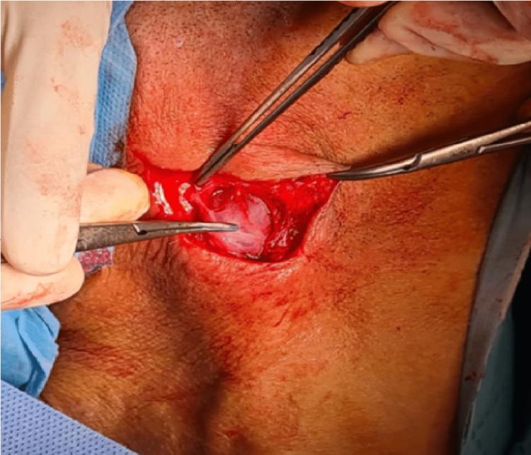
This clinical photograph displays an intraoperative view of an adenectomy, specifically a centered arcuate incision on the neck to address cervical lymph node tuberculosis (LNTB). The surgical field shows a small, transverse skin incision held open by surgical retractors and forceps. Centered within the incision is a prominent, whitish, ovoid structure consistent with an enlarged, encapsulated lymph node (adenopathy). The node exhibits a smooth, slightly translucent surface with minor superficial vascularization. The surrounding subcutaneous tissue appears erythematous and inflamed, with a small amount of surgical bleeding visible. The procedure illustrates the surgical management of a single large adenopathy or 'cold abscess' related to Mycobacterium tuberculosis infection. This image serves as an educational resource for ENT surgeons and medical students to identify the macroscopic appearance of tuberculous lymphadenitis and the technical approach for diagnostic or therapeutic excision.
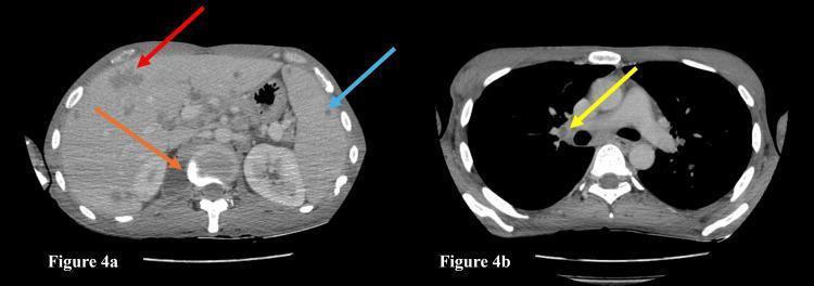
This diagnostic visual consists of two axial contrast-enhanced computed tomography (CECT) images demonstrating multi-organ involvement in a case of disseminated tuberculosis. Figure 4a (left) shows an axial section of the upper abdomen. Key findings include multiple hypodense lesions in the liver (indicated by the red arrow) and several small, low-attenuation lesions within the spleen (blue arrow), consistent with hepatosplenic microabscesses. Additionally, an orange arrow points to a paravertebral abscess with peripheral enhancement adjacent to the vertebral body, suggestive of spinal involvement. Figure 4b (right) displays an axial section of the chest in a soft tissue window. A yellow arrow highlights a necrotic right hilar lymph node, characterized by a central area of low density (necrosis) with a surrounding rim of enhancement. The lymph node is located anterior to the right main bronchus and adjacent to the pulmonary artery. The composite image serves as a clinical teaching tool for identifying extra-pulmonary manifestations of tuberculosis, specifically the radiographic characteristics of visceral lesions, nodal necrosis, and cold abscesses in the setting of disseminated disease.
Classification of Abscess
A. Based on Duration / Clinical Behavior
1. Acute (Hot) Abscess
- Rapid onset with all classic signs of acute inflammation
- Calor (heat), rubor (redness), dolor (pain), tumor (swelling), functio laesa (loss of function) - Celsus' pentad
- Caused by pyogenic organisms, predominantly Staphylococcus aureus
- Pus is composed of dead and dying neutrophils destroyed by bacterial toxins
- The abscess draws fluid in due to hyperosmolar contents, raising pressure and causing pain
- Tracks along planes of least resistance and points toward the skin if undrained
2. Chronic (Cold) Abscess
- Develops slowly over weeks to months
- Lacks the classic signs of inflammation - no heat, no redness
- Fluctuant, painless mass - hence the term "cold"
- Classically due to tuberculosis (Mycobacterium tuberculosis), also Actinomyces
- Microscopically shows lymphocytes, plasma cells, caseation, and later calcification
- May lead to sinus or fistula formation if untreated
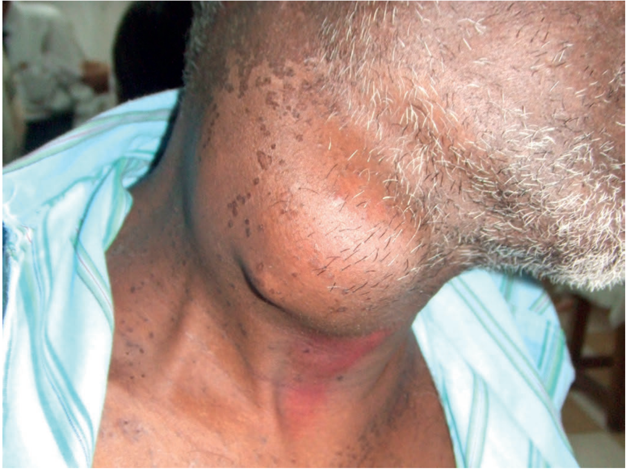
Cold abscess - clinical example (cervical tuberculous):
B. Based on Causative Organism
| Type | Organism | Features |
|---|---|---|
| Pyogenic | S. aureus, Streptococcus, coliforms | Classic hot abscess with creamy pus |
| Tuberculous | M. tuberculosis | Cold abscess; caseous "cheesy" pus |
| Amoebic | Entamoeba histolytica | Liver; "anchovy sauce" pus (brown, odourless) |
| Actinomycotic | Actinomyces israelii | Chronic; "sulphur granules" in pus; jaw/neck/abdomen |
| Fungal | Aspergillus, Candida | Immunocompromised host |
C. Based on Depth / Location from Skin
| Type | Description |
|---|---|
| Superficial (Cutaneous) | Within skin or subcutaneous tissue; easy to see and diagnose |
| Deep | Within body cavities or organs (liver, brain, lung, abdomen); harder to diagnose; may only present with systemic sepsis |
D. Based on Anatomical Location
1. Superficial Skin Abscesses
- Furuncle (Boil) - single hair follicle + surrounding tissue; S. aureus
- Carbuncle - multiple coalescing hair follicles; back of neck, thighs; severe systemic features
- Pilonidal abscess - natal cleft; driven by a hair nidus; high recurrence
- Hidradenitis suppurativa - recurrent apocrine gland abscesses; axillae/groin
- Wound/Incisional abscess - post-surgical; typically 7-10 days after operation
2. Head and Neck
- Dental/Periapical - at root of tooth
- Periodontal - in gum tissue
- Peritonsillar (Quinsy) - between tonsil capsule and superior constrictor; most common deep neck abscess
- Retropharyngeal - in retropharyngeal space; airway emergency, more common in children
- Ludwig's angina - bilateral submandibular space; airway-threatening
- Brain abscess - focal suppuration in brain parenchyma; from otitis media, sinusitis, haematogenous spread
3. Thoracic
- Lung abscess - cavitating necrotic pneumonia; aspiration or haematogenous
- Empyema (pleural abscess) - pus in pleural space
- Breast abscess - lactational (commonest) or non-lactational/subareolar
- Mediastinal abscess - from oesophageal perforation or descending neck infections
4. Abdominal / Pelvic
- Subphrenic abscess - below diaphragm, post-op or perforated viscus
- Liver abscess - pyogenic (portal pyemia) or amoebic
- Psoas abscess - primary (TB) or secondary (Crohn's, vertebral osteomyelitis)
- Appendiceal abscess - walled-off appendix perforation
- Pericolic/Diverticular - perforated diverticulum
- Tubo-ovarian abscess - complication of PID (N. gonorrhoeae, Chlamydia, anaerobes)
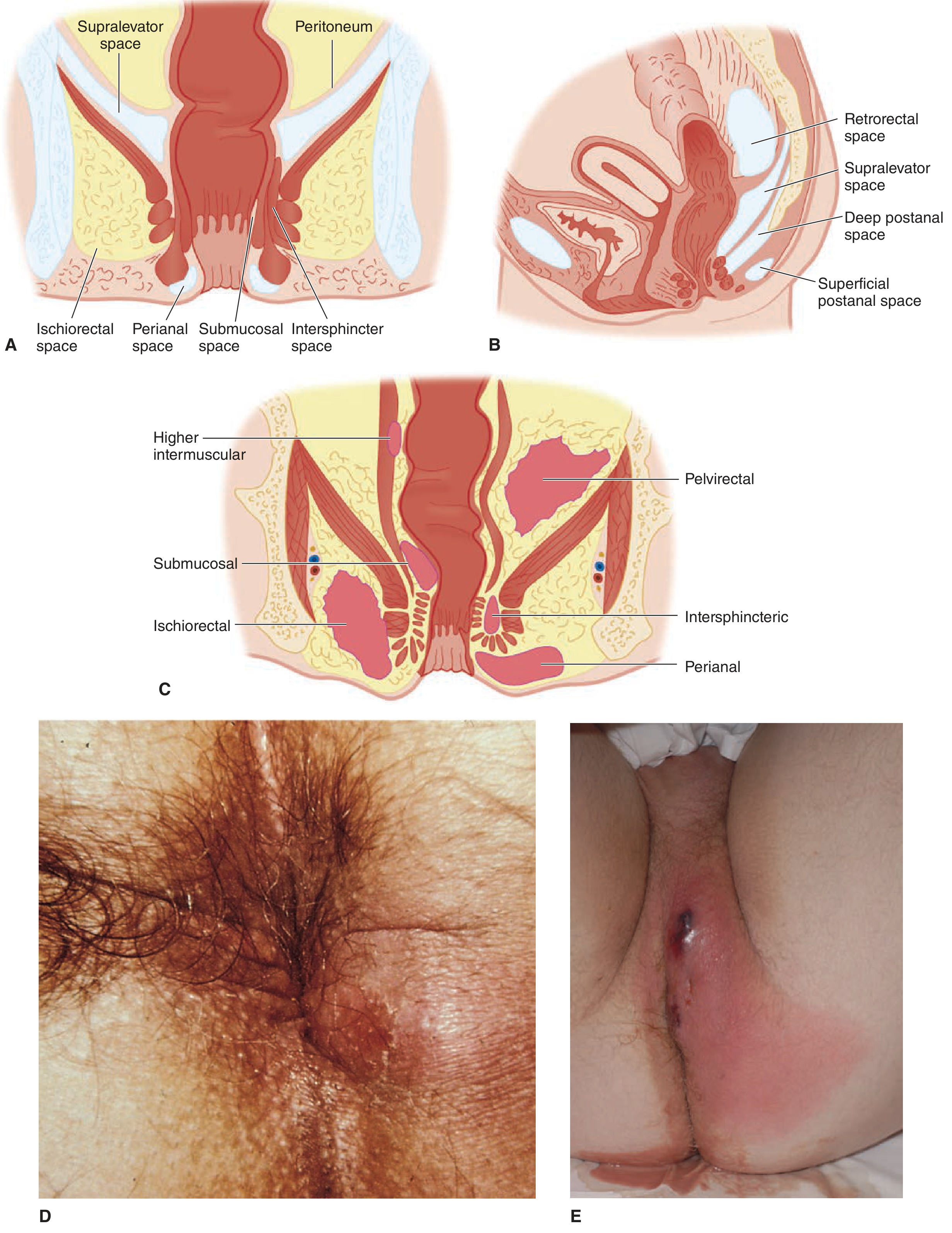
5. Anorectal Abscesses (Parks Classification)
| Type | Location | Frequency |
|---|---|---|
| Perianal | Subcutaneous, adjacent to anal verge | ~60% - most common |
| Ischiorectal (Ischioanal) | Ischiorectal fossa, lateral to external sphincter | ~20% |
| Intersphincteric | Between internal and external sphincters | ~15% |
| Submucosal | Submucosa of anal canal above dentate line | Uncommon |
| Pelvirectal (Supralevator) | Above levator ani | ~5% - rarest, most complex |
| Postanal / Retrorectal | Behind rectum | Uncommon |
E. Special Named Abscesses
| Name | Location / Feature |
|---|---|
| Collar-stud abscess | Bilocular (dumbbell-shaped) TB abscess; deep portion bursts through deep fascia into superficial fascia, connected by a narrow neck; classic in TB cervical lymphadenitis |
| Brodie's abscess | Chronic bone abscess in metaphysis (usually distal tibia); subacute osteomyelitis in children |
| Bezold's abscess | Extension of acute mastoiditis through mastoid tip into neck |
| Paget's abscess | Apparently healed abscess that recurs |
| Metastatic (pyaemic) abscess | Multiple small abscesses in multiple organs from bacteraemia / septic emboli |
| Antibioma | Partially sterilized abscess from empirical antibiotics; no frank pus; mass of fibrous tissue |
| Bartholin's abscess | Bartholin's gland of vulva; polymicrobial or STI-related |
F. Based on Route of Infection
| Route | Example |
|---|---|
| Direct inoculation | Wound abscess, injection site abscess (IV drug use) |
| Contiguous spread | Peritonsillar abscess from tonsillitis; subphrenic from perforated peptic ulcer |
| Haematogenous (metastatic) | Liver abscess from portal pyemia; brain abscess from endocarditis |
| Lymphatic | Axillary abscess from infected hand wound |
Summary Table
| Classification Basis | Categories |
|---|---|
| Duration | Acute (hot) vs. Chronic (cold) |
| Organism | Pyogenic, Tuberculous, Amoebic, Actinomycotic, Fungal |
| Depth | Superficial vs. Deep |
| Location | Skin, Head/Neck, Thoracic, Abdominal/Pelvic, Anorectal |
| Route | Inoculation, Contiguous, Haematogenous, Lymphatic |
| Morphology | Simple vs. Collar-stud (bilocular) |
- Bailey and Love's Short Practice of Surgery 28th Ed, p. 4226-4262, 6771-6787
- Tintinalli's Emergency Medicine, Fig. 85-11