
1. Give an account of cytogenetic disorders involving sex chromosomes. And discuss sex linked disorders. 2. Fragile X syndrome 3. Gaucher’s disease 4. Fluorescence in situ hybridisation 5. Klinefelters syndrome 6. Glycogen storage diseases 7. Gene mutations 8. Gene therapy 9. Down’s syndrome 10. Lysosomal storage diseases 11. Nieman pick disease 12. Single gene disorders
Reading File
Now I'll run parallel DB searches across all 12 topics:
Finding Sources
Finding Sources
Finding Sources
Finding Sources
Finding Sources
Finding Sources
Finding Sources
Finding Sources
Finding Sources
I have 1 DB query left. Let me use it wisely to get the most important missing results, then read all found files in parallel:
Finding Sources
Reading File
Reading File
Reading File
Reading File
Reading File
I have the key textbook content. Now let me read the remaining important blocks in parallel:
Reading File
Reading File
Reading File
Reading File
Reading File
Excellent - I now have rich textbook content. Let me read the critical remaining sections on cytogenetic/sex chromosome disorders, FISH, Gaucher/Niemann-Pick, and more:
Reading File
Reading File
Reading File
Now let me fetch the critical section on sex chromosome disorders, Klinefelter, Gaucher/Niemann-Pick, FISH, and gene mutations from Robbins:
Reading File
Reading File
I now have comprehensive textbook content. Let me get a few more critical sections:
Reading File
Reading File
Excellent - I now have very comprehensive textbook content across all topics. Let me read two more critical remaining sections on sex chromosome disorders (Klinefelter, Turner), FISH, gene mutations, and single gene disorders:
Reading File
Reading File
I now have comprehensive content for all 12 topics. Let me get two final sections - FISH, gene mutations, single gene disorders, and gene therapy from the main Robbins text:
Reading File
Reading File
I now have comprehensive content for all 12 topics. Let me get the FISH section and the gene mutation/single gene disorder sections:
Reading File
Reading File
I now have thorough textbook content for all 12 topics. Let me get the FISH section and Gaucher disease section now:
Reading File
Reading File
I now have comprehensive content from Robbins & Kumar Basic Pathology and Thompson & Thompson Genetics. Let me get the FISH section:
Reading File
I now have extremely thorough content. Let me get the FISH technique section quickly:
Reading File
Here is a comprehensive account of all 12 topics, drawn primarily from Robbins & Kumar Basic Pathology and Thompson & Thompson Genetics and Genomics in Medicine.
1. Cytogenetic Disorders Involving Sex Chromosomes & Sex-Linked Disorders
Background: Lyonization (X-Inactivation)
A number of abnormal karyotypes involving sex chromosomes, ranging from 45,X to 49,XXXXY, are compatible with life - a flexibility not seen with autosomes. Two factors explain this:
- Lyon hypothesis (lyonization): In females, only one X chromosome is genetically active. X inactivation occurs ~16 days post-conception. Either the paternal or maternal X is randomly inactivated in each cell, and the same X remains inactive in all daughter cells. This is mediated by the XIST long non-coding RNA, which "coats" the inactive X chromosome and silences its genes.
- However, ~30% of genes on Xp and 3% on Xq escape X-inactivation - important for understanding Turner syndrome.
- Y chromosome: Carries minimal genetic information beyond male sex determination, so extra Y chromosomes are well tolerated.
General Features of Sex Chromosome Disorders
- Often produce subtle changes, sometimes undetected at birth
- Infertility is a common feature, not diagnosable until adolescence
- Loss of sex chromosomal material is better tolerated than loss of autosomal material
- Extra X chromosomes cause progressively more severe phenotypes with increasing number
Klinefelter Syndrome (47,XXY) - see Topic 5 below
Turner Syndrome (45,X)
- Karyotype: ~57% have 45,X (complete monosomy); 14% have structural X abnormalities (isochromosome Xq, deletions); 29% are mosaics (45,X/46,XX)
- Pathogenesis: During normal development, ovaries develop normally in early embryogenesis but the absence of the second X leads to accelerated loss of oocytes. By age 2, the ovaries are reduced to fibrous "streak ovaries" - "menopause occurs before menarche"
- SHOX gene: The short stature homeobox (SHOX) gene at Xp22.33 escapes X-inactivation; normally two active copies exist in both sexes. Loss of one copy in Turner syndrome explains short stature
- Clinical features:
- Short stature (most consistent somatic feature)
- Primary amenorrhea and infertility (streak ovaries)
- Webbing of the neck (pterygium colli)
- Low posterior hairline
- Cubitus valgus (wide carrying angle of arms)
- Broad shield-shaped chest with widely spaced nipples
- Congenital heart defects (coarctation of aorta, bicuspid aortic valve)
- Lymphedema of hands and feet at birth
- Renal anomalies (horseshoe kidney)
- Intelligence is usually normal; some specific cognitive deficits (spatial reasoning)
- Mosaics (45,X/46,XX): May have minimal findings, present only with primary amenorrhea
Other Sex Chromosome Disorders
| Karyotype | Phenotype | Features |
|---|---|---|
| 47,XYY | Male | Tall stature; normal intelligence; normal fertility in most |
| 47,XXX | Female (Triple X) | Usually normal phenotype; mild intellectual deficit possible; fertile |
| 48,XXXY / 49,XXXXY | Male | Increasingly severe intellectual disability, hypogonadism |
Sex-Linked Disorders
Sex-linked (X-linked) disorders result from mutations in genes on the X chromosome.
X-linked recessive disorders (much more common):
- Affect males almost exclusively (hemizygous - only one X)
- Females are carriers (heterozygous) and usually unaffected; may show mild features due to random X-inactivation
- Examples: Haemophilia A (Factor VIII deficiency), Haemophilia B (Factor IX deficiency), Duchenne muscular dystrophy, G6PD deficiency, colour blindness, Fragile X syndrome
X-linked dominant disorders (rare):
- Both males and females affected, but males more severely
- Lethal in hemizygous males in some conditions (e.g., incontinentia pigmenti)
- Example: Fragile X syndrome (moderate penetrance in females)
Pedigree patterns for X-linked recessive:
- No male-to-male transmission
- All daughters of affected males are obligate carriers
- 50% of sons of carrier females are affected; 50% of daughters are carriers
2. Fragile X Syndrome
Genetics: Fragile X syndrome (FXS) is the most common inherited cause of intellectual disability and the second most common genetic cause overall (after Down syndrome). Frequency: 1 in 1550 males, 1 in 8000 females.
Molecular Basis: Caused by an unstable trinucleotide (CGG) repeat expansion in the 5' UTR of the FMR1 gene at Xq27.3.
| Repeat Count | Status |
|---|---|
| Up to 55 repeats | Normal |
| 55-200 repeats | Premutation (carrier) |
| >200 repeats | Full mutation - FXS |
The full mutation leads to:
- Hypermethylation of the FMR1 promoter CpG island
- Histone deacetylation
- Transcriptional silencing of FMR1
- Absent FMRP protein (a ~80 kDa RNA-binding protein expressed in neurons and non-neural tissues)
Inheritance (Sherman paradox): Unlike typical X-linked recessive disorders, the risk of full mutation increases in successive generations (anticipation). Premutation alleles expand to full mutations almost exclusively during female gametogenesis (not male). Carrier males (premutation) do not pass on expanded alleles to their daughters; however, their daughters' children are at risk. This explained why "normal transmitting males" existed.
Clinical Features:
- Moderate intellectual disability (cognitive impairment is the cardinal feature)
- Autism spectrum disorder (~2-3% of ASD patients have FXS)
- Long, narrow face with prominent forehead and jaw
- Large, protruding ears
- Macroorchidism (post-pubertal testicular enlargement)
- Hyperextensible joints, flat feet
- Behavioural features: hyperactivity, hand-flapping, temper tantrums, perseverative speech, poor eye contact
Sex difference: Affected females have milder features due to random X-inactivation - roughly half their cells express the normal allele.
Premutation carriers are at risk for:
- FXTAS (Fragile X-associated tremor/ataxia syndrome) - late-onset in males
- FXPOI (Fragile X-associated premature ovarian insufficiency) - in females
Diagnosis: PCR-based triplet repeat analysis and Southern blot for methylation status.
Treatment: Trofinetide (an IGF-1 analogue) has shown encouraging results in clinical trials. Treatment remains primarily supportive.
3. Gaucher's Disease
Definition: Gaucher disease is the most common lysosomal storage disease. It results from mutations in the gene encoding glucocerebrosidase (acid beta-glucosidase), leading to accumulation of glucocerebroside (glucosylceramide) in mononuclear phagocytic cells (macrophages).
Inheritance: Autosomal recessive.
Pathogenesis: Macrophages normally degrade glycolipids derived from senescent blood cells. In Gaucher disease, degradation stops at glucocerebroside. "Gaucher cells" - enlarged macrophages with characteristic cytoplasm resembling crumpled tissue paper (due to glucocerebroside accumulation) - accumulate in spleen, liver, bone marrow, and lungs.
Three Clinical Types
| Type | Name | CNS Involvement | Onset | Course |
|---|---|---|---|---|
| Type 1 | Non-neuronopathic | None | Any age (most adult) | Chronic; most common |
| Type 2 | Acute neuronopathic | Severe, early | Infantile | Fatal by age 3 |
| Type 3 | Chronic neuronopathic | Late, milder | Childhood/adolescence | Slowly progressive |
Type 1 Clinical Features (most common):
- Hepatosplenomegaly (splenomegaly often massive)
- Bone marrow infiltration causing anaemia, thrombocytopenia, leukopenia
- Bone pain and pathological fractures (Erlenmeyer flask deformity of distal femur)
- No neurologic involvement
Parkinson disease link: Gaucher disease mutation (heterozygous) is the strongest genetic risk factor for Parkinson disease; patients with Gaucher disease have a 20-fold higher Parkinson risk.
Diagnosis: Glucocerebroside level in leukocytes or cultured fibroblasts; DNA testing.
Treatment:
- Enzyme replacement therapy (ERT): Lifelong IV infusion of recombinant glucocerebrosidase (imiglucerase/velaglucerase)
- Substrate reduction therapy: Oral inhibitor of glucosylceramide synthase (miglustat, eliglustat) - reduces glucocerebroside production
- Emerging: gene therapy via hematopoietic stem cell transplant expressing normal glucocerebrosidase
4. Fluorescence In Situ Hybridisation (FISH)
Principle: FISH is a molecular cytogenetic technique that uses fluorescently labelled DNA probes that hybridise to complementary sequences on chromosomes in cell preparations. The bound probes are visualised under a fluorescence microscope, allowing detection of specific chromosomal sequences.
Steps:
- Prepare metaphase spreads or interphase nuclei on a slide
- Denature both probe and chromosomal DNA (strand separation)
- Allow fluorescently-labelled probe to hybridise to its target sequence
- Wash away unhybridised probe
- Visualise under fluorescence microscope - positive hybridisation appears as fluorescent spots
Types of FISH probes:
- Locus-specific probes: Detect a specific gene or chromosomal region (e.g., HER2 amplification in breast cancer)
- Centromeric/alpha-satellite probes: Detect chromosome number using centromere sequences (e.g., chromosome enumeration in aneuploidy)
- Whole chromosome painting probes: Contain sequences from an entire chromosome; used for detecting translocations
- Telomere probes: Detect cryptic subtelomeric rearrangements
Applications:
- Detection of chromosomal aneuploidy (e.g., trisomy 21, 18, 13; sex chromosome abnormalities)
- Detection of microdeletions (e.g., 22q11.2 deletion syndrome - DiGeorge/velocardiofacial syndrome - cannot be detected by conventional karyotype; requires FISH)
- Detection of translocations (e.g., BCR-ABL in CML)
- Gene amplification in cancer (e.g., HER2/neu, MYCN in neuroblastoma)
- Prenatal diagnosis on uncultured amniocytes (rapid)
- Preimplantation genetic diagnosis
Advantages over conventional karyotype:
- Can detect submicroscopic deletions and duplications (<5 Mb) invisible on G-banding
- Can be performed on interphase cells (no need for cell culture/metaphase)
- Higher resolution for specific loci
Limitations:
- Only identifies abnormalities at targeted loci (not a genome-wide screen)
- Cannot detect point mutations
- Requires prior knowledge of the suspected abnormality
Related technique - Comparative Genomic Hybridisation (CGH): Uses two differentially labelled genomes (test and reference) to scan the entire genome for copy number changes.
5. Klinefelter Syndrome
Definition: Male hypogonadism in an individual with at least two X chromosomes and one or more Y chromosomes.
Karyotype: Most common is 47,XXY. Mosaics (~15%): 46,XY/47,XXY (milder phenotype). More severe variants: 48,XXXY, 49,XXXXY.
Incidence: 1 in 500-1000 male births; one of the most common causes of hypogonadism in males.
Pathogenesis: Results from nondisjunction of sex chromosomes during meiosis. Maternal and paternal nondisjunction contribute equally.
Clinical Features
Body habitus:
- Tall stature with increased soles-to-pubic bone length (elongated legs)
- Gynecomastia
- Reduced facial, body, and pubic hair
- Female fat distribution
Gonadal:
- Small, firm testes (often only 2 cm) - most consistent feature
- Azoospermia/severe oligospermia → sterility (rare fertility in mosaics)
- Low serum testosterone; elevated FSH and LH
- Histology: hyalinisation of seminiferous tubules (ghost-like structures); prominent Leydig cells (apparent hyperplasia)
Cognitive/Neurologic:
- IQ ranges from average to below average; verbal IQ deficit more common
- Increased risk of learning difficulties and language delay
Comorbidities:
- Type 2 diabetes and metabolic syndrome (insulin resistance)
- Mitral valve prolapse (~50% of adults)
- 20-30-fold increased risk of extragonadal germ cell tumours (especially mediastinal teratomas)
- Increased risk of breast cancer
- Increased risk of autoimmune diseases (SLE)
Diagnosis: Confirmed by karyotyping. Buccal smear shows Barr body (inactive X) in most cells.
Treatment:
- Testosterone replacement from puberty onwards (improves virilisation, energy, bone density)
- Gynecomastia may require surgery
- Assisted reproduction (testicular sperm extraction + ICSI) possible in some patients
6. Glycogen Storage Diseases (GSD)
Definition: A group of at least 12 rare inherited disorders of glycogen metabolism, each caused by deficiency of a specific enzyme. Categorised numerically in chronological order of discovery.
Pathophysiology: Liver glycogen is converted to glucose during fasting (energy for whole body). Skeletal muscle glycogen is used locally (lacks glucose-6-phosphatase). Enzyme deficiencies cause either qualitative or quantitative defects in glycogen storage.
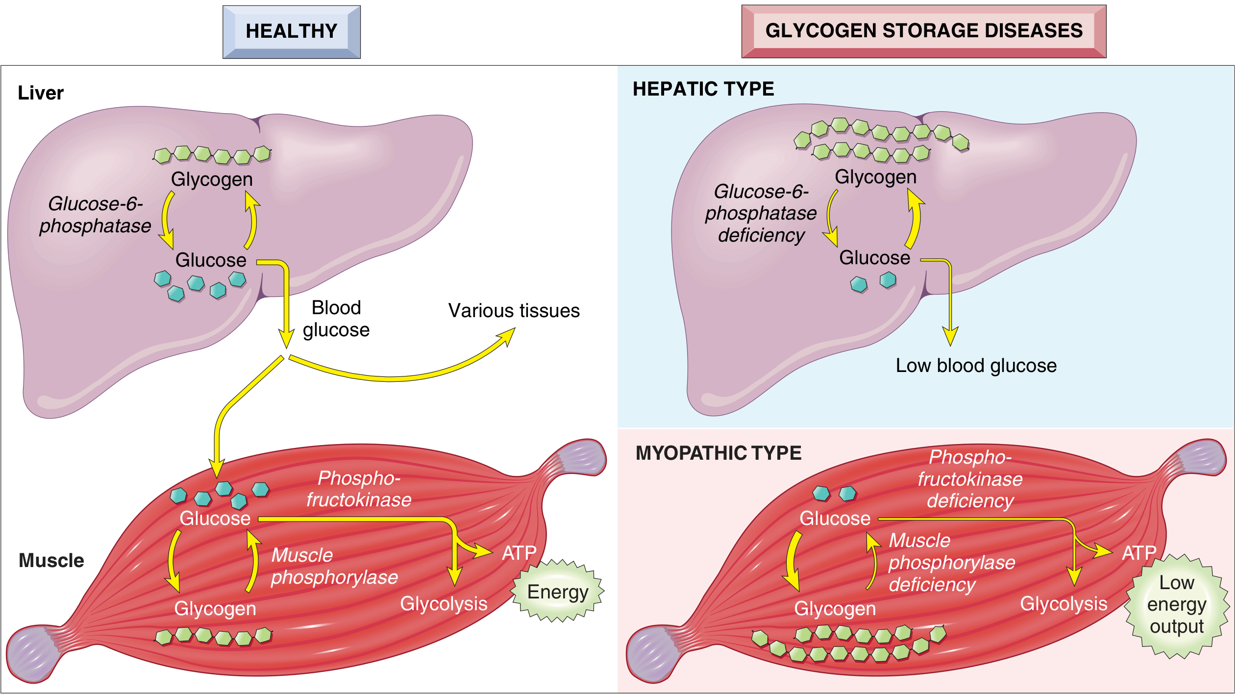
Major Types
| Type | Eponym | Enzyme Deficient | Tissues | Key Features |
|---|---|---|---|---|
| I (Ia) | von Gierke | Glucose-6-phosphatase | Liver, kidney | Severe fasting hypoglycaemia, hepatomegaly, lactic acidosis, hyperuricaemia, hyperlipidaemia |
| I (Ib) | von Gierke Ib | G6P translocase | Liver, neutrophils | As type Ia + neutropenia, recurrent infections, inflammatory bowel disease |
| II | Pompe | Acid alpha-glucosidase (lysosomal) | All tissues esp. heart, muscle | Cardiomegaly, hypotonia, respiratory failure; also adult form with proximal myopathy |
| III | Cori/Forbes | Debranching enzyme (amylo-1,6-glucosidase) | Liver, muscle | Milder hypoglycaemia; hepatomegaly; muscle weakness |
| IV | Andersen | Branching enzyme | Liver, widespread | Cirrhosis, progressive liver failure; abnormal branched glycogen |
| V | McArdle | Muscle phosphorylase | Skeletal muscle | Exercise intolerance, painful cramps, myoglobinuria; no rise in blood lactate with exercise |
| VI | Hers | Liver phosphorylase | Liver | Mild hepatomegaly, mild hypoglycaemia |
| IX | - | Phosphorylase kinase | Liver (± muscle) | Mild hepatomegaly |
Hepatic forms (Types I, III, IV, VI, IX) - ~80% of total: hepatomegaly + hypoglycaemia.
Myopathic forms (Types II, V, VII): muscle weakness/cramps ± myoglobinuria.
Treatment
- GSD I: Frequent feedings + uncooked cornstarch (slow glucose release) to maintain normoglycaemia; avoid sucrose/fructose/galactose; multivitamins, calcium, Vitamin D. Empagliflozin (SGLT2 inhibitor) for GSD Ib (reduces neutrophil dysfunction). Liver transplantation for multiple or malignant adenomas.
- GSD II (Pompe): Enzyme replacement therapy - alglucosidase alfa (recombinant acid alpha-glucosidase); newer avalglucosidase alfa has higher mannose-6-phosphate tags for better uptake. Multidisciplinary team: physiotherapy, speech therapy, ventilatory support.
- GSD III: Uncooked cornstarch + high-protein diet (up to 3g/kg/day) to utilise intact gluconeogenesis; liver or cardiac transplantation in advanced disease.
- GSD V (McArdle): Aerobic exercise training; avoid intense exercise; sucrose supplementation before exercise.
7. Gene Mutations
Definition: A mutation is a permanent change in the DNA sequence. Germline mutations are transmitted to offspring (inherited diseases). Somatic mutations are not heritable but cause cancers and some congenital disorders.
Types of Gene Mutations
1. Point Mutations (Single Nucleotide Substitutions)
- Missense mutation: One nucleotide substituted by another, changing one amino acid to another. Example: β-globin point mutation (GAG→GTG) changing glutamate to valine → Haemoglobin S → sickle cell disease.
- Nonsense mutation: Point mutation creates a stop codon (UAA, UAG, UGA) → truncated/absent protein. Example: many mutations causing Duchenne muscular dystrophy.
- Silent mutation: Nucleotide change does not alter amino acid (due to degeneracy of genetic code) - no phenotypic effect.
2. Frameshift Mutations
- Insertion or deletion of one or two base pairs (not multiples of 3) → alters reading frame of all downstream codons → usually non-functional protein.
- Example: Most mutations in Duchenne muscular dystrophy are deletions causing frameshift.
3. Trinucleotide (Triplet) Repeat Mutations
- Amplification of a 3-nucleotide sequence beyond a threshold number.
- Dynamic mutations: Degree of amplification increases with transmission (especially in certain sexes) → anticipation.
- Examples:
- Fragile X syndrome: CGG repeat in FMR1 (normal <55; full mutation >200)
- Huntington disease: CAG repeat in HTT (normal <35; disease >40)
- Myotonic dystrophy: CTG repeat in DMPK
- Spinocerebellar ataxias: Various CAG repeats
- ~50 diseases caused by trinucleotide repeats; all involve neurodegeneration.
4. Splice Site Mutations
- Mutations at intron-exon boundaries disrupt pre-mRNA splicing → exon skipping, intron retention → abnormal protein.
5. Promoter/Regulatory Region Mutations
- Affect transcription factor binding → altered gene expression level without changing protein structure.
6. Large Deletions/Insertions
- Deletion of an entire exon or multiple exons.
- Example: Deletion mutations in dystrophin gene in Duchenne/Becker MD.
7. Copy Number Variants (CNVs)
- Amplifications or deletions of chromosomal segments containing multiple genes.
- Example: HER2 amplification in breast cancer; 22q11.2 microdeletion.
Functional Consequences
- Loss-of-function (LOF): Reduced or absent protein activity. Most autosomal recessive diseases result from LOF mutations in both alleles.
- Gain-of-function (GOF): Abnormal new function or overactivity. Example: dominant negative effect, constitutive receptor activation (e.g., RET mutations in MEN2).
- Dominant negative: Mutant protein interferes with the function of the normal protein from the other allele. Example: p53 mutations in Li-Fraumeni syndrome.
8. Gene Therapy
Definition: The introduction of a biologically active gene into a cell to achieve a therapeutic benefit. The goal is to transfer a therapeutic gene early enough to prevent or reverse pathological events.
Approved/Clinical Applications (Table from Thompson & Thompson)
| Disease | Defective Gene | Vector | Outcome |
|---|---|---|---|
| X-linked SCID | IL2RG | Retroviral vector, HSCs | Significant improvement in 27/32 patients |
| SCID (ADA deficiency) | ADA | Retroviral vector, HSCs | 29/40 off enzyme replacement therapy |
| Spinal muscular atrophy | SMN1 | AAV (IV injection) | Marked improvement; FDA approved |
| Haemophilia B | Factor IX (F9) | AAV (single IV injection) | Stable factor IX expression; >20 patients stopped prophylaxis |
| X-linked adrenoleukodystrophy | ABCD1 | Lentiviral vector, HSCs | Arrest of cerebral demyelination in 17/19 boys |
| Leber congenital amaurosis | RPE65 | AAV (subretinal injection) | Improved visual function |
Vectors
Viral Vectors:
- Retroviruses/Lentiviruses: Integrate into host genome → stable long-term expression. Risk of insertional mutagenesis (activating oncogenes). Used for haematopoietic stem cells.
- Adeno-associated virus (AAV): Does not integrate (forms episome); low immunogenicity; long-term expression in post-mitotic cells (neurons, hepatocytes, myocytes). Most widely used vector. Risk: limited cargo size (~4.7 kb).
- Adenoviruses: High efficiency transduction; episomal; strong immune response limits repeat dosing.
Non-viral vectors: Liposomes, nanoparticles - lower efficiency but less immunogenic.
Strategies
- Gene addition: Introduce functional copy of a defective gene (most common; for LOF diseases)
- Gene correction: Edit the defective gene in its native chromosomal context (using CRISPR-Cas9)
- Gene silencing: RNA interference (RNAi) or antisense oligonucleotides to suppress GOF mutations (e.g., Huntington disease)
Somatic vs. Germline Gene Therapy
- Somatic gene therapy: Targets specific cells (lung, liver, HSCs); changes are not passed to offspring. Currently approved and in clinical use.
- Germline gene therapy: Changes reproductive cells; would be inherited by future generations. No country currently permits this. Raises major ethical concerns regarding consent of future persons, enhancement vs. therapy, unpredictable long-term effects.
Induced Pluripotent Stem Cells (iPSCs) in Gene Therapy
Patient's own somatic cells (e.g., fibroblasts) are:
- Gene-corrected (by gene editing or gene therapy)
- Induced to pluripotency → differentiated into the required cell type
- Transplanted into the patient
Advantages: autologous (no rejection), genetically corrected. Hurdles: safety of iPSC methodology, epigenetic stability.
9. Down's Syndrome (Trisomy 21)
Incidence: 1 in 700 live births - the most common chromosomal disorder.
Karyotypes
| Type | Karyotype | Frequency | Notes |
|---|---|---|---|
| Trisomy 21 | 47,XX/XY,+21 | 95% | Sporadic; meiotic nondisjunction |
| Translocation | 46,XX,der(14;21)(q10;q10),+21 | 4% | Often familial; carrier parent has Robertsonian translocation |
| Mosaic | 46,XX/47,XX,+21 | 1% | Mitotic nondisjunction; milder phenotype |
Pathogenesis
The most common cause is meiotic nondisjunction (95% maternal in origin). Maternal age strongly influences incidence:
- <20 years: 1 in 1550 births
-
45 years: 1 in 25 births
The amyloid precursor protein (APP) gene is on chromosome 21 - explains why virtually all Down syndrome patients >40 years develop Alzheimer disease neuropathology.
Clinical Features
Dysmorphic facies (usually apparent at birth):
- Flat facial profile
- Oblique (upward-slanting) palpebral fissures
- Epicanthic folds
- Small, low-set ears
- Brushfield spots on iris
- Protruding tongue, small mouth (relative macroglossia)
Neurologic/Cognitive:
- Leading cause of severe intellectual disability; IQ 25-50 in ~80%
- Hypotonia (neonatal)
- Alzheimer disease in virtually all patients >40 years (APP gene on chr 21)
Cardiac (~40%):
- Endocardial cushion defects (most common)
- Atrial septal defects
- Ventricular septal defects
- Accounts for majority of early deaths
Haematologic:
- 10-20-fold increased risk of acute leukaemia (both ALL and AML)
Other:
- Atlantoaxial instability (~10-20%)
- Hypothyroidism (increased incidence)
- Gastrointestinal: duodenal atresia, Hirschsprung disease
- Ophthalmologic: cataracts, refractive errors
- Recurrent infections (impaired T-cell function, thyroid autoimmunity)
- Cryptorchidism, hypospadias
Prenatal Screening
- First trimester: maternal serum β-hCG (elevated), PAPP-A (decreased), nuchal translucency ultrasound
- Cell-free fetal DNA in maternal blood (liquid biopsy): sensitive and specific, but confirmed by conventional cytogenetics (amniocentesis)
10. Lysosomal Storage Diseases (LSDs)
Definition: LSDs are a group of inherited metabolic disorders caused by deficiency of specific lysosomal hydrolases (or, in some cases, non-enzymatic lysosomal proteins), resulting in accumulation of undegraded substrates within lysosomes. Most are autosomal recessive. Over 50 different LSDs are recognised.
General Mechanism
Lysosomes receive macromolecules via endocytosis, autophagy, or phagocytosis. Specific enzymes sequentially degrade these molecules. A deficiency in any one enzyme leads to accumulation of the substrate within lysosomes → cell swelling and dysfunction → organ damage.
Classification by Substrate Accumulated
| Category | Examples | Substrate Stored |
|---|---|---|
| Sphingolipidoses | Gaucher, Niemann-Pick A/B, Tay-Sachs, Fabry, Krabbe | Sphingolipids |
| Mucopolysaccharidoses | Hurler (MPS I), Hunter (MPS II), Sanfilippo, Morquio | Glycosaminoglycans |
| Glycoproteinoses | Mannosidosis, Fucosidosis | Glycoproteins |
| Mucolipidoses | ML II (I-cell disease) | Multiple |
| Glycogen | Pompe (GSD II) | Glycogen (lysosomal) |
Common Features
- Hepatosplenomegaly (phagocytic cells accumulate storage material)
- Progressive neurodegeneration (for disorders with CNS involvement)
- Coarse facial features (especially mucopolysaccharidoses)
- Skeletal abnormalities (dysostosis multiplex in MPS)
- Corneal clouding (MPS, mucolipidoses)
- Onset in infancy/childhood; progressive deterioration
Treatment Approaches
- Enzyme replacement therapy (ERT): IV infusion of recombinant enzyme. Effective for Gaucher type 1, Pompe, Fabry, some MPS types. Cannot cross the blood-brain barrier → ineffective for CNS disease.
- Substrate reduction therapy: Reduce substrate production (miglustat for Gaucher and Niemann-Pick type C).
- Haematopoietic stem cell transplantation: Provides donor macrophages expressing normal enzyme. Effective if done early (before neurologic damage); used for Hurler syndrome.
- Chaperone therapy: Small molecule chaperones stabilise misfolded enzymes (clinical trials for Gaucher, Fabry, some Tay-Sachs variants).
- Gene therapy: Emerging; clinical trials ongoing.
11. Niemann-Pick Disease
Niemann-Pick disease encompasses several distinct disorders. The classic types are:
Type A and Type B - Sphingomyelinase Deficiency
Enzyme deficiency: Acid sphingomyelinase (encoded on chromosome 11p15.1/15.4 - imprinted gene, preferentially expressed from maternal chromosome)
Substrate accumulated: Sphingomyelin → failure to cleave sphingomyelin into ceramide and phosphorylcholine
Ethnic predisposition: Increased incidence in Ashkenazi Jewish populations
Type A (Severe, Infantile Neuronopathic)
- Complete sphingomyelinase deficiency
- Sphingomyelin accumulates in phagocytic cells and neurons
- Macrophages become stuffed with lipid droplets ("foam cells") → fine vacuolation/foaminess - "zebra bodies" on electron microscopy (concentric lamellar myelin figures)
- Organs most affected: Spleen, liver, bone marrow, lymph nodes, lungs (high phagocytic cell content)
- CNS: All levels affected (spinal cord, ganglia, brain); neurons enlarged and vacuolated
- Clinical: Progressive hepatosplenomegaly, failure to thrive, neurologic deterioration from infancy
- Cherry red spot in retina (similar to Tay-Sachs, as both involve ganglion cell swelling)
- Fatal: Death usually within first 3 years of life
Type B (Non-Neuronopathic)
- Mutant sphingomyelinase with some residual activity
- Organomegaly (hepatosplenomegaly) present
- No neurologic manifestations (unlike Type A)
- Patients survive into adulthood
- Diagnosis: Sphingomyelinase activity in leukocytes; molecular genetic testing
Type C - Cholesterol Transport Defect
Mechanism: Mutations in NPC1 (majority) or NPC2 genes → defective transport of free cholesterol from lysosomes to cytoplasm → accumulation of cholesterol and gangliosides (GM1, GM2) within cells.
Distinct from Types A and B (different gene, mechanism, and substrate).
Most common form overall (more common than Types A + B combined)
Clinical features: Ataxia, vertical supranuclear gaze palsy (characteristic), dystonia, dysarthria, psychomotor regression. Onset in childhood (most common form).
Treatment: Miglustat (substrate reduction therapy) approved to stabilise neurological manifestations in Type C.
12. Single Gene Disorders
Definition: Disorders resulting from mutations in a single gene, following Mendelian patterns of inheritance (or in some cases, non-Mendelian patterns).
Inheritance Patterns
Autosomal Dominant (AD)
- One abnormal allele sufficient to cause disease
- Heterozygote affected; vertical transmission through generations
- 50% risk to offspring
- Often due to gain-of-function (GOF) or dominant negative mutations; haploinsufficiency in some
- Examples: Marfan syndrome, Huntington disease, familial hypercholesterolaemia (heterozygous), neurofibromatosis, achondroplasia, BRCA1/2-related cancer syndromes
Autosomal Recessive (AR)
- Both alleles must be abnormal (homozygous or compound heterozygous)
- Parents are typically unaffected carriers
- 25% risk to offspring of two carriers
- Usually enzyme/structural protein deficiencies
- Examples: Cystic fibrosis, sickle cell disease, phenylketonuria, Tay-Sachs disease, Gaucher disease, all glycogen storage diseases (except some forms)
X-Linked Recessive
- Gene on X chromosome; affects hemizygous males
- Females (heterozygous) are usually carriers
- No male-to-male transmission
- Examples: Haemophilia A and B, Duchenne MD, G6PD deficiency, Fragile X (X-linked with modified dominance)
X-Linked Dominant (rare)
- Both males and females affected; males often more severely
- Examples: Fragile X syndrome (reduced penetrance in females), incontinentia pigmenti, Rett syndrome
Non-Mendelian Single Gene Disorders
1. Triplet Repeat Disorders
- Fragile X, Huntington disease, myotonic dystrophy, spinocerebellar ataxias
- Dynamic mutations, anticipation (see Topics 2 and 7)
2. Mitochondrial (Maternal) Inheritance
- Mutations in mitochondrial DNA (mtDNA)
- Strictly maternal transmission (mitochondria are maternally inherited)
- Heteroplasmy: cells contain a mixture of normal and mutant mitochondria - variable expressivity
- Threshold effect: disease manifests only when mutant mtDNA exceeds a threshold proportion
- Examples: MELAS (mitochondrial encephalomyopathy, lactic acidosis, stroke-like episodes), MERRF, Leber hereditary optic neuropathy (LHON)
- Tissues most affected: those with highest energy demands (neurons, muscle, retina)
3. Genomic Imprinting
- Epigenetic phenomenon where the expression of a gene depends on whether it was inherited from the mother or father
- Imprinted genes are "stamped" with the parental sex of origin during gametogenesis (DNA methylation)
- Example: Prader-Willi syndrome (loss of paternal 15q11-13 expression) vs. Angelman syndrome (loss of maternal 15q11-13 expression) - same chromosomal region, different clinical phenotypes depending on parent of origin
Important Concepts
Penetrance: Proportion of individuals with a given genotype who manifest the phenotype. Fragile X has reduced penetrance (~79% in males, ~35% in females).
Expressivity: Degree to which the phenotype is manifest among those who carry the genotype. Variable expressivity is common in AD disorders (e.g., neurofibromatosis).
Pleiotropy: One gene affecting multiple organs/systems (e.g., Marfan syndrome: skeleton, eyes, cardiovascular).
Genetic heterogeneity: Same phenotype caused by mutations at different loci (e.g., retinitis pigmentosa, non-syndromic hearing loss).
Modifier genes: Other genetic loci influence severity of a known single-gene disorder (e.g., cystic fibrosis severity modified by genes outside CFTR).
Primary Sources: Robbins & Kumar Basic Pathology, 10th ed.; Thompson & Thompson Genetics and Genomics in Medicine, 9th ed.; Goldman-Cecil Medicine; Tietz Textbook of Laboratory Medicine, 7th ed.
This is a shared conversation. Sign in to Orris to start your own chat.