
Write me all about limb ischemia in detailed manner from Bailey dogar and other standard books make me a complete topic
Reading File
Finding Sources
Finding Sources
Now let me also check for Bailey & Love specifically, and peripheral arterial disease sections:
Finding Sources
Finding Sources
Finding Sources
Now let me read the key blocks in parallel - Sabiston (most comprehensive surgical text), Goldman-Cecil, and Harrison's:
Reading File
Reading File
Reading File
Excellent content from Sabiston, Goldman-Cecil, and Harrison's. Now let me read the continuation of the Sabiston text (covering management, thromboembolectomy, bypass) and the chronic limb ischemia sections:
Reading File
Reading File
Reading File
Excellent data. Now let me read more from Sabiston on management continuation + chronic limb ischemia, and also search for images and PubMed:
Reading File
Searching Images
acute limb ischemia clinical appearance
Searching PubMed
"limb ischemia" AND management
Now let me read more from Sabiston on management (the critical part with thromboembolectomy and thrombolysis details), and the chronic limb ischemia section:
Reading File
Reading File
Reading File
Reading File
I now have comprehensive data from multiple major textbooks. Let me fetch the key clinical images and read the Sabiston section on reperfusion injury and fasciotomy:
Loading Image
Loading Image
Reading File
I now have all the material needed. Let me also get the reperfusion injury and compartment syndrome section from Sabiston, plus read the Harrison's treatment section continuation:
Reading File
Reading File
I now have excellent comprehensive content from multiple authoritative textbooks. Let me update todos and compile the full write-up:# LIMB ISCHEMIA — Complete Topic
Sources: Sabiston Textbook of Surgery (21e), Goldman-Cecil Medicine (International Ed.), Harrison's Principles of Internal Medicine (22e, 2025), Current Surgical Therapy (14e), with supplementary PubMed evidence
1. DEFINITION AND OVERVIEW
Limb ischemia is defined as inadequate arterial blood supply to a limb, resulting in tissue hypoxia that may threaten limb viability. It exists on a spectrum from mild intermittent claudication to irreversible gangrene. It is divided into two major categories:
- Acute Limb Ischemia (ALI) — symptoms present for < 2 weeks, representing a vascular emergency
- Chronic Limb Ischemia (CLI) / Chronic Limb-Threatening Ischemia (CLTI) — symptoms of rest pain or tissue loss present > 2 weeks, with corresponding diagnostic evidence of arterial insufficiency
There are approximately 1.5 cases of ALI per 10,000 persons per year. Perioperative mortality in lower extremity ALI is 20–40%, with limb loss rates of 12–50%.
— Sabiston Textbook of Surgery, 21e
2. ETIOLOGY AND PATHOPHYSIOLOGY
2.1 Acute Limb Ischemia
ALI results from abrupt interruption of blood flow with downstream tissue hypoperfusion. Irreversible muscle and nerve injury can occur after 6 hours of severe arterial insufficiency.
Principal causes:
| Cause | Key Features |
|---|---|
| Embolism | ~80% originate in the heart; lodge at bifurcations |
| Thrombosis in situ | In already diseased/atherosclerotic vessels or bypass grafts |
| Arterial dissection | Intimal flap obstructs lumen |
| Trauma | Direct disruption or formation of acute thrombus |
| Iatrogenic | Post-arterial puncture or catheter placement |
| Rare | Popliteal entrapment, thoracic outlet compression, hypercoagulable states, polycythemia, paradoxical embolism via PFO |
Cardiac sources of embolism include:
- Atrial fibrillation (most common)
- Recent myocardial infarction with mural thrombus
- Ventricular aneurysm, cardiomyopathy
- Infective and marantic endocarditis
- Prosthetic heart valves (risk 0.6–2.3%/patient-year; mitral > aortic by 2–3×)
- Atrial myxoma
Non-cardiac embolic sources: aortic, femoral, and popliteal aneurysms; atherosclerotic plaque rupture (microemboli → "blue toe syndrome").
Emboli lodge preferentially at arterial bifurcations — in the lower limb: femoral artery > iliac artery > aorta > popliteal/tibioperoneal.
— Harrison's Principles of Internal Medicine 22e; Goldman-Cecil Medicine
2.2 Embolism vs. Thrombosis (critical distinction for management)
| Feature | Embolism | Thrombosis in situ |
|---|---|---|
| Onset | Sudden, dramatic | More gradual |
| Prior PAD history | Absent | Usually present |
| Contralateral limb | Normal pulses | Often abnormal |
| Atrial fibrillation/cardiac disease | Often present | May be absent |
| Collateral circulation | Poor (no time to develop) | Better established |
| Severity | Often more severe | May be less severe |
— Sabiston Textbook of Surgery
2.3 Chronic Limb Ischemia / CLTI
CLTI develops most commonly from atherosclerosis causing multilevel disease along the length of the limb. Other causes include:
- Thromboangiitis obliterans (Buerger's disease)
- Vasculitis
- Thromboembolic disease
- Cystic adventitial disease
- Popliteal artery entrapment
- Hypercoagulable states
Inflammatory mechanism: Inflammation drives atherosclerosis progression. Elevated CRP, IL-6, TNF-α, and platelet activation markers are increased compared with controls. Approximately 20% of the risk for low ABI is attributable to heritability.
Natural history: < 5% of patients with PAD progress to CLTI. However, if CLTI is left untreated, major amputation rates reach 22% at 1 year.
— Goldman-Cecil Medicine; Current Surgical Therapy 14e
3. CLINICAL FEATURES
3.1 The "6 Ps" of Acute Limb Ischemia
Pain — Pallor — Pulselessness — Poikilothermia (coolness) — Paresthesia — Paralysis
- Pain: Severe, sudden onset; affects major muscle groups below the occlusion
- Pallor: Early sign; transitions to cyanosis and mottling over time
- Pulselessness: Loss of pulses distal to occlusion; helps localize the level
- Poikilothermia: Coolness of the limb; particularly significant if contralateral limb is warm; transition level correlates with occlusion site
- Paresthesia: Numbness, tingling — indicates sensory nerve ischemia
- Paralysis: Late, grave sign — indicates motor nerve and muscle ischemia; motor deficits progress from distal to proximal (intrinsic foot muscles first → complete paralysis = late/irreversible)
Note: In microembolism or cholesterol embolism, distal pulses may remain palpable despite limb ischemia (emboli lodge in small distal vessels).
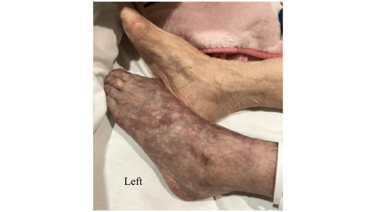
3.2 Chronic Limb Ischemia — Symptom Spectrum
| Stage | Symptoms |
|---|---|
| Intermittent claudication | Reproducible muscle pain on exertion, relieved by rest; cramping, tightness |
| Rest pain | Severe burning pain at rest, typically in toes/forefoot; worsened supine, relieved by dependency |
| Tissue loss | Non-healing ulcers, gangrene |
Claudication must be distinguished from pseudoclaudication (spinal stenosis — relieved by sitting/position change) and venous/compartment causes.
4. CLASSIFICATION OF ACUTE LIMB ISCHEMIA
Rutherford Classification of ALI (the gold standard)
| Category | Description/Prognosis | Sensory Loss | Muscle Weakness | Arterial Doppler | Venous Doppler |
|---|---|---|---|---|---|
| I — Viable | Not immediately threatened | None | None | Audible | Audible |
| IIa — Marginally Threatened | Salvageable if promptly treated | Minimal (toes) or none | None | Inaudible | Audible |
| IIb — Immediately Threatened | Salvageable with immediate revascularization | More than toes; rest pain | Mild–moderate | Inaudible | Audible |
| III — Irreversible | Major tissue loss / permanent nerve damage inevitable | Profound, anesthetic | Profound paralysis, rigor | Inaudible | Inaudible |
Source: Rutherford RB et al. J Vasc Surg. 1997;26:517–538; reproduced in Sabiston, Goldman-Cecil, and Harrison's
Key point: Rutherford III with rigor (muscle stiffening) is irreversible — revascularization risks multi-organ failure without functional limb salvage. Primary amputation is preferred.
5. CHRONIC LIMB ISCHEMIA — CLASSIFICATION
Fontaine Classification
| Stage | Symptoms |
|---|---|
| I | Asymptomatic |
| IIa | Mild claudication (>200 m) |
| IIb | Moderate-severe claudication (<200 m) |
| III | Ischaemic rest pain |
| IV | Ulceration or gangrene |
WIfI Classification (Society for Vascular Surgery)
A modern system combining three factors for risk stratification in CLTI:
- Wound characteristics
- Ischemia (degree of pedal perfusion)
- foot Infection extent
Higher WIfI scores correlate with increased risk of major amputation and guide goal-directed therapy.
ABI (Ankle-Brachial Index)
- Normal: 1.0–1.4
- Borderline: 0.91–0.99
- Mild PAD: 0.70–0.90
- Moderate PAD: 0.50–0.69
- Severe/CLTI: < 0.50
- Rest pain typically occurs at ABI < 0.40
- Non-compressible (calcified) vessels: ABI > 1.4 (diabetics, elderly)
6. INVESTIGATIONS
Bedside/Immediate
- Continuous-wave Doppler: Assess peripheral signals — multiphasic (normal) vs. monophasic/absent. Rutherford classification relies on presence/absence of arterial and venous Doppler signals
- ABI measurement
- ECG: Identify atrial fibrillation, recent MI
Vascular Imaging
| Modality | Use |
|---|---|
| Duplex ultrasound | Confirm location, rule out popliteal aneurysm as source, assess DVT |
| CT Angiography (CTA) | Fast, widely available; best for guiding open/endovascular/hybrid planning |
| MR Angiography (MRA) | No radiation; excellent soft-tissue detail; useful in renal impairment |
| Catheter-based arteriography | Gold standard; allows simultaneous intervention |
Workup for Underlying Etiology (ALI)
- Echocardiogram: Identify wall motion abnormalities, intracardiac thrombus, vegetations, atrial myxoma
- CTA chest/abdomen/pelvis: Aortic atheroma, aneurysm, primary aortic thrombus
- Hypercoagulable panel: If no cardiac or atherosclerotic source identified
- Venous duplex: Rule out paradoxical embolism source (DVT + PFO)
Chronic Ischemia Workup
- Segmental limb pressures — localize level of disease
- Pulse volume recordings (PVR)
- Transcutaneous oxygen (TcPO₂): Predicts wound healing and guides amputation level
- Toe pressures / toe-brachial index (TBI) — more reliable in calcified vessels
7. MANAGEMENT
7.1 Acute Limb Ischemia
Immediate General Measures
- IV heparin immediately upon suspicion: 75–100 units/kg bolus, then 15–18 units/kg/min infusion
- Prevents thrombus propagation and maintains collateral patency
- Continue until definitive treatment and transition to oral anticoagulation
- Alternative in heparin-contraindicated patients: lepirudin (renal metabolism) or argatroban (hepatic metabolism)
- Analgesia
- Admit, stabilize medically, assess severity (Rutherford classification)
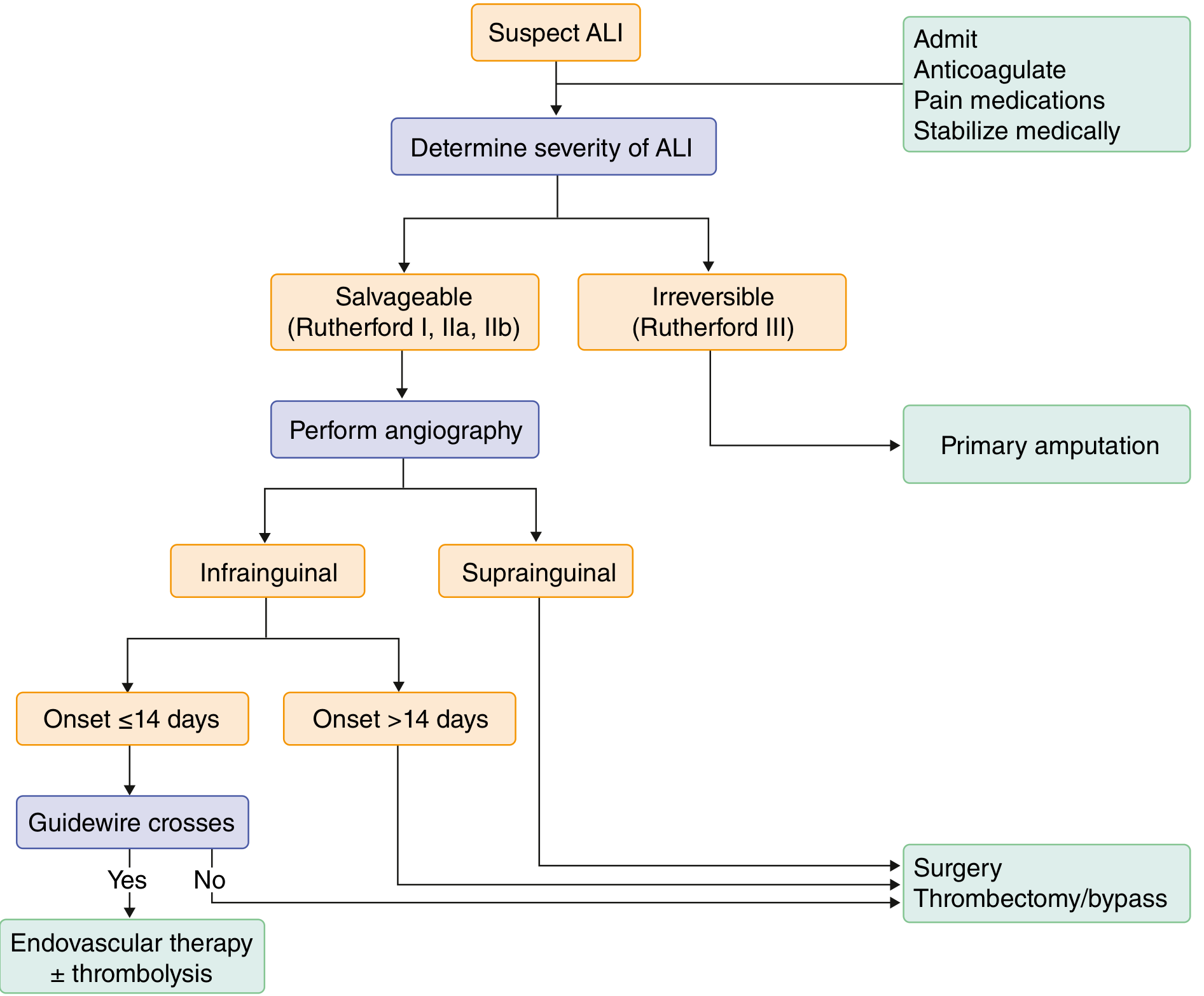
Treatment algorithm:
7.2 Interventional Approaches
Three primary strategies — no high-quality data demonstrates superiority of any one technique. Selection is based on ischemia severity, time to reperfusion, occlusion location, etiology, and comorbidities.
A. Endovascular Approach (Rutherford I and IIa; thrombotic occlusion; high surgical risk)
Catheter-Directed Thrombolysis (CDT)
- Agents: recombinant tPA, reteplase, tenecteplase
- Most effective for: recent thrombotic occlusion in atherosclerotic vessel, bypass graft, or occluded stent; distal emboli where surgical access is limited
- Ultrasound-emitting catheters improve thrombus permeability
- Disadvantage: longer time to reperfusion; bleeding risk
Percutaneous Mechanical Thrombectomy (PMT)
- Devices using hydrodynamic forces or rotating baskets to fragment/aspirate thrombus
- Reduces infusion time; fewer procedures needed
- Often combined with pharmacologic thrombolysis
Absolute contraindications to pharmacologic thrombolysis:
- Active bleeding
- Stroke or neurosurgical procedure within 3 months
- Malignant intracranial neoplasm
- History of hemorrhagic stroke
- Recent GI bleed
Relative contraindications: Severe hypertension, CNS tumors, major surgery within 3 weeks, trauma
B. Open Surgical Approach (Rutherford IIb; contraindication to thrombolysis)
Fogarty Balloon Catheter Embolectomy — preferred for immediately threatened limb
- Balloon-tipped catheter passed beyond the thrombus, inflated, then withdrawn to extract clot
- Femoral embolectomy: indicated for absent/water-hammer ipsilateral femoral pulse with normal contralateral pulse
- Bilateral femoral embolectomies: for absence of both femoral pulses (aortic thrombus/saddle embolus)
- Performed in a hybrid OR to allow completion angiography and adjunct endovascular treatment
- After embolectomy in thrombotic disease, underlying stenosis must be treated (angioplasty, endarterectomy, stenting, or bypass) to prevent re-thrombosis
Arterial Bypass:
- For extensive thrombotic disease or when embolectomy fails
- Autogenous vein (great saphenous) preferred for infrainguinal bypass (BASIL trial)
- Prosthetic grafts used for suprainguinal or when vein unavailable
C. Primary Amputation (Rutherford III — irreversible ischemia)
- Indicated in: profound paralysis with rigor, anesthetic limb, inaudible arterial AND venous Doppler signals, livedo reticularis
- Revascularizing irreversible ischemia risks multi-organ failure from rhabdomyolysis, hyperkalemia, metabolic acidosis
- Also consider in: poor overall health, devastating tissue loss, non-ambulatory status with poor prognosis
7.3 Post-Revascularization: Compartment Syndrome and Reperfusion Injury
Reperfusion injury occurs when previously ischemic tissue swells within the confined fascial compartments after blood flow is restored.
Fasciotomy — Four-compartment lower limb fasciotomy:
- Prophylactically performed in Rutherford IIb ischemia (or if ischemia time > 6 hours)
- Medial incision (2–3 cm posterior to medial tibial border): releases both posterior compartments — superficial posterior, then divide soleus attachments to release deep posterior
- Lateral incision (between anterior crest of tibia and fibula): releases anterior and lateral compartments via parallel fascia incisions; care to avoid superficial peroneal nerve
- Signs of evolving compartment syndrome requiring immediate release:
- Tense compartments
- Pain with passive stretch
- Decreased sensation at dorsal first webspace
- Motor weakness
Postoperative monitoring:
- Hemodynamic monitoring and fluid resuscitation
- Correct acidosis and electrolyte imbalance (K⁺ and H⁺ release from damaged cells)
- Serial neurovascular exams
- Continued therapeutic anticoagulation (risk of re-thrombosis up to 30%)
7.4 Chronic Limb Ischemia (CLI/CLTI) — Management
The approach targets both limb preservation and reduction of systemic cardiovascular risk.
A. Risk Factor Modification
- Smoking cessation: Most important modifiable risk factor; counselling + pharmacotherapy (nicotine replacement, varenicline, bupropion)
- Lipid management: High-intensity statin therapy to LDL ≤ 70 mg/dL reduces amputation and death. PCSK9 inhibitors further reduce cardiovascular and major limb events in those already on statin therapy
- Blood pressure control: Target < 130/80 mmHg; β-blockers are NOT contraindicated in PAD
- Diabetes control: Crucial especially with coexisting CLTI
- Exercise prescription: Supervised exercise program preferred
B. Exercise Therapy
- Supervised exercise therapy (SET) is the first-line treatment for intermittent claudication
- Improves walking distance more than unsupervised home exercise
- RCT evidence: supervised exercise > primary stent therapy or home walking + cilostazol for aortoiliac PAD
- Combination of SET + balloon angioplasty superior to SET alone for ABI, walking distance, and quality of life
C. Antiplatelet and Antithrombotic Therapy
- Aspirin 75–325 mg/day: Established role in secondary prevention of MI, stroke, and vascular death in all PAD patients
- Clopidogrel 75 mg/day: Indicated if aspirin not tolerated
- Dual pathway inhibition: Low-dose rivaroxaban 2.5 mg twice daily + aspirin 100 mg daily reduces major cardiovascular events, limb events, and all-cause mortality (at expense of increased bleeding) — COMPASS trial evidence
- Vorapaxar (PAR-1 antagonist): May reduce acute limb ischemia incidence and revascularization need
- Long-term anticoagulation indicated when ALI caused by cardiac thromboembolism
D. Pharmacotherapy for Claudication
- Cilostazol 50–100 mg twice daily (phosphodiesterase inhibitor): Improves maximal walking distance by 40–50% vs. placebo; reduces restenosis after femoral revascularization; contraindicated in heart failure
- Oral vasodilating prostaglandins, vitamin E, chelation therapy with EDTA — not effective
E. Revascularization for Chronic Ischemia
Indicated when:
- Lifestyle-limiting claudication unresponsive to exercise + pharmacotherapy
- Rest pain
- Tissue loss (ulcer/gangrene) — CLTI
Endovascular vs. Surgical:
- Endovascular first preferred for amenable lesions (lower peri-procedural mortality/morbidity)
- Angioplasty with or without stenting
- Drug-eluting stents (DES) and drug-coated balloons (DCB) provide superior, more durable benefit for femoropopliteal disease vs. angioplasty alone (equivalent to each other)
- Angioplasty more cost-effective than surgery if expected 5-year patency ≥ 30%
- Open surgical bypass preferred for:
- Extensive multi-segment disease not amenable to endovascular treatment
- Failed endovascular approach
- BASIL trial: bypass vs. angioplasty — similar mortality, amputation, and patency at 4 years in femoropopliteal disease
- BASIL-2 trial (2023): In CLTI requiring infrapopliteal intervention — vein bypass vs. best endovascular treatment first; vein bypass showed improved outcomes in patients with suitable vein
- BEST-CLI trial (2022): Surgery or endovascular therapy — surgery superior in patients with single-segment great saphenous vein available
- Hybrid procedures: Combine open and endovascular in same setting
Wound care and infection control: Meticulous care for tissue loss; infection management critical for limb salvage
8. ATHEROEMBOLISM (Special Variant)
- Multiple small deposits of fibrin, platelets, and cholesterol debris from proximal atherosclerotic lesions/aneurysms lodge in distal small vessels
- Distal pulses remain palpable (unlike classic ALI)
- Presentations: "Blue toe syndrome," petechiae, splinter hemorrhages, livedo reticularis, localized tenderness
- Skin/muscle biopsy: cholesterol crystals (pathognomonic)
- May follow intraarterial procedures
- Difficult to treat: Local foot care ± amputation; analgesics; statins + antiplatelet therapy for cardiovascular risk; thrombolysis and surgical revascularization are NOT helpful due to multiplicity and distal location
- Endovascular/surgical exclusion of the source vessel/aneurysm may be needed for recurrent atheroembolism
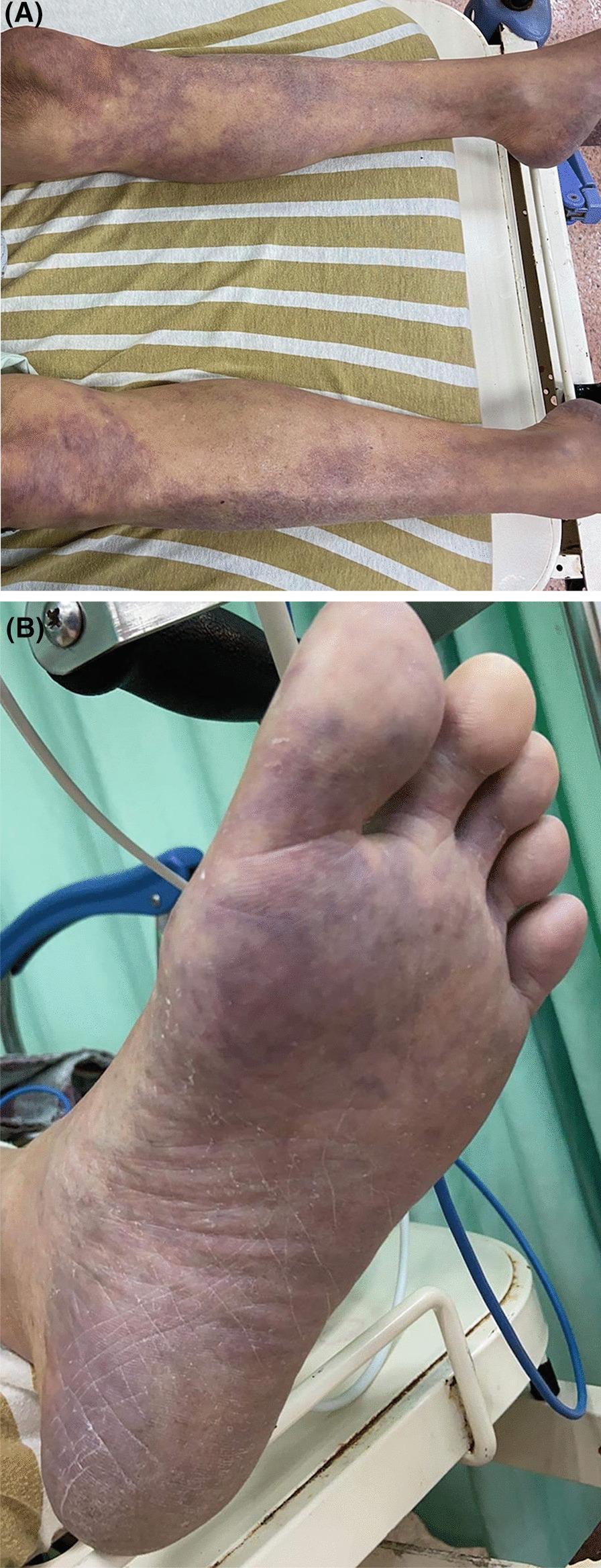
9. COMPLICATIONS OF LIMB ISCHEMIA
| Complication | Mechanism / Notes |
|---|---|
| Gangrene | Dry (arterial) or wet (superadded infection); may require amputation |
| Compartment syndrome | Post-reperfusion swelling in fascial compartments; requires emergency fasciotomy |
| Rhabdomyolysis | Myocyte necrosis → myoglobinuria → acute kidney injury |
| Hyperkalemia / metabolic acidosis | Release from ischemic muscle cells; can cause cardiac arrhythmias |
| Multi-organ failure | From reperfusion injury metabolites systemic release |
| Re-occlusion / re-thrombosis | Up to 30% risk post-revascularization; prevented by anticoagulation |
| Amputation | Major limb loss; 30-day mortality 8.9% (below-knee) to 27.7% (above-knee) |
10. PROGNOSIS
- ALI: Perioperative mortality 20–40%; limb loss 12–50% for lower extremity ALI
- CLTI untreated: Major amputation rate ~22% at 1 year
- Below-knee amputation: 30-day mortality ~8.9%
- Above-knee amputation: 30-day mortality ~27.7%
- Quality of life is profoundly impaired in CLTI; significant improvement follows successful revascularization
- Patients with PAD carry high systemic cardiovascular risk: MI, stroke, and vascular death are the primary causes of death
11. RECENT EVIDENCE (PubMed, 2023–2026)
| Study | Finding |
|---|---|
| Ramesh et al., Cardiovasc Revasc Med 2025 [PMID 40087129] | Meta-analysis: endovascular intervention vs. bypass surgery for CLTI — comparable outcomes, supporting individualized approach |
| Zhou et al., J Vasc Surg 2026 [PMID 41338295] | Network meta-analysis: endovascular treatments for lower extremity arterial disease stratified by lesion location and severity — significant variation in outcomes by lesion type |
| Zhang et al., Ann Vasc Surg 2025 [PMID 39096950] | Meta-analysis: prediabetes independently associated with increased risk of PAD, supporting early metabolic intervention |
SUMMARY MNEMONICS
6 Ps of Acute Limb Ischemia:
Pain, Pallor, Pulselessness, Poikilothermia, Paresthesia, Paralysis
Rutherford I → IIa → IIb → III
Viable → Marginally threatened → Immediately threatened → Irreversible
Management of ALI:
Heparin immediately → Assess Rutherford → Image (CTA/angio) → Revascularize (endovascular IIa, open IIb) or Amputate (III)
Embolus vs. Thrombosis:
Embolus = sudden, no PAD, cardiac source, Fogarty Thrombosis = background PAD, Thrombolysis or bypass
*Sources: Sabiston Textbook of Surgery (21e); Goldman-Cecil Medicine International Edition; Harrison's Principles of Internal Medicine, 22e (2025); Current Surgical Therapy, 14e; supplemented by PubMed meta-analyses (2025–2026)*This is a comprehensive, exam-ready write-up on Limb Ischemia synthesized from Sabiston Textbook of Surgery (21e), Goldman-Cecil Medicine, Harrison's Principles of Internal Medicine (22e, 2025), and Current Surgical Therapy (14e), supplemented with current PubMed evidence (2025–2026).
What's covered:
- Definition & Overview — ALI vs. CLI definitions, incidence, mortality figures
- Etiology & Pathophysiology — Embolism vs. thrombosis distinction (clinically critical), cardiac/non-cardiac sources, atherosclerotic mechanisms, inflammatory role in CLTI
- Clinical Features — The 6 Ps (Pain, Pallor, Pulselessness, Poikilothermia, Paresthesia, Paralysis); claudication spectrum; differential diagnosis table
- Rutherford Classification — Full table with Doppler findings and prognosis for each grade (I, IIa, IIb, III)
- Chronic Ischemia Classification — Fontaine stages, WIfI system, ABI thresholds
- Investigations — Bedside Doppler, ABI, duplex, CTA, MRA, arteriography, echocardiography
- Management of ALI — Immediate heparin, Fogarty embolectomy, catheter-directed thrombolysis, mechanical thrombectomy, bypass, primary amputation; contraindications to thrombolysis; treatment algorithm image
- Compartment Syndrome & Reperfusion Injury — Four-compartment fasciotomy technique and indications
- Chronic Limb Ischemia Management — Risk factor modification, supervised exercise, cilostazol, aspirin/rivaroxaban dual pathway, endovascular vs. bypass (BASIL, BEST-CLI, BASIL-2 trial evidence)
- Atheroembolism — Blue toe syndrome, special management considerations
- Complications, Prognosis, Recent Evidence
- Mnemonics for quick revision
Also add from bailey and love
Finding Sources
Finding Sources
Finding Sources
You've run out of free credits. Sign up for a free account to continue using Orris.
This is a shared conversation. Sign in to Orris to start your own chat.