
IT fracture clarification s explain and eloborate all
Reading File
Intertrochanteric (IT) fractures are extracapsular hip fractures occurring between the greater and lesser trochanters of the femur. They are among the most common fractures in elderly patients, typically caused by low-energy falls in osteoporotic bone.
Searching sources...
Finding Sources
Finding Sources
Reading File
Reading File
Finding Sources
Reading File
Reading File
Loading Image
Reading File
Reading File
Loading Image
Reading File
Loading Image
Reading File
Loading Image
Now I have comprehensive content from multiple authoritative sources (Rosen's Emergency Medicine, Rockwood & Green's Fractures 10th ed 2025, Miller's Review of Orthopaedics 9th ed, Campbell's Operative Orthopaedics 15th ed 2026). Let me compile the full answer.
Intertrochanteric (IT) Fracture - Complete Guide
1. Definition and Anatomy
An intertrochanteric fracture is a break in the proximal femur whose fracture line extends between the greater and lesser trochanters. These are classified as extracapsular fractures - the fracture lies outside the hip joint capsule, within the cancellous (trabecular) bone of the trochanteric region.
Key anatomical points:
- The trochanteric region has abundant blood supply from the surrounding cancellous bone, so avascular necrosis (AVN) is NOT a major concern (unlike femoral neck fractures)
- The calcar femorale (Adam's arch) - a dense posteromedial bone plate - is the most critical structural support. When it is comminuted, the fracture becomes unstable
- The short external rotators remain attached to the distal fragment; the internal rotators attach to the proximal fragment
- The gluteus medius and short external rotators abduct and externally rotate the greater trochanter; adductors and hamstrings pull the shaft posteromedially - causing the classic shortening and varus deformity
2. Epidemiology and Risk Factors
- One of the most common fractures in the elderly (along with femoral neck fractures)
- Predominantly caused by low-energy falls in patients with osteoporotic bone
- 1-year mortality rate up to 30% - survival is most strongly related to patient age and preexisting comorbidities
- Life expectancy returns to normal among patients who survive that first year
- Only about 50% of patients regain their original level of ambulation
Risk factors (from Rockwood & Green's):
| Category | Factors |
|---|---|
| Patient factors | Increased age, osteoporosis, female sex |
| Medical | Concurrent illness, previous stroke, visual impairment |
| Functional | Disorders of gait/balance, physical disability, mental impairment |
| Medications | Tranquilizers, antihypertensives, multiple medications, alcohol |
| Lifestyle | No regular exercise, undernourishment, greater dependence on others |
3. Classification
A. Number-of-Parts Classification (Practical/Simple)
This is the most clinically intuitive system:
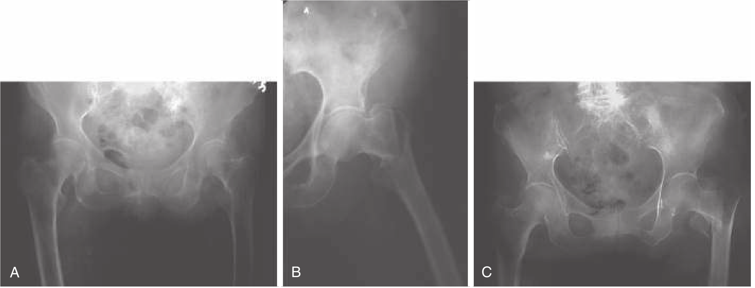
(A) Two-part: head/neck fragment + shaft fragment. (B) Three-part: one trochanter is also fractured, adding instability. (C) Four-part: both trochanters are fractured.
- 2-part: One part connected to femoral head, one to shaft - relatively stable
- 3-part: Greater or lesser trochanter is additionally fractured - the muscles still pulling on that fragment create instability
- 4-part: Both trochanters fractured - most unstable
B. OTA/AO Classification (Gold Standard - Rockwood & Green's 2025)
This is the classification incorporated into national hip fracture guidelines in Europe and the United States. The three broad groups (A1, A2, A3) have shown acceptable intra- and interobserver variation and are the most clinically useful:
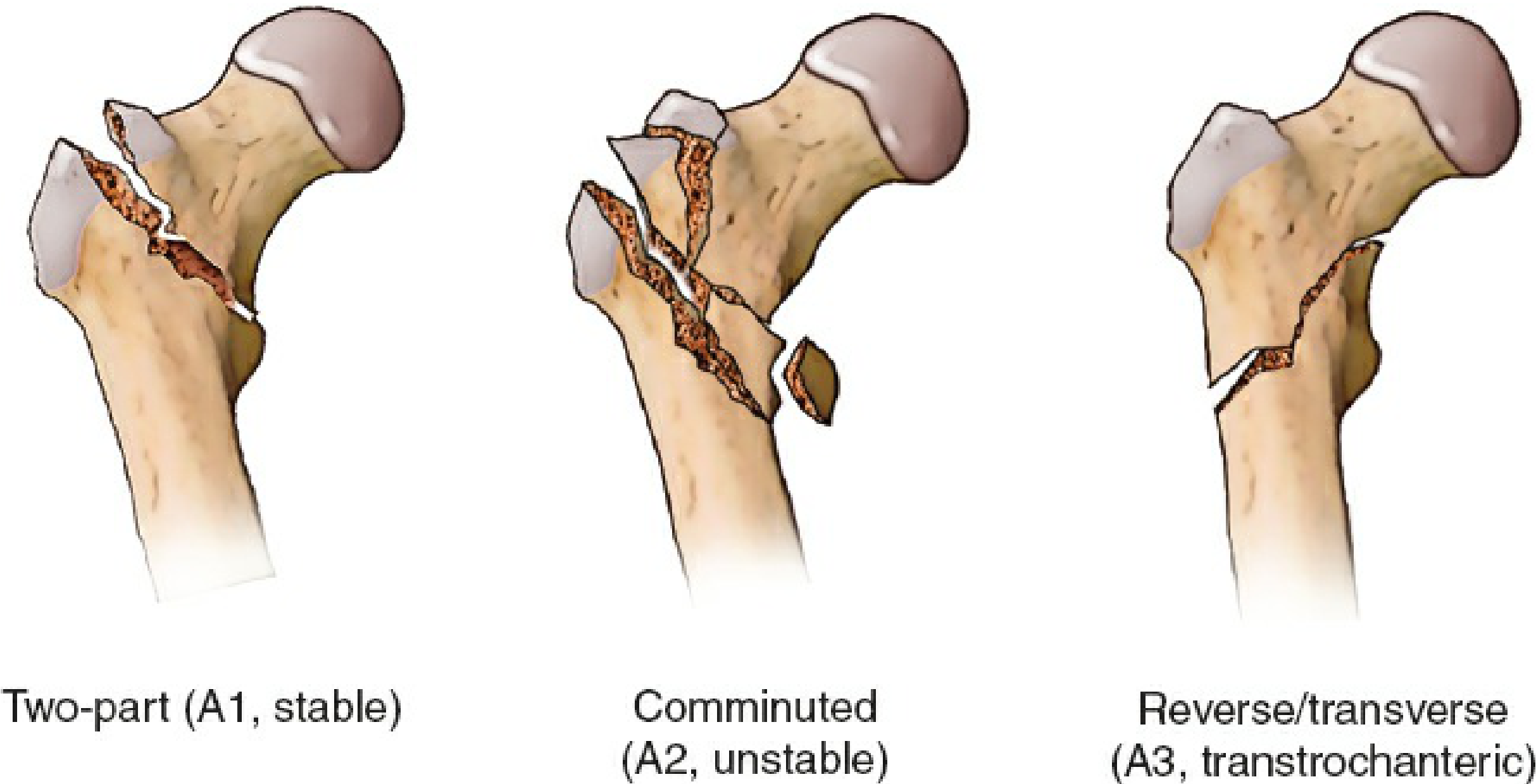
| Type | Description | Stability | Key Feature |
|---|---|---|---|
| 31.A1 | Simple two-part fracture, intact lateral wall | Stable | Fracture line runs through the trochanters; medial cortex interrupted at one place; lesser trochanter may be on either fragment |
| 31.A2 | Comminuted fracture, lateral wall essentially intact | Unstable | One or two coronal fragments at greater trochanter level (posterior/anterior); lesser trochanter detached - minimum 3-4 parts |
| 31.A3 | Reverse oblique or horizontal fracture | Very Unstable (transtrochanteric) | Fracture line from lesser trochanter extends to lateral cortex, rendering the lateral wall unstable; may extend into subtrochanteric region |
Important: The A3 reverse oblique pattern is easily missed on plain X-ray. CT scanning or fluoroscopy on the traction table may be needed to identify it. It is critical to recognize because it mandates an intramedullary (IM) nail rather than a sliding hip screw.
Note on the additional OTA subdivisions (each group is further split into 3 subgroups): Studies have shown these finer subdivisions do NOT reliably guide treatment and have unacceptable inter/intraobserver variation - they are therefore not routinely used clinically.
C. Evans Classification (Historical)
Introduced in 1949, distinguishes stable vs. unstable based on medial cortex comminution. Historically important but has poor reproducibility and has largely been superseded by the OTA/AO system.
4. The Lateral Wall - A Critical Concept
The lateral wall refers to the lateral femoral cortex from where the cortical bone narrows into the greater trochanter. Its integrity is critical for surgical planning:
- If the lateral wall is intact (>20.5 mm thick): a Sliding Hip Screw (SHS) is appropriate
- If the lateral wall is <20.5 mm or deficient: risk of iatrogenic lateral wall fracture is high with SHS; a cephalomedullary (IM) nail is preferred
- A3-type fractures inherently have an unstable lateral wall and require IM nailing
5. Clinical Presentation
Symptoms:
- Acute pain around the hip and thigh with inability to stand or walk
- Pain may radiate to the ipsilateral knee
Examination findings:
- Shortened and externally rotated leg (the strong iliopsoas action on the lesser trochanter pulls the proximal fragment into this position)
- Contusion or hematoma around the hip
- Passive range of motion is painful
- In nondisplaced fractures: axial compression pain may be the only sign (limb may appear normal)
Associated injuries (~4% of patients):
- Ipsilateral distal radius fracture (most common, ~2%)
- Ipsilateral proximal humerus fracture (~1%)
- Head trauma (suspect in all; CT head warranted in ~21% of low-energy fracture patients)
- A second contralateral hip fracture occurs in up to 1 in 3 patients within ~1.5 years
6. Diagnosis
Imaging
| Modality | Role |
|---|---|
| AP pelvis X-ray | First-line; include both hips. Contralateral hip helps judge reduction/varus/length intraoperatively |
| Lateral hip X-ray | Assesses displacement, angulation, posterior comminution |
| Internal rotation AP view (+10°) | When diagnosis is uncertain on standard views |
| MRI | Best for occult fractures not visible on X-ray; also shows bone bruise, soft tissue injury |
| CT scan | Practical alternative when MRI not available; helps characterize comminution, lateral wall, A3 reverse oblique patterns |
7. Management
A. General Principles
- Goal: stable fixation allowing early weight bearing
- Modifiable comorbidities should be corrected
- Surgery should be performed within the first 48 hours - early repair (24-48h) improves 1-year mortality
- Operating on the day of injury (before resuscitation) increases mortality risk - patients need preoperative optimization
- Multidisciplinary team approach (orthogeriatrics model) reduces 1-year mortality
B. Nonoperative Treatment
Reserved for a very small subgroup:
- Nondisplaced fractures in patients able to comply with non-weight-bearing
- Displaced fractures in nonambulatory patients or those with prohibitive operative risk
- Bedbound/end-of-life patients (frank discussion required)
- Management: touch-down weight bearing for 6-8 weeks with analgesic nerve blocks
- Associated with higher mortality compared to surgical management
C. Operative Treatment - Implant Selection
This is the cornerstone of management. Implant choice is driven by fracture stability:
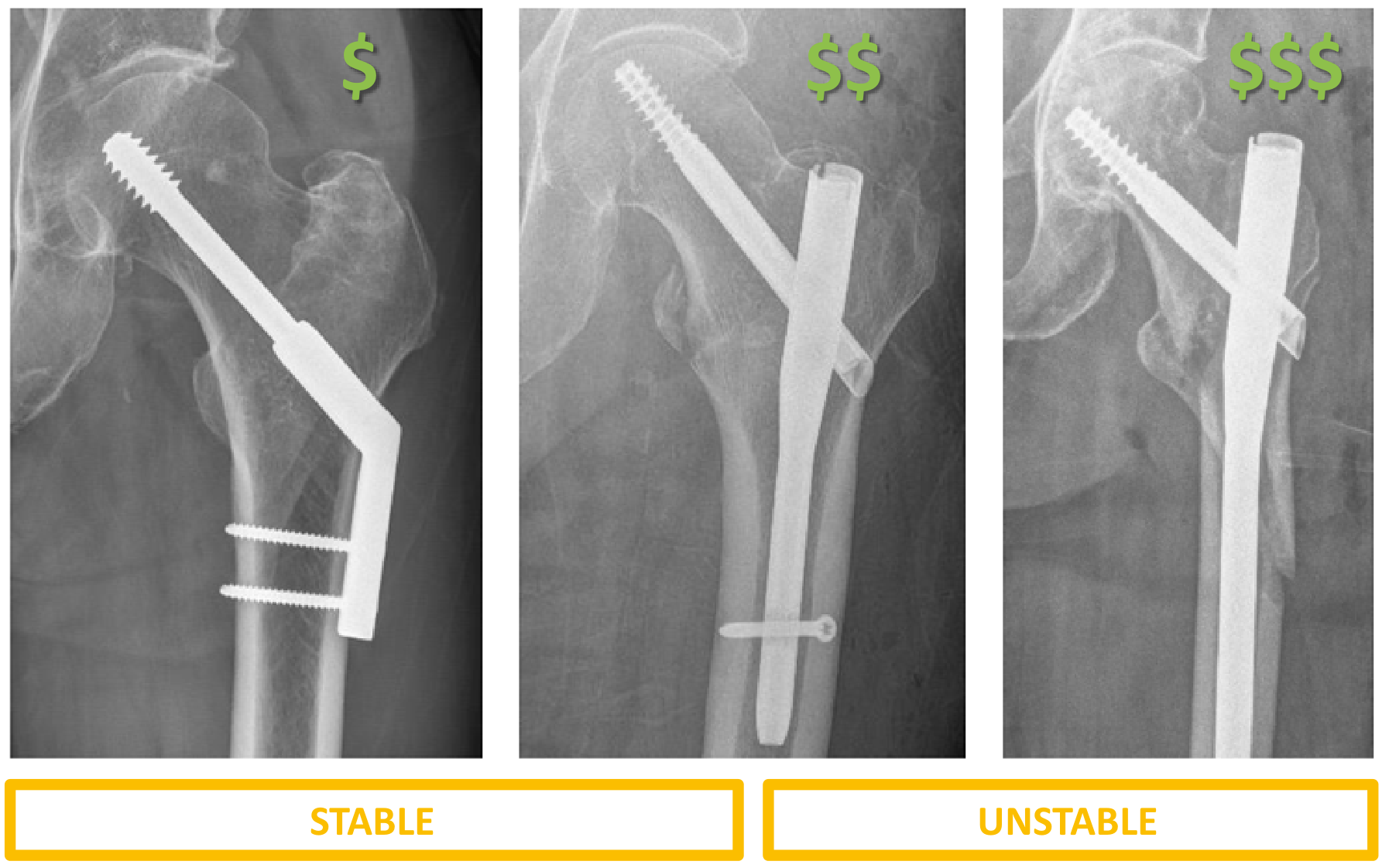
1. Sliding Hip Screw (SHS / Dynamic Hip Screw - DHS)
- Indicated for STABLE fractures (A1) with an intact lateral wall (>20.5 mm)
- A 2-hole side plate is sufficient for stable fractures
- Lag screw must be placed in center-center position in the femoral head
- Tip-Apex Distance (TAD) <25 mm is the key predictor of lowest screw cut-out rate (see diagram below)
- Lower cost than cephalomedullary nails
- Not suitable for unstable fractures - associated with more collapse, limb shortening, and medialization; higher revision rates in transverse/reverse oblique fractures
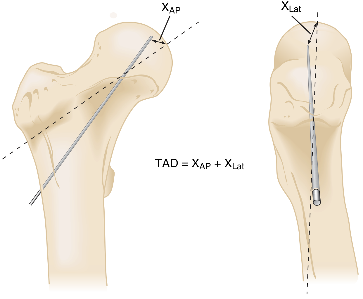
TAD = distance from screw tip to apex of femoral head on AP view + same measurement on lateral view. Target <25 mm.
2. Cephalomedullary (Intramedullary) Nail
- Valid for ALL IT fractures - the preferred device for unstable patterns
- Mandatory for:
- Reverse oblique fractures (A3)
- Subtrochanteric extension
- Fractures with comminution
- Lateral wall deficient/intact <20.5 mm
- Provides an IM buttress effect - less collapse than SHS
- Short nails - for standard oblique fractures (distal interlocking optional)
- Long nails - for standard oblique, reverse oblique, and subtrochanteric fractures
- Single lag screw design: aim for center-center with TAD <25 mm to minimize cut-out
3. Fixed-Angle Devices (95° blade plate / Locking Proximal Femoral Plate)
- Indicated for: reverse oblique fractures, highly comminuted fractures, and nonunion repair
8. Complications
| Complication | Notes |
|---|---|
| Screw cut-out | Most common mechanical failure; strongly predicted by TAD >25 mm; more common with varus malreduction |
| Mechanical failure / nonunion | Much more common in unstable fractures and inadequately reduced fractures; IM implant failure rate lower than with SHS in unstable patterns |
| Implant migration | Lag screw backing out or cutting out superomedially |
| Limb shortening & varus | Especially with SHS in unstable fractures; reduces abductor moment arm causing functional deficit |
| Iatrogenic lateral wall fracture | Risk increases with SHS if lateral wall is <20.5 mm |
| Distal anterior cortical perforation | Risk with IM nails, due to mismatch between anterior femoral bow and nail; also associated with a posterior starting point |
| Infection | More common with nails than plates (but low with current nail designs) |
| DVT/PE | Anticoagulation prophylaxis (LMWH or DOACs) for 7-28 days post-injury/surgery recommended |
| 1-year mortality ~30% | Related to patient age and comorbidities, not directly to fracture type |
9. Quick Summary Table
| Feature | Stable IT Fracture | Unstable IT Fracture |
|---|---|---|
| OTA/AO type | A1 | A2, A3 |
| Lateral wall | Intact (>20.5 mm) | Compromised (<20.5 mm) or absent |
| Implant of choice | SHS (DHS) | Cephalomedullary IM nail |
| TAD goal | <25 mm | <25 mm |
| Collapse risk | Low | High with SHS; reduced with IM nail |
| Cost | Lower (SHS cheapest) | Higher (long nail most expensive) |
Sources: - Rosen's Emergency Medicine, 9th Ed | - Rockwood and Green's Fractures in Adults, 10th Ed (2025) | - Miller's Review of Orthopaedics, 9th Ed | - Campbell's Operative Orthopaedics, 15th Ed (2026)
This is a shared conversation. Sign in to Orris to start your own chat.