Give me all set of image related questions for breastfeeding and breastmilk quiz

Reading File
Finding Sources
Searching Images

breastfeeding positions latch technique
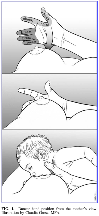
Summary : This illustration demonstrates the "dancer hand position" technique for breastfeeding, as viewed from the mother's perspective. The figure consists of three panels showing the step-by-step hand placement and support for effective latch and feeding.

illustration:
# Scene Overview :
  • The main subject is a mother’s hand supporting her breast and the infant’s head during breastfeeding.
  • The perspective is from the mother’s viewpoint, focusing on the hand, breast, and infant.
  • The illustration is in grayscale, with clear anatomical labeling in the top panel.

# Technical Details :
  • No scale bar or magnification is present.
  • The top panel labels anatomical landmarks: "cheek," "chin," and "breast."
  • The hand is shown with fingers supporting the breast and the thumb positioned near the infant’s cheek and chin.
  • The middle panel shows the hand maintaining support as the infant approaches the breast.
  • The bottom panel depicts the infant latched onto the breast, with the mother’s hand still in the dancer position.

# Spatial Relationships :
  • The hand is consistently placed under the breast, with the thumb and fingers providing support and gentle pressure.
  • The infant’s head is cradled by the mother’s hand, ensuring alignment of the chin and cheek with the breast for optimal latch.
  • The sequence moves from preparation (top), approach (middle), to active feeding (bottom).

# Analysis :
  • The illustration visually instructs on the dancer hand position, emphasizing anatomical alignment and support for successful breastfeeding.
  • The stepwise depiction clarifies the technique, showing how the hand stabilizes both the breast and the infant’s head to facilitate feeding.
  • The labeled anatomical points guide correct finger placement for mothers learning this method.

Summary : This illustration demonstrates the "dancer hand position" technique for breastfeeding, as viewed from the mother's perspective. The figure consists of three panels showing the step-by-step hand placement and support for effective latch and feeding. illustration: # Scene Overview : • The main subject is a mother’s hand supporting her breast and the infant’s head during breastfeeding. • The perspective is from the mother’s viewpoint, focusing on the hand, breast, and infant. • The illustration is in grayscale, with clear anatomical labeling in the top panel. # Technical Details : • No scale bar or magnification is present. • The top panel labels anatomical landmarks: "cheek," "chin," and "breast." • The hand is shown with fingers supporting the breast and the thumb positioned near the infant’s cheek and chin. • The middle panel shows the hand maintaining support as the infant approaches the breast. • The bottom panel depicts the infant latched onto the breast, with the mother’s hand still in the dancer position. # Spatial Relationships : • The hand is consistently placed under the breast, with the thumb and fingers providing support and gentle pressure. • The infant’s head is cradled by the mother’s hand, ensuring alignment of the chin and cheek with the breast for optimal latch. • The sequence moves from preparation (top), approach (middle), to active feeding (bottom). # Analysis : • The illustration visually instructs on the dancer hand position, emphasizing anatomical alignment and support for successful breastfeeding. • The stepwise depiction clarifies the technique, showing how the hand stabilizes both the breast and the infant’s head to facilitate feeding. • The labeled anatomical points guide correct finger placement for mothers learning this method.
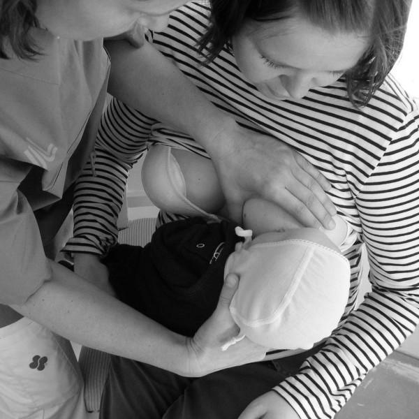
This clinical photograph captures a hands-on teaching interaction between a nurse and a mother within a neonatal or maternity care setting. The image demonstrates a breastfeeding support technique where the nurse is actively involved in positioning the infant for latching. The nurse, wearing professional medical scrubs, is positioned on the mother's left side. Her right hand is shown supporting the infant’s head and neck to maintain alignment, while her left hand is used to stabilize the mother’s breast to facilitate a proper latch. The mother is seated, wearing a striped shirt, and holds the infant close to her chest, focusing on the baby. The infant is wearing a bonnet and is swaddled, a common practice for maintaining warmth in neonatal care units. This visual illustrates the 'hands-on' approach to breastfeeding education, which is a clinical concept used to demonstrate physical positioning and latching techniques for new mothers, particularly in the context of preterm or sick infants requiring specialized support.

This clinical photograph captures a hands-on teaching interaction between a nurse and a mother within a neonatal or maternity care setting. The image demonstrates a breastfeeding support technique where the nurse is actively involved in positioning the infant for latching. The nurse, wearing professional medical scrubs, is positioned on the mother's left side. Her right hand is shown supporting the infant’s head and neck to maintain alignment, while her left hand is used to stabilize the mother’s breast to facilitate a proper latch. The mother is seated, wearing a striped shirt, and holds the infant close to her chest, focusing on the baby. The infant is wearing a bonnet and is swaddled, a common practice for maintaining warmth in neonatal care units. This visual illustrates the 'hands-on' approach to breastfeeding education, which is a clinical concept used to demonstrate physical positioning and latching techniques for new mothers, particularly in the context of preterm or sick infants requiring specialized support.

A black-and-white clinical photograph documenting an infant breastfeeding, serving as an educational example of maternal-child health and nutrition in global health settings. The image illustrates the 'cradle hold' positioning, with the infant's face turned toward the breast and a deep latch visible. The adult provides manual support to the infant's occiput to facilitate stable positioning during feeding. Notable cultural markers include stacked brass neck rings and wrist coils on the adult, which are characteristic of specific ethnic groups in Southeast Asia. This visual emphasizes the clinical significance of breastfeeding as a critical intervention for infant survival and immune support, particularly in resource-limited or emergency environments like refugee camps. Key educational concepts include lactation management, infant latch technique, and the sociocultural context of neonatal care.
A black-and-white clinical photograph documenting an infant breastfeeding, serving as an educational example of maternal-child health and nutrition in global health settings. The image illustrates the 'cradle hold' positioning, with the infant's face turned toward the breast and a deep latch visible. The adult provides manual support to the infant's occiput to facilitate stable positioning during feeding. Notable cultural markers include stacked brass neck rings and wrist coils on the adult, which are characteristic of specific ethnic groups in Southeast Asia. This visual emphasizes the clinical significance of breastfeeding as a critical intervention for infant survival and immune support, particularly in resource-limited or emergency environments like refugee camps. Key educational concepts include lactation management, infant latch technique, and the sociocultural context of neonatal care.

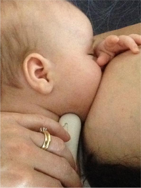
A clinical photograph depicting an infant latched onto a mother's breast during breastfeeding. The image provides a close-up, lateral view of the infant's head and neck in relation to the breast tissue. The infant is positioned with the chin pressed firmly into the breast, demonstrating an asymmetrical latch. The infant's upper body is oriented horizontally, and the nose appears clear of the breast tissue. A caregiver's hand is visible at the bottom of the frame, providing support to the infant's neck and base of the skull to maintain positioning. The photograph illustrates post-intervention breastfeeding mechanics following a 'gestalt' clinical approach, which focuses on optimizing maternal-infant positioning and attachment to address issues like nipple pain and poor milk transfer. The educational focus is on the visual assessment of latch quality, specifically the deep attachment and chin-breast contact required for effective suckling and maternal comfort.
A clinical photograph depicting an infant latched onto a mother's breast during breastfeeding. The image provides a close-up, lateral view of the infant's head and neck in relation to the breast tissue. The infant is positioned with the chin pressed firmly into the breast, demonstrating an asymmetrical latch. The infant's upper body is oriented horizontally, and the nose appears clear of the breast tissue. A caregiver's hand is visible at the bottom of the frame, providing support to the infant's neck and base of the skull to maintain positioning. The photograph illustrates post-intervention breastfeeding mechanics following a 'gestalt' clinical approach, which focuses on optimizing maternal-infant positioning and attachment to address issues like nipple pain and poor milk transfer. The educational focus is on the visual assessment of latch quality, specifically the deep attachment and chin-breast contact required for effective suckling and maternal comfort.

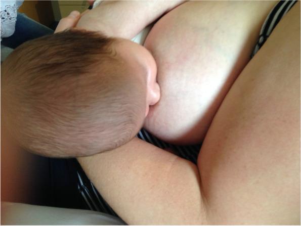
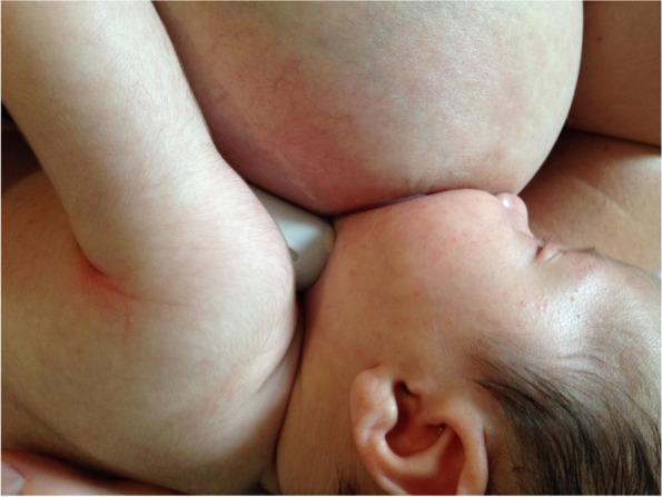
This clinical photograph demonstrates an infant breastfeeding, specifically illustrating an optimized latch following a gestalt intervention. The infant is positioned in close physical contact with the mother, demonstrating effective positioning and attachment. The infant's mouth is wide open and deeply latched onto the maternal breast, covering a significant portion of the areola. The infant's nose is in close proximity to the breast, and the chin is tucked into the breast tissue, supporting a stable, asymmetrical latch. The infant is supported by the mother's arm, ensuring a belly-to-belly orientation that facilitates proper alignment of the head, neck, and spine. This image serves as a clinical example of breastfeeding mechanics and positional stability, relevant for lactation consultancy and pediatric primary care. It highlights the visible contact points and body alignment necessary to maximize intra-oral breast tissue volume and potentially reduce maternal nipple pain associated with breastfeeding difficulties like tongue-tie or shallow latch.
This clinical photograph demonstrates an infant breastfeeding, specifically illustrating an optimized latch following a gestalt intervention. The infant is positioned in close physical contact with the mother, demonstrating effective positioning and attachment. The infant's mouth is wide open and deeply latched onto the maternal breast, covering a significant portion of the areola. The infant's nose is in close proximity to the breast, and the chin is tucked into the breast tissue, supporting a stable, asymmetrical latch. The infant is supported by the mother's arm, ensuring a belly-to-belly orientation that facilitates proper alignment of the head, neck, and spine. This image serves as a clinical example of breastfeeding mechanics and positional stability, relevant for lactation consultancy and pediatric primary care. It highlights the visible contact points and body alignment necessary to maximize intra-oral breast tissue volume and potentially reduce maternal nipple pain associated with breastfeeding difficulties like tongue-tie or shallow latch.

Clinical photograph demonstrating an incorrect, shallow breastfeeding latch in an infant. The image shows the infant's mouth attached to the breast with a narrow angle of opening, rather than a wide-gaped seal. A significant portion of the maternal areola is visible above the infant's upper lip, while very little areolar tissue is captured below the lower lip, indicating that the infant is primarily grasping the nipple rather than the underlying breast tissue. This positioning is characteristic of a 'shallow hold,' which can lead to nipple trauma for the mother and inefficient milk transfer for the infant. The photograph serves as an educational tool in lactation consultancy and pediatrics to distinguish between suboptimal and optimal latch mechanics and their impact on craniofacial development and breastfeeding success.
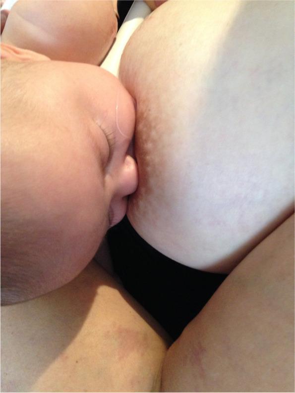
Clinical photograph demonstrating an incorrect, shallow breastfeeding latch in an infant. The image shows the infant's mouth attached to the breast with a narrow angle of opening, rather than a wide-gaped seal. A significant portion of the maternal areola is visible above the infant's upper lip, while very little areolar tissue is captured below the lower lip, indicating that the infant is primarily grasping the nipple rather than the underlying breast tissue. This positioning is characteristic of a 'shallow hold,' which can lead to nipple trauma for the mother and inefficient milk transfer for the infant. The photograph serves as an educational tool in lactation consultancy and pediatrics to distinguish between suboptimal and optimal latch mechanics and their impact on craniofacial development and breastfeeding success.

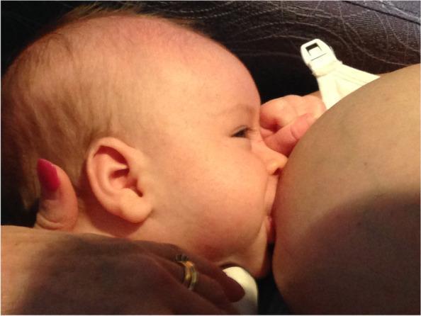
A clinical photograph depicting an infant latched onto a mother's breast, illustrating a post-gestalt intervention breastfeeding position. The image provides a lateral view of the infant's face in close proximity to the breast tissue. The infant's mouth is wide open and fully sealed around the nipple and a significant portion of the lower areola, demonstrating an asymmetrical, deep latch. The infant's nose is positioned lightly against the breast, and the chin is tucked firmly into the breast tissue. The cheeks appear rounded without visible dimpling, suggesting effective intra-oral vacuum and stability. The areola shows slight stretching and distortion consistent with active breastfeeding. This visual serves as an educational example of optimal breastfeeding mechanics and positioning designed to maximize milk transfer and minimize maternal nipple pain by increasing intra-oral breast tissue volume.
A clinical photograph depicting an infant latched onto a mother's breast, illustrating a post-gestalt intervention breastfeeding position. The image provides a lateral view of the infant's face in close proximity to the breast tissue. The infant's mouth is wide open and fully sealed around the nipple and a significant portion of the lower areola, demonstrating an asymmetrical, deep latch. The infant's nose is positioned lightly against the breast, and the chin is tucked firmly into the breast tissue. The cheeks appear rounded without visible dimpling, suggesting effective intra-oral vacuum and stability. The areola shows slight stretching and distortion consistent with active breastfeeding. This visual serves as an educational example of optimal breastfeeding mechanics and positioning designed to maximize milk transfer and minimize maternal nipple pain by increasing intra-oral breast tissue volume.

Clinical photograph of a 4-week-old infant breastfeeding using the 'gestalt' method, emphasizing optimal latching mechanics and positional stability. The image shows the infant in a cross-cradle or similar hold, with the head tilted slightly upward (asymmetric latch). The infant's mouth is wide open and deeply attached to the breast, though the nipple itself is not visible. Notably, the infant's nose is in close contact with the breast tissue, and the chin is tucked firmly into the breast. A white nipple shield is partially visible at the point of attachment. The infant's skin is fair with fine vellus hair, and the scalp has dark hair. The maternal breast appears full and rounded, with visible skin texture and faint vascular patterns. This image demonstrates post-intervention positioning intended to address nipple pain and infant fussiness associated with latch difficulties or oral connective tissue restrictions.
Clinical photograph of a 4-week-old infant breastfeeding using the 'gestalt' method, emphasizing optimal latching mechanics and positional stability. The image shows the infant in a cross-cradle or similar hold, with the head tilted slightly upward (asymmetric latch). The infant's mouth is wide open and deeply attached to the breast, though the nipple itself is not visible. Notably, the infant's nose is in close contact with the breast tissue, and the chin is tucked firmly into the breast. A white nipple shield is partially visible at the point of attachment. The infant's skin is fair with fine vellus hair, and the scalp has dark hair. The maternal breast appears full and rounded, with visible skin texture and faint vascular patterns. This image demonstrates post-intervention positioning intended to address nipple pain and infant fussiness associated with latch difficulties or oral connective tissue restrictions.

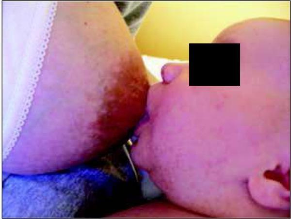
A clinical photograph depicting an 8-week-old infant breastfeeding, labeled as a 'pre-gestalt intervention' state. The image illustrates the physical dynamics of infant latching and maternal positioning. The infant is shown in a lateral profile with a relatively shallow latch; the mouth is open but primarily encompasses the nipple and only a minimal portion of the areola. The infant's chin is pressed against the breast tissue, while the nose remains close to the breast surface but clear for respiration. There is visible drawing of the breast tissue into the oral cavity. The mother's breast appears full, and her hand is visible supporting the infant's neck/head. A breastfeeding bra with a white plastic clasp is visible in the background. This clinical image is used in a medical educational context to demonstrate latch characteristics associated with maternal nipple pain and difficult attachment prior to corrective lactation support or positional interventions.
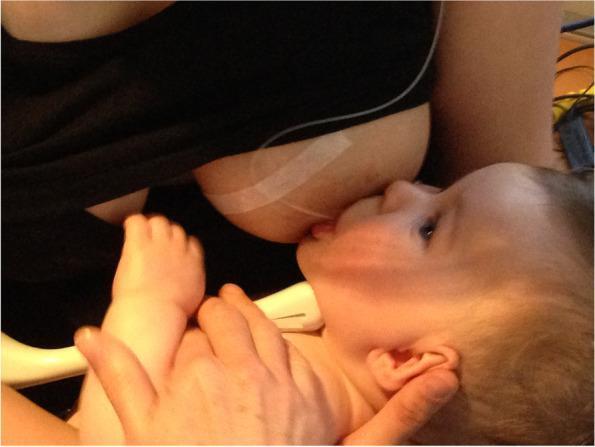
A clinical photograph depicting an 8-week-old infant breastfeeding, labeled as a 'pre-gestalt intervention' state. The image illustrates the physical dynamics of infant latching and maternal positioning. The infant is shown in a lateral profile with a relatively shallow latch; the mouth is open but primarily encompasses the nipple and only a minimal portion of the areola. The infant's chin is pressed against the breast tissue, while the nose remains close to the breast surface but clear for respiration. There is visible drawing of the breast tissue into the oral cavity. The mother's breast appears full, and her hand is visible supporting the infant's neck/head. A breastfeeding bra with a white plastic clasp is visible in the background. This clinical image is used in a medical educational context to demonstrate latch characteristics associated with maternal nipple pain and difficult attachment prior to corrective lactation support or positional interventions.

This clinical photograph captures a lateral view of an infant attempting to latch onto the maternal breast during a breastfeeding assessment. The infant is positioned in a supine or slightly reclined posture, supported by a caregiver's hand near the base of the skull and neck. The infant's mouth is partially open, engaging with the nipple and areola, with visible head extension and an upward gaze. A distinctive white adhesive marker or tape is applied to the superior aspect of the breast, serving as a landmark for simultaneous ultrasound imaging as indicated by the presence of an ultrasound transducer positioned submentally against the infant's jawline. The breast tissue appears moderate in size with normal skin pigmentation, though the nipple is elongated within the latch. This image serves as a procedural reference for evaluating breastfeeding mechanics, particularly the 'fit and hold' and intra-oral breast tissue volume before clinical intervention in a case involving maternal nipple pain and post-frenotomy complications.

This clinical photograph captures a lateral view of an infant attempting to latch onto the maternal breast during a breastfeeding assessment. The infant is positioned in a supine or slightly reclined posture, supported by a caregiver's hand near the base of the skull and neck. The infant's mouth is partially open, engaging with the nipple and areola, with visible head extension and an upward gaze. A distinctive white adhesive marker or tape is applied to the superior aspect of the breast, serving as a landmark for simultaneous ultrasound imaging as indicated by the presence of an ultrasound transducer positioned submentally against the infant's jawline. The breast tissue appears moderate in size with normal skin pigmentation, though the nipple is elongated within the latch. This image serves as a procedural reference for evaluating breastfeeding mechanics, particularly the 'fit and hold' and intra-oral breast tissue volume before clinical intervention in a case involving maternal nipple pain and post-frenotomy complications.

Searching Images

breast milk composition colostrum foremilk hindmilk
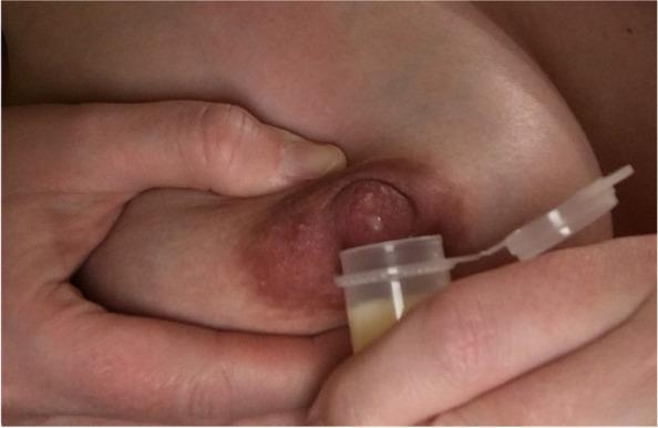
A close-up clinical photograph demonstrating the Marmet technique for hand-expressing antenatal milk or colostrum. The image shows a person's hands positioned on a lactating breast. One hand supports the breast tissue, with the thumb and index finger placed approximately 2-3 cm behind the nipple at the edge of the areola, applying rhythmic compression to the underlying milk ducts. The areola appears physiologically hyperpigmented and slightly engorged, typical of late pregnancy or the early postpartum period. A small, clear, flip-top collection vial is held directly beneath the nipple to capture the expressed fluid. The vial contains a small volume of yellowish, viscous fluid, characteristic of colostrum. This visual serves as an educational guide for lactation support, specifically teaching manual expression and collection techniques for individuals in the late third trimester (antenatal milk expression) or early postpartum phase.
A close-up clinical photograph demonstrating the Marmet technique for hand-expressing antenatal milk or colostrum. The image shows a person's hands positioned on a lactating breast. One hand supports the breast tissue, with the thumb and index finger placed approximately 2-3 cm behind the nipple at the edge of the areola, applying rhythmic compression to the underlying milk ducts. The areola appears physiologically hyperpigmented and slightly engorged, typical of late pregnancy or the early postpartum period. A small, clear, flip-top collection vial is held directly beneath the nipple to capture the expressed fluid. The vial contains a small volume of yellowish, viscous fluid, characteristic of colostrum. This visual serves as an educational guide for lactation support, specifically teaching manual expression and collection techniques for individuals in the late third trimester (antenatal milk expression) or early postpartum phase.

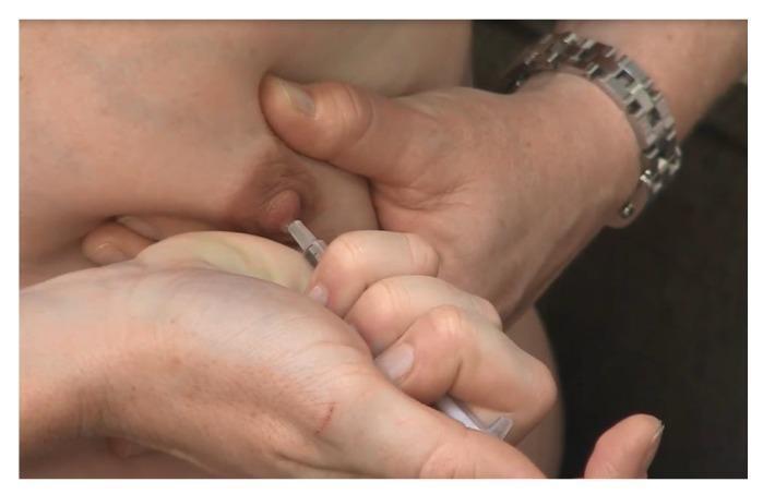
This clinical photograph captures a close-up procedural demonstration of Antenatal Colostrum Expression (ACE). The image illustrates the 'C-hold' hand technique, where the breast tissue is compressed between the thumb and fingers, positioned approximately 2-3 cm behind the nipple at the edge of the areola. A second hand is shown holding a small oral syringe directly at the nipple tip to collect the expressed droplets of colostrum. The procedural focus is on the manual stimulation and compression of the lactiferous sinuses to facilitate milk removal. This visual serves as an educational tool for lactation consultants and expectant mothers, highlighting the correct hand placement and the use of a syringe for hygienic colostrum harvesting and storage during late pregnancy. The setting is likely an antenatal clinic or educational session designed to improve breastfeeding confidence and knowledge.
This clinical photograph captures a close-up procedural demonstration of Antenatal Colostrum Expression (ACE). The image illustrates the 'C-hold' hand technique, where the breast tissue is compressed between the thumb and fingers, positioned approximately 2-3 cm behind the nipple at the edge of the areola. A second hand is shown holding a small oral syringe directly at the nipple tip to collect the expressed droplets of colostrum. The procedural focus is on the manual stimulation and compression of the lactiferous sinuses to facilitate milk removal. This visual serves as an educational tool for lactation consultants and expectant mothers, highlighting the correct hand placement and the use of a syringe for hygienic colostrum harvesting and storage during late pregnancy. The setting is likely an antenatal clinic or educational session designed to improve breastfeeding confidence and knowledge.

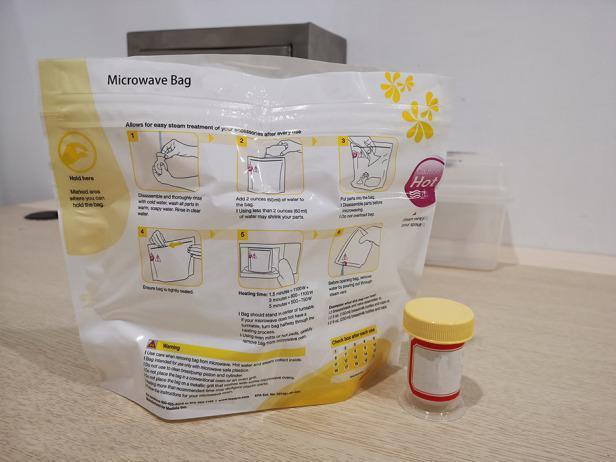
This clinical photograph displays a medical-grade disinfection system used for neonatal care accessories, specifically for the steam sterilization of breast milk collection equipment. The image features a specialized microwave steam sterilization bag alongside a sterile collection cup. The bag is a flexible, translucent white pouch with yellow instructional graphics and a 'Hold here' safety zone. It displays a six-step illustrated guide for cleaning, adding water (60ml), and microwave heating times based on wattage. A tracking grid at the bottom is included to monitor the bag's 20-use lifecycle. Next to the bag is a cylindrical, transparent collection cup with a yellow screw-top lid and a red-bordered label for patient identification. This visual represents standard protocols for infection control in a Neonatal Intensive Care Unit (NICU) or home care setting, demonstrating the process of ensuring the microbiological safety of colostrum and breast milk storage containers through rapid steam treatment.
This clinical photograph displays a medical-grade disinfection system used for neonatal care accessories, specifically for the steam sterilization of breast milk collection equipment. The image features a specialized microwave steam sterilization bag alongside a sterile collection cup. The bag is a flexible, translucent white pouch with yellow instructional graphics and a 'Hold here' safety zone. It displays a six-step illustrated guide for cleaning, adding water (60ml), and microwave heating times based on wattage. A tracking grid at the bottom is included to monitor the bag's 20-use lifecycle. Next to the bag is a cylindrical, transparent collection cup with a yellow screw-top lid and a red-bordered label for patient identification. This visual represents standard protocols for infection control in a Neonatal Intensive Care Unit (NICU) or home care setting, demonstrating the process of ensuring the microbiological safety of colostrum and breast milk storage containers through rapid steam treatment.

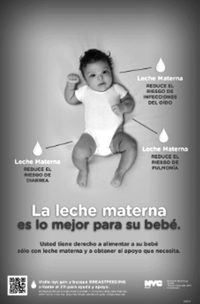
Summary : This is a public health promotional poster advocating breastfeeding, highlighting the health benefits of breast milk for infants, with supporting text and official branding.

photo:
Scene Overview :
  • Main subject: An infant lying on a light background, wearing a white onesie.
  • Perspective: Overhead view, centered composition.
  • Colour palette: Grayscale (black and white).
  • Lighting: Even, soft, no harsh shadows.
  • On-image text: Multiple Spanish-language health messages and official logos.

Technical Details :
  • No scale bar or magnification.
  • Three labeled droplet icons point to the infant, each with a health benefit:
    • "Leche Materna REDUCE EL RIESGO DE DIARREA"
    • "Leche Materna REDUCE EL RIESGO DE INFECCIONES DEL OÍDO"
    • "Leche Materna REDUCE EL RIESGO DE PULMONÍA"
  • Central bold text: "La leche materna es lo mejor para su bebé."
  • Additional text: "Usted tiene derecho a alimentar a su bebé sólo con leche materna y a obtener el apoyo que necesita."
  • QR code and website link at the bottom for more information.
  • NYC Health Department logo and contact details in the lower right.

Spatial Relationships :
  • The infant is centrally placed, with health benefit icons and text radiating outward.
  • Main message is directly below the infant, occupying the lower third of the poster.
  • Official branding and QR code are at the bottom edge.

Analysis :
  • The poster visually and textually emphasizes the health advantages of breastfeeding, specifically reducing risks of diarrhea, ear infections, and pneumonia.
  • The layout is designed to draw attention to the infant and the associated health benefits, reinforcing the message that breast milk is the optimal choice for babies.
  • Official branding and contact information provide credibility and resources for further support.
Summary : This is a public health promotional poster advocating breastfeeding, highlighting the health benefits of breast milk for infants, with supporting text and official branding. photo: Scene Overview : • Main subject: An infant lying on a light background, wearing a white onesie. • Perspective: Overhead view, centered composition. • Colour palette: Grayscale (black and white). • Lighting: Even, soft, no harsh shadows. • On-image text: Multiple Spanish-language health messages and official logos. Technical Details : • No scale bar or magnification. • Three labeled droplet icons point to the infant, each with a health benefit: • "Leche Materna REDUCE EL RIESGO DE DIARREA" • "Leche Materna REDUCE EL RIESGO DE INFECCIONES DEL OÍDO" • "Leche Materna REDUCE EL RIESGO DE PULMONÍA" • Central bold text: "La leche materna es lo mejor para su bebé." • Additional text: "Usted tiene derecho a alimentar a su bebé sólo con leche materna y a obtener el apoyo que necesita." • QR code and website link at the bottom for more information. • NYC Health Department logo and contact details in the lower right. Spatial Relationships : • The infant is centrally placed, with health benefit icons and text radiating outward. • Main message is directly below the infant, occupying the lower third of the poster. • Official branding and QR code are at the bottom edge. Analysis : • The poster visually and textually emphasizes the health advantages of breastfeeding, specifically reducing risks of diarrhea, ear infections, and pneumonia. • The layout is designed to draw attention to the infant and the associated health benefits, reinforcing the message that breast milk is the optimal choice for babies. • Official branding and contact information provide credibility and resources for further support.

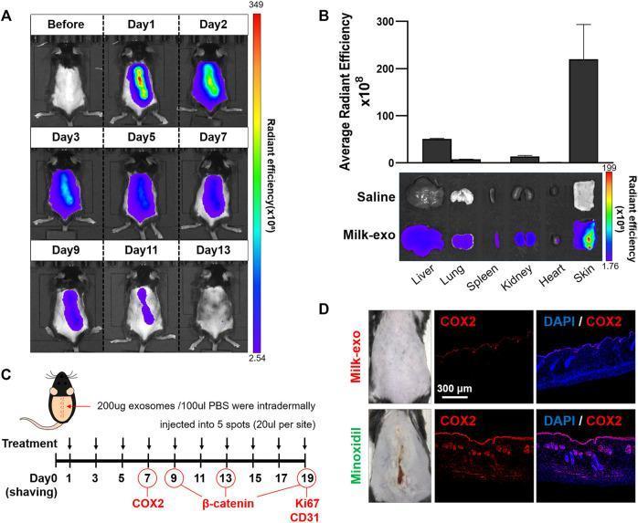
This composite image illustrates the in vivo and ex vivo biodistribution and inflammatory profile of colostrum-derived milk exosomes (Milk-exo) in a murine model. Panel A presents a longitudinal series of in vivo fluorescence imaging frames showing Cy5.5-NHS labeled exosomes over 13 days. Initially, a high-intensity signal (red/yellow radiant efficiency) is localized at the intradermal injection site, which progressively dissipates and shifts to lower intensity (blue/purple) until complete clearance by Day 13. Panel B includes a bar graph and corresponding ex vivo organ imaging comparing Saline and Milk-exo groups at 2 days post-injection. The Milk-exo group shows significantly higher radiant efficiency, predominantly localized in the skin, with secondary distribution to the liver, lungs, and kidneys. Panel C displays the experimental timeline and injection protocol. Panel D provides clinical photographs and immunofluorescence histology sections of mouse skin treated with Milk-exo vs. Minoxidil. The staining compares COX2 expression (red) with DAPI nuclear counterstaining (blue), demonstrating that Milk-exo treatment maintains low COX2 expression compared to the high inflammatory response observed in the Minoxidil group.
This composite image illustrates the in vivo and ex vivo biodistribution and inflammatory profile of colostrum-derived milk exosomes (Milk-exo) in a murine model. Panel A presents a longitudinal series of in vivo fluorescence imaging frames showing Cy5.5-NHS labeled exosomes over 13 days. Initially, a high-intensity signal (red/yellow radiant efficiency) is localized at the intradermal injection site, which progressively dissipates and shifts to lower intensity (blue/purple) until complete clearance by Day 13. Panel B includes a bar graph and corresponding ex vivo organ imaging comparing Saline and Milk-exo groups at 2 days post-injection. The Milk-exo group shows significantly higher radiant efficiency, predominantly localized in the skin, with secondary distribution to the liver, lungs, and kidneys. Panel C displays the experimental timeline and injection protocol. Panel D provides clinical photographs and immunofluorescence histology sections of mouse skin treated with Milk-exo vs. Minoxidil. The staining compares COX2 expression (red) with DAPI nuclear counterstaining (blue), demonstrating that Milk-exo treatment maintains low COX2 expression compared to the high inflammatory response observed in the Minoxidil group.

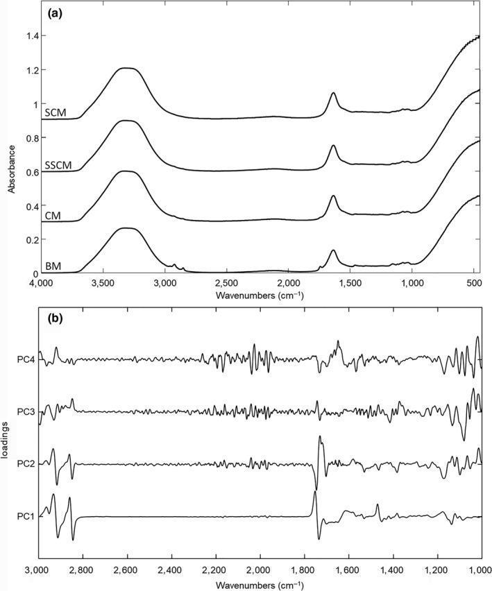
Educational analytical chart displaying spectroscopic data for the evaluation of human breast milk (BM) purity. Panel (a) illustrates Fourier-Transform Infrared (FTIR) spectra comparing pure BM with whole cow milk (CM), semi-skimmed cow milk (SSCM), and skimmed cow milk (SCM). The x-axis represents wavenumbers (cm⁻¹), and the y-axis shows absorbance. Key peaks include the 3,300-3,500 cm⁻¹ range (H2O stretching), 2,800-3,000 cm⁻¹ (lipid CH2 stretching), and ~1,743 cm⁻¹ (lipid C=O stretching), alongside protein-related bands near 1,650 cm⁻¹ (Amide I) and 1,550 cm⁻¹ (Amide II). Panel (b) presents a Principal Component Analysis (PCA) loadings plot for PCs 1 through 4, identifying the spectral regions most responsible for variance among samples. Significant loading peaks and troughs are observed in the protein and lipid-specific regions (1,000–3,000 cm⁻¹). This visualization is used in neonatal nutrition and food science to demonstrate methods for detecting breast milk adulteration and verifying macronutrient composition.
Educational analytical chart displaying spectroscopic data for the evaluation of human breast milk (BM) purity. Panel (a) illustrates Fourier-Transform Infrared (FTIR) spectra comparing pure BM with whole cow milk (CM), semi-skimmed cow milk (SSCM), and skimmed cow milk (SCM). The x-axis represents wavenumbers (cm⁻¹), and the y-axis shows absorbance. Key peaks include the 3,300-3,500 cm⁻¹ range (H2O stretching), 2,800-3,000 cm⁻¹ (lipid CH2 stretching), and ~1,743 cm⁻¹ (lipid C=O stretching), alongside protein-related bands near 1,650 cm⁻¹ (Amide I) and 1,550 cm⁻¹ (Amide II). Panel (b) presents a Principal Component Analysis (PCA) loadings plot for PCs 1 through 4, identifying the spectral regions most responsible for variance among samples. Significant loading peaks and troughs are observed in the protein and lipid-specific regions (1,000–3,000 cm⁻¹). This visualization is used in neonatal nutrition and food science to demonstrate methods for detecting breast milk adulteration and verifying macronutrient composition.

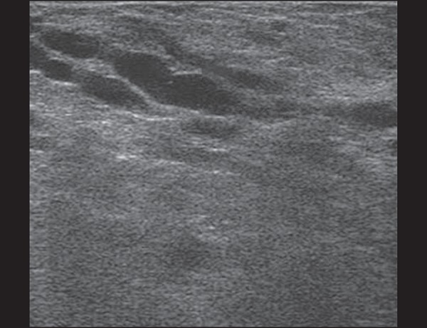
**Imaging Modality:** B-mode grayscale ultrasound.

**Anatomical Region:** Female breast tissue.

**Observed Findings:**
*   **Parenchyma:** The breast tissue demonstrates a diffusely hyperechoic and heterogeneous echo texture, characteristic of physiologic changes in the lactating breast (lactational adenosis).
*   **Ductal Structures:** Multiple dilated, prominent, and branching tubular structures are visible in the subareolar and mid-parenchymal regions. These represent dilated lactiferous ducts.
*   **Internal Echoes:** The dilated ducts contain anechoic to low-level internal echoes, consistent with the accumulation of milk or colostrum.
*   **Tissue Architecture:** There is increased vascularity and glandular prominence with a decrease in visible subcutaneous and retromammary fat, reflecting the functional state of lactation.

**Diagnostic Features:** Diffuse hyperechogenicity of the glandular elements combined with prominent ductal dilatation (ductectasia) in a clinical context of lactation. No discrete solid masses or suspicious architectural distortions are visualized in this section.

**Clinical Significance:** This appearance is a normal physiologic variation of the breast during pregnancy and the postpartum period.
**Imaging Modality:** B-mode grayscale ultrasound. **Anatomical Region:** Female breast tissue. **Observed Findings:** * **Parenchyma:** The breast tissue demonstrates a diffusely hyperechoic and heterogeneous echo texture, characteristic of physiologic changes in the lactating breast (lactational adenosis). * **Ductal Structures:** Multiple dilated, prominent, and branching tubular structures are visible in the subareolar and mid-parenchymal regions. These represent dilated lactiferous ducts. * **Internal Echoes:** The dilated ducts contain anechoic to low-level internal echoes, consistent with the accumulation of milk or colostrum. * **Tissue Architecture:** There is increased vascularity and glandular prominence with a decrease in visible subcutaneous and retromammary fat, reflecting the functional state of lactation. **Diagnostic Features:** Diffuse hyperechogenicity of the glandular elements combined with prominent ductal dilatation (ductectasia) in a clinical context of lactation. No discrete solid masses or suspicious architectural distortions are visualized in this section. **Clinical Significance:** This appearance is a normal physiologic variation of the breast during pregnancy and the postpartum period.

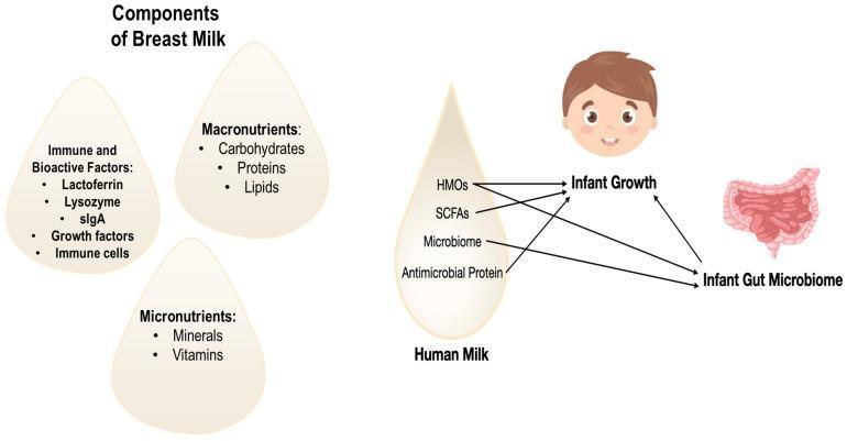
An educational infographic and signaling diagram illustrating the components of human breast milk and their physiological impact on neonatal development. The left side categorizes 'Components of Breast Milk' into three milk-droplet-shaped panels: 'Immune and Bioactive Factors' (including Lactoferrin, Lysozyme, sIgA, growth factors, and immune cells), 'Macronutrients' (Carbohydrates, Proteins, Lipids), and 'Micronutrients' (Minerals, Vitamins). The right side features a flow diagram detailing how specific human milk constituents influence clinical outcomes. Human Milk Oligosaccharides (HMOs), Short-Chain Fatty Acids (SCFAs), and Antimicrobial Proteins are shown to directly impact both 'Infant Growth' and the 'Infant Gut Microbiome.' The 'Microbiome' component within the milk is depicted as specifically influencing the infant's gut microbiome. The diagram highlights the bidirectional relationship between the developing gut microbiome and general infant growth, emphasizing the role of maternal milk in shaping metabolic and immunological health during early development.

An educational infographic and signaling diagram illustrating the components of human breast milk and their physiological impact on neonatal development. The left side categorizes 'Components of Breast Milk' into three milk-droplet-shaped panels: 'Immune and Bioactive Factors' (including Lactoferrin, Lysozyme, sIgA, growth factors, and immune cells), 'Macronutrients' (Carbohydrates, Proteins, Lipids), and 'Micronutrients' (Minerals, Vitamins). The right side features a flow diagram detailing how specific human milk constituents influence clinical outcomes. Human Milk Oligosaccharides (HMOs), Short-Chain Fatty Acids (SCFAs), and Antimicrobial Proteins are shown to directly impact both 'Infant Growth' and the 'Infant Gut Microbiome.' The 'Microbiome' component within the milk is depicted as specifically influencing the infant's gut microbiome. The diagram highlights the bidirectional relationship between the developing gut microbiome and general infant growth, emphasizing the role of maternal milk in shaping metabolic and immunological health during early development.

Searching Images

breastfeeding anatomy mammary gland milk production
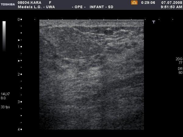
This diagnostic ultrasound image demonstrates the anatomy of a human lactating breast in the 12 o'clock position following breastfeeding. The imaging captures the superficial skin and subcutaneous layers overlying the mammary parenchyma. The skin appears as a thin, hyperechoic line at the superior margin, maintaining uniform thickness. The underlying glandular tissue exhibits a generally homogeneous echotexture with a notable reduction in thickness and overall echogenicity post-feed. Several localized hypoechoic (darker) regions are visible within the parenchyma, particularly in the upper right quadrant, representing fluid-filled structures or small milk ducts. The image provides clinical insight into the physiological changes of the mammary gland during lactation, specifically illustrating how milk removal affects the density and visual appearance of glandular tissue on high-resolution ultrasound. No pathological skin thickening or significant masses are observed.
This diagnostic ultrasound image demonstrates the anatomy of a human lactating breast in the 12 o'clock position following breastfeeding. The imaging captures the superficial skin and subcutaneous layers overlying the mammary parenchyma. The skin appears as a thin, hyperechoic line at the superior margin, maintaining uniform thickness. The underlying glandular tissue exhibits a generally homogeneous echotexture with a notable reduction in thickness and overall echogenicity post-feed. Several localized hypoechoic (darker) regions are visible within the parenchyma, particularly in the upper right quadrant, representing fluid-filled structures or small milk ducts. The image provides clinical insight into the physiological changes of the mammary gland during lactation, specifically illustrating how milk removal affects the density and visual appearance of glandular tissue on high-resolution ultrasound. No pathological skin thickening or significant masses are observed.

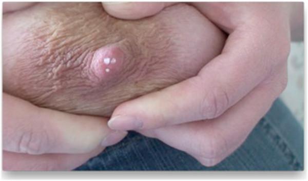
This clinical photograph shows a close-up view of the nipple-areola complex (NAC) during manual milk expression. The central focus is the nipple, a raised pinkish papilla mammaria, which exhibits several small, discrete white droplets of breast milk at the ductal orifices. The surrounding areola mammae displays a light tan or pinkish pigmentation with a textured, wrinkled surface marked by skin folds and creases. Fingers are visible at the upper and lower margins of the image, demonstrating the technique of manual compression to elicit milk flow. The image serves as an educational tool for identifying the functional anatomy of the mammary gland, specifically the location and distribution of milk ducts that transport milk to the surface during lactation. It illustrates clinical signs associated with breastfeeding and the physiological process of lactation in a female patient.
This clinical photograph shows a close-up view of the nipple-areola complex (NAC) during manual milk expression. The central focus is the nipple, a raised pinkish papilla mammaria, which exhibits several small, discrete white droplets of breast milk at the ductal orifices. The surrounding areola mammae displays a light tan or pinkish pigmentation with a textured, wrinkled surface marked by skin folds and creases. Fingers are visible at the upper and lower margins of the image, demonstrating the technique of manual compression to elicit milk flow. The image serves as an educational tool for identifying the functional anatomy of the mammary gland, specifically the location and distribution of milk ducts that transport milk to the surface during lactation. It illustrates clinical signs associated with breastfeeding and the physiological process of lactation in a female patient.

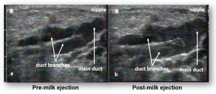
This diagnostic image consists of two side-by-side ultrasound frames (a and b) illustrating the functional anatomy of the lactating human breast during the milk ejection reflex. The imaging modality is B-mode ultrasound, used to visualize the mammary ductal system in real-time. In both images, the milk ducts are identified as prominent anechoic (black) tubular structures, providing high contrast against the surrounding hyperechoic, granular connective and adipose tissue. Figure (a), labeled 'Pre-milk ejection', shows the main duct and duct branches in a relatively narrow state. Figure (b), labeled 'Post-milk ejection', demonstrates significant ductal expansion and distention due to the presence of breast milk following the ejection reflex. Annotated labels indicate the 'nipple' position at the top right, with arrows highlighting the 'main duct' and 'duct branches'. This comparison serves as educational material to demonstrate that mammary ducts function primarily as transport vessels that dilate during milk flow rather than serving as large static storage reservoirs, contributing to contemporary understanding of breastfeeding physiology and mammary gland function.
This diagnostic image consists of two side-by-side ultrasound frames (a and b) illustrating the functional anatomy of the lactating human breast during the milk ejection reflex. The imaging modality is B-mode ultrasound, used to visualize the mammary ductal system in real-time. In both images, the milk ducts are identified as prominent anechoic (black) tubular structures, providing high contrast against the surrounding hyperechoic, granular connective and adipose tissue. Figure (a), labeled 'Pre-milk ejection', shows the main duct and duct branches in a relatively narrow state. Figure (b), labeled 'Post-milk ejection', demonstrates significant ductal expansion and distention due to the presence of breast milk following the ejection reflex. Annotated labels indicate the 'nipple' position at the top right, with arrows highlighting the 'main duct' and 'duct branches'. This comparison serves as educational material to demonstrate that mammary ducts function primarily as transport vessels that dilate during milk flow rather than serving as large static storage reservoirs, contributing to contemporary understanding of breastfeeding physiology and mammary gland function.

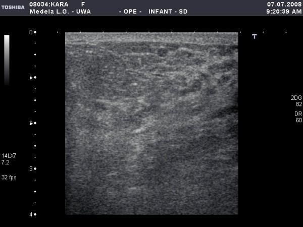
This diagnostic ultrasound image displays a radial or anti-radial section of a human lactating breast, specifically documented at the 12 o'clock position before a breastfeeding event. The image shows the characteristic heterogeneous echotexture of active mammary tissue. Prominent features include a dense, hyperechoic glandular parenchyma interspersed with darker, hypoechoic regions representing adipose tissue and small, branching milk ducts. Subtle linear hypoechoic structures are visible throughout the stroma, which correspond to non-distended lactiferous ducts. The subcutaneous fat layer and Cooper's ligaments contribute to the mixed echogenicity. This imaging provides educational value for understanding the normal physiological changes of the mammary gland during lactation, highlighting the increased glandular density and the presence of small, compressible ductal systems typical of a breast ready for milk ejection.
This diagnostic ultrasound image displays a radial or anti-radial section of a human lactating breast, specifically documented at the 12 o'clock position before a breastfeeding event. The image shows the characteristic heterogeneous echotexture of active mammary tissue. Prominent features include a dense, hyperechoic glandular parenchyma interspersed with darker, hypoechoic regions representing adipose tissue and small, branching milk ducts. Subtle linear hypoechoic structures are visible throughout the stroma, which correspond to non-distended lactiferous ducts. The subcutaneous fat layer and Cooper's ligaments contribute to the mixed echogenicity. This imaging provides educational value for understanding the normal physiological changes of the mammary gland during lactation, highlighting the increased glandular density and the presence of small, compressible ductal systems typical of a breast ready for milk ejection.

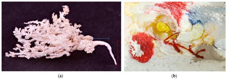
This composite educational image features corrosion casts of mammary gland anatomy. Figure (a) displays a white glandular cast against a black background, illustrating the complex branching architecture of the ductal system. It shows a primary ascendant milk duct (vertical red marker) transitioning into a wider transversal duct (horizontal blue marker) before arborizing into numerous secondary ducts (white arrowhead) and distal clusters representing terminal alveoli (white arrow). The absence of a prominent teat cistern is a notable anatomical variation. Figure (b) demonstrates a cross-sectional view of resin-perfused ducts within preserved tissue. A primary duct is filled with yellow resin, showing the hollow structure of the teat canal, while an adjacent underdeveloped, blind-ended duct is filled with red resin. These casts are used in anatomical research to study ductal morphology, lobation patterns, and developmental variations such as atrophic or rudimentary ducts, providing insight into physiological milk drainage and the potential pathways for disseminated infections.
This composite educational image features corrosion casts of mammary gland anatomy. Figure (a) displays a white glandular cast against a black background, illustrating the complex branching architecture of the ductal system. It shows a primary ascendant milk duct (vertical red marker) transitioning into a wider transversal duct (horizontal blue marker) before arborizing into numerous secondary ducts (white arrowhead) and distal clusters representing terminal alveoli (white arrow). The absence of a prominent teat cistern is a notable anatomical variation. Figure (b) demonstrates a cross-sectional view of resin-perfused ducts within preserved tissue. A primary duct is filled with yellow resin, showing the hollow structure of the teat canal, while an adjacent underdeveloped, blind-ended duct is filled with red resin. These casts are used in anatomical research to study ductal morphology, lobation patterns, and developmental variations such as atrophic or rudimentary ducts, providing insight into physiological milk drainage and the potential pathways for disseminated infections.

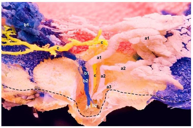
This clinical photograph displays a sagittal plane of a resin-casted mammary gland, illustrating the complex internal anatomy of milk ducts and glandular compartments. The specimen features three distinct color-coded resin applications (a, b, c) injected into separate teat orifices to map ductal pathways. Compartment 'a' (white) shows multiple functional ducts (a1, a2) with significant branching that leads to glandular lobes, alongside a blind-ended duct (a3). Compartment 'b' (blue) demonstrates multiple ducts (b1, b2) originating from two orifices that coalesce at a sharp curvature point before ascending. Compartment 'c' (yellow) illustrates a single, linear duct draining a single compartment without extensive branching or attached alveoli. A dashed line indicates the preserved superficial skin layer. This anatomical preparation highlights the high degree of variation in ductal morphology, showing that teat ducts can be solitary or split into multiple sections, and demonstrating the in situ spatial relationships between different glandular compartments which may overlap without mixing their ductal systems.
This clinical photograph displays a sagittal plane of a resin-casted mammary gland, illustrating the complex internal anatomy of milk ducts and glandular compartments. The specimen features three distinct color-coded resin applications (a, b, c) injected into separate teat orifices to map ductal pathways. Compartment 'a' (white) shows multiple functional ducts (a1, a2) with significant branching that leads to glandular lobes, alongside a blind-ended duct (a3). Compartment 'b' (blue) demonstrates multiple ducts (b1, b2) originating from two orifices that coalesce at a sharp curvature point before ascending. Compartment 'c' (yellow) illustrates a single, linear duct draining a single compartment without extensive branching or attached alveoli. A dashed line indicates the preserved superficial skin layer. This anatomical preparation highlights the high degree of variation in ductal morphology, showing that teat ducts can be solitary or split into multiple sections, and demonstrating the in situ spatial relationships between different glandular compartments which may overlap without mixing their ductal systems.

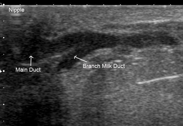
This diagnostic ultrasound image demonstrates the ductal anatomy of a human lactating breast. The image displays a longitudinal view of a main milk duct and a smaller branch milk duct. Both structures appear as hypoechoic (dark), fluid-filled tubular lumens contrasted against the more echogenic (brighter) surrounding glandular parenchyma and connective tissue. The main duct is shown with a diameter of approximately 2.4 mm, while the secondary branch milk duct measures 1.7 mm. The branching point is visible, showing the smaller duct draining glandular tissue and merging into the main collecting duct near the nipple region, which is labeled at the top left of the frame. This image illustrates the normal physiological state of milk ducts during lactation, emphasizing the lack of a prominent lactiferous sinus and the characteristic branching pattern close to the nipple. The clear differentiation between the hypoechoic ductal fluid and the hyperechoic breast tissue highlights the utility of high-resolution ultrasound in assessing mammary gland anatomy and potential obstructions.
This diagnostic ultrasound image demonstrates the ductal anatomy of a human lactating breast. The image displays a longitudinal view of a main milk duct and a smaller branch milk duct. Both structures appear as hypoechoic (dark), fluid-filled tubular lumens contrasted against the more echogenic (brighter) surrounding glandular parenchyma and connective tissue. The main duct is shown with a diameter of approximately 2.4 mm, while the secondary branch milk duct measures 1.7 mm. The branching point is visible, showing the smaller duct draining glandular tissue and merging into the main collecting duct near the nipple region, which is labeled at the top left of the frame. This image illustrates the normal physiological state of milk ducts during lactation, emphasizing the lack of a prominent lactiferous sinus and the characteristic branching pattern close to the nipple. The clear differentiation between the hypoechoic ductal fluid and the hyperechoic breast tissue highlights the utility of high-resolution ultrasound in assessing mammary gland anatomy and potential obstructions.

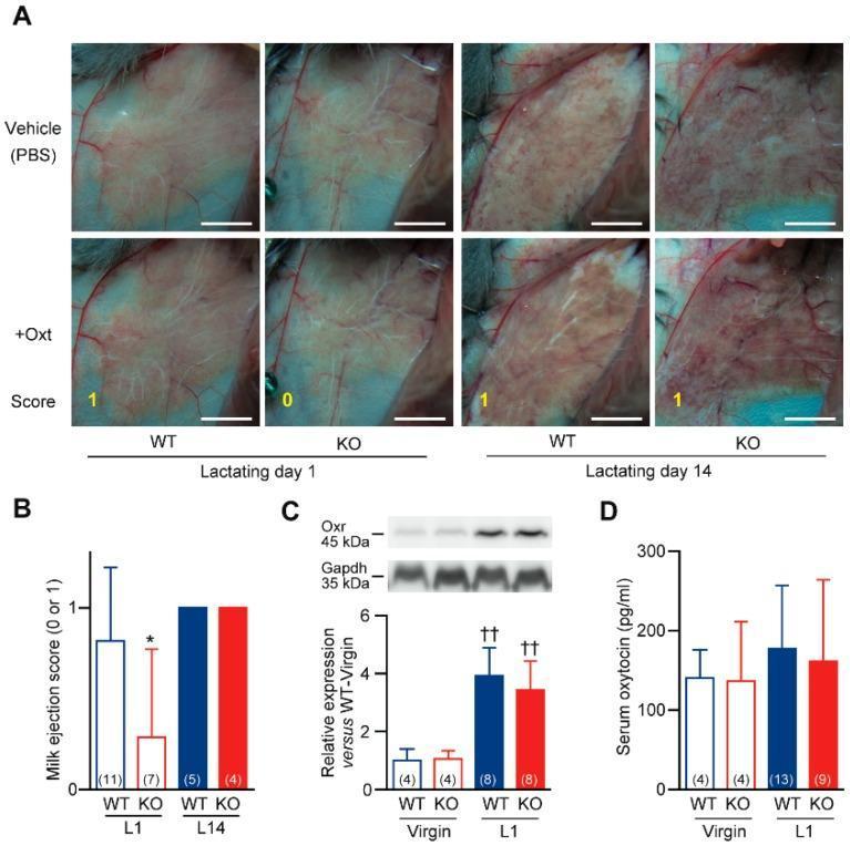
Multi-panel figure illustrating impaired milk ejection in cystathionine gamma-lyase deficient (Cth-/- or KO) mouse models. Panel A: Clinical photographs of surgically exposed abdominal mammary glands in wild-type (WT) and KO mice at lactating day 1 (L1) and day 14 (L14). Images compare milk ejection following vehicle (PBS) or oxytocin (+Oxt) application. In WT L1, oxytocin induces visible milk accumulation in larger ducts (Score 1), whereas KO L1 mice show a failure of milk ejection (Score 0). By L14, both genotypes exhibit Score 1. Panel B: Bar graph quantifying milk ejection scores (0 or 1), showing significant impairment specifically in L1 KO mice. Panel C: Western blot analysis of oxytocin receptor (Oxr) expression at 45 kDa, with Gapdh as a loading control at 35 kDa. The quantitative bar graph confirms significant upregulation of Oxr in both WT and KO mice during lactation compared to the virgin state. Panel D: Bar graph comparing serum oxytocin levels (pg/ml) between virgin and L1 stages, showing no significant difference between genotypes. This figure demonstrates that breastfeeding failure in Cth-/- models is due to peripheral oxytocin resistance rather than receptor deficiency or low circulating hormone levels.

Multi-panel figure illustrating impaired milk ejection in cystathionine gamma-lyase deficient (Cth-/- or KO) mouse models. Panel A: Clinical photographs of surgically exposed abdominal mammary glands in wild-type (WT) and KO mice at lactating day 1 (L1) and day 14 (L14). Images compare milk ejection following vehicle (PBS) or oxytocin (+Oxt) application. In WT L1, oxytocin induces visible milk accumulation in larger ducts (Score 1), whereas KO L1 mice show a failure of milk ejection (Score 0). By L14, both genotypes exhibit Score 1. Panel B: Bar graph quantifying milk ejection scores (0 or 1), showing significant impairment specifically in L1 KO mice. Panel C: Western blot analysis of oxytocin receptor (Oxr) expression at 45 kDa, with Gapdh as a loading control at 35 kDa. The quantitative bar graph confirms significant upregulation of Oxr in both WT and KO mice during lactation compared to the virgin state. Panel D: Bar graph comparing serum oxytocin levels (pg/ml) between virgin and L1 stages, showing no significant difference between genotypes. This figure demonstrates that breastfeeding failure in Cth-/- models is due to peripheral oxytocin resistance rather than receptor deficiency or low circulating hormone levels.

Searching Images

mastitis engorgement nipple problems breastfeeding complications

This clinical photograph set displays four distinct pathological conditions of the human breast and nipple in lactating patients across various skin tones. Panel A demonstrates breast engorgement featuring an inverted nipple with a light-colored central depression and taut, shiny skin. Panel B shows a breast abscess characterized by significant periareolar erythema, localized swelling, and a dark necrotic-appearing lesion at the nipple-areolar complex suggestive of nipple damage or a draining sinus. Panel C illustrates granulomatous mastitis with visible skin wrinkling, nipple inversion, and generalized breast inflammation. Panel D depicts acute nipple damage on dark-pigmented skin, highlighted by a prominent, raised, fluid-filled blood blister (hemorrhagic bleb) located on the lateral aspect of the nipple. This series serves as an educational comparison for differentiating mastitis, abscess, engorgement, and mechanical nipple trauma, while highlighting common visual pitfalls that can lead to clinical misclassification.
This clinical photograph set displays four distinct pathological conditions of the human breast and nipple in lactating patients across various skin tones. Panel A demonstrates breast engorgement featuring an inverted nipple with a light-colored central depression and taut, shiny skin. Panel B shows a breast abscess characterized by significant periareolar erythema, localized swelling, and a dark necrotic-appearing lesion at the nipple-areolar complex suggestive of nipple damage or a draining sinus. Panel C illustrates granulomatous mastitis with visible skin wrinkling, nipple inversion, and generalized breast inflammation. Panel D depicts acute nipple damage on dark-pigmented skin, highlighted by a prominent, raised, fluid-filled blood blister (hemorrhagic bleb) located on the lateral aspect of the nipple. This series serves as an educational comparison for differentiating mastitis, abscess, engorgement, and mechanical nipple trauma, while highlighting common visual pitfalls that can lead to clinical misclassification.

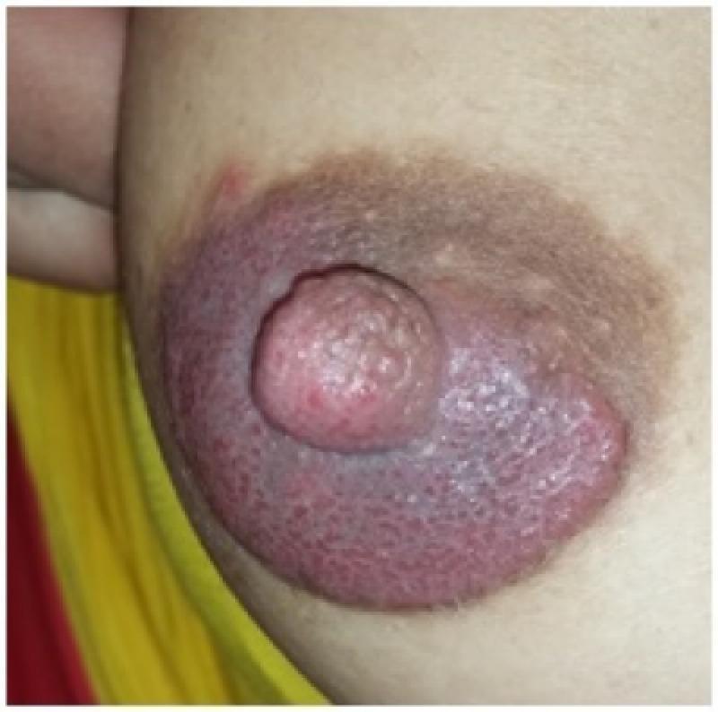
This clinical photograph displays a close-up view of the right female breast, focusing on the nipple-areolar complex (NAC). The primary finding is prominent erythematous hyperkeratosis of the areola and nipple. The affected area exhibits diffuse redness and a thickened, irregular, and verrucous texture, with visible scaling and crusting indicative of chronic inflammation. The nipple itself appears enlarged and edematous with surface cobblestoning. A distinct hyperpigmented ring borders the erythematous zone, and there are subtle indications of nipple discharge. These visual manifestations are characteristic of inflammatory or infectious conditions of the breast skin, such as cutaneous candidiasis or mammary Paget's disease. In the documented clinical context of a breastfeeding mother with a history of recurrent yeast infections, the image demonstrates the clinical presentation of areolar hyperkeratosis secondary to a Candida albicans infection. The image serves as an educational reference for dermatological and obstetric evaluation of infectious mastitis and associated skin lesions during lactation.
This clinical photograph displays a close-up view of the right female breast, focusing on the nipple-areolar complex (NAC). The primary finding is prominent erythematous hyperkeratosis of the areola and nipple. The affected area exhibits diffuse redness and a thickened, irregular, and verrucous texture, with visible scaling and crusting indicative of chronic inflammation. The nipple itself appears enlarged and edematous with surface cobblestoning. A distinct hyperpigmented ring borders the erythematous zone, and there are subtle indications of nipple discharge. These visual manifestations are characteristic of inflammatory or infectious conditions of the breast skin, such as cutaneous candidiasis or mammary Paget's disease. In the documented clinical context of a breastfeeding mother with a history of recurrent yeast infections, the image demonstrates the clinical presentation of areolar hyperkeratosis secondary to a Candida albicans infection. The image serves as an educational reference for dermatological and obstetric evaluation of infectious mastitis and associated skin lesions during lactation.

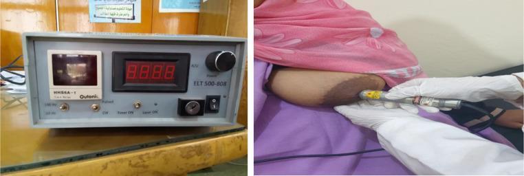
This composite clinical photograph illustrates the application of Photobiomodulation (PBM) therapy for lactation-related nipple pathology. The left panel shows a low-level diode laser device (808 nm) featuring a digital control panel with a four-digit LED display, a power adjustment knob labeled 'ELT 500-808', a safety key switch, and toggle switches for pulse and timer functions. The right panel displays the procedural application of the treatment. A healthcare professional wearing white clinical gloves uses a handheld laser probe to deliver targeted irradiation to the areola and nipple of a patient. The clinical focus is the management of inflammatory conditions, such as nipple fissures or mastitis, in breastfeeding individuals. The procedure aims to utilize low-level laser therapy (LLLT) to reduce pain (as measured by the Visual Analogue Scale), accelerate tissue repair, and decrease inflammation (erythema and heat) associated with breastfeeding trauma.
This composite clinical photograph illustrates the application of Photobiomodulation (PBM) therapy for lactation-related nipple pathology. The left panel shows a low-level diode laser device (808 nm) featuring a digital control panel with a four-digit LED display, a power adjustment knob labeled 'ELT 500-808', a safety key switch, and toggle switches for pulse and timer functions. The right panel displays the procedural application of the treatment. A healthcare professional wearing white clinical gloves uses a handheld laser probe to deliver targeted irradiation to the areola and nipple of a patient. The clinical focus is the management of inflammatory conditions, such as nipple fissures or mastitis, in breastfeeding individuals. The procedure aims to utilize low-level laser therapy (LLLT) to reduce pain (as measured by the Visual Analogue Scale), accelerate tissue repair, and decrease inflammation (erythema and heat) associated with breastfeeding trauma.

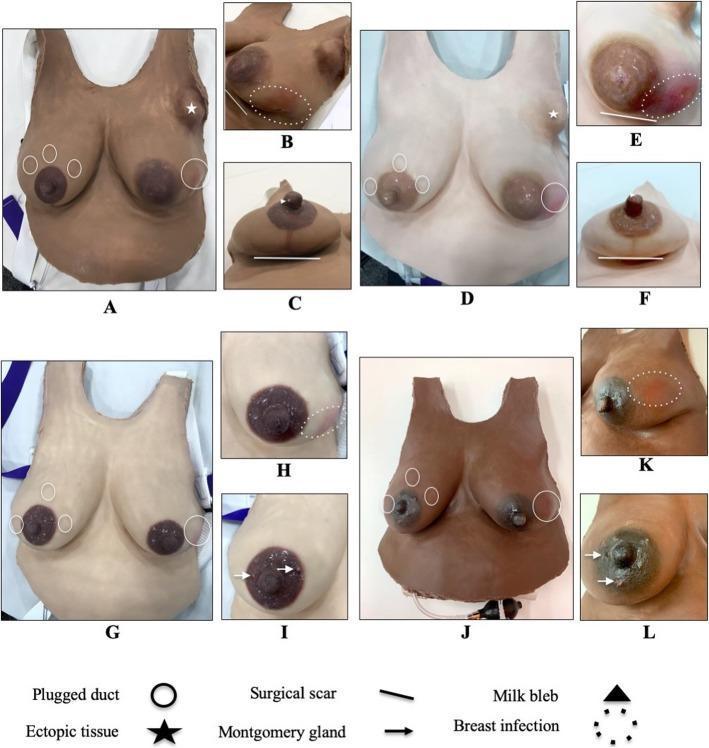
This clinical comparison chart displays various Lactation Simulation Models (LSM) designed for medical education, shown in multiple skin tones. The visual provides examples of breast anatomy, lactation-related pathologies, and surgical history relevant to obstetrics, gynecology, and breastfeeding medicine. Key anatomical variations depicted include round, pinched, flat, and bulbous nipple morphologies. Pathological signs illustrated include mastitis (breast infection), indicated by erythema; plugged ducts, represented by palpable nodules; milk blebs; and Montgomery glands. Additionally, the models demonstrate surgical markers such as augmentation and reduction scars, as well as axillary ectopic breast tissue. Annotation symbols (circles, stars, lines, and arrows) are used throughout the panels to identify specific clinical findings. The collection serves as an educational tool for training healthcare providers in breast examination, lactation assessment, and the identification of both normal variations and common breastfeeding complications.
This clinical comparison chart displays various Lactation Simulation Models (LSM) designed for medical education, shown in multiple skin tones. The visual provides examples of breast anatomy, lactation-related pathologies, and surgical history relevant to obstetrics, gynecology, and breastfeeding medicine. Key anatomical variations depicted include round, pinched, flat, and bulbous nipple morphologies. Pathological signs illustrated include mastitis (breast infection), indicated by erythema; plugged ducts, represented by palpable nodules; milk blebs; and Montgomery glands. Additionally, the models demonstrate surgical markers such as augmentation and reduction scars, as well as axillary ectopic breast tissue. Annotation symbols (circles, stars, lines, and arrows) are used throughout the panels to identify specific clinical findings. The collection serves as an educational tool for training healthcare providers in breast examination, lactation assessment, and the identification of both normal variations and common breastfeeding complications.

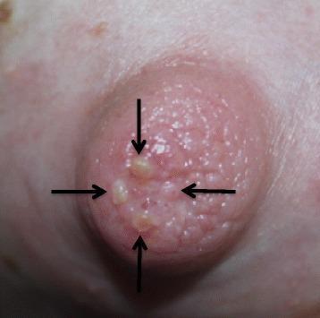
This clinical photograph shows a close-up view of a human female nipple exhibiting trauma secondary to breastfeeding. The nipple is visibly erythematous and has a rough, pebbled texture. Several small, discrete, raised, fluid-filled vesicles (blisters) are located on the nipple surface, particularly near the apex. These lesions are approximately 2-4 mm in diameter, yellowish-white in color, and indicated by four black arrows in the image. The surrounding areolar skin appears unremarkable without significant inflammation or secondary skin lesions. Clinically, this presentation illustrates mechanical skin trauma, specifically suction-induced friction blisters resulting from high intra-oral vacuum and the use of nipple shields during lactation. This image serves as an educational example of maternal breastfeeding complications and the physiological impact of infant latch mechanics on maternal tissue integrity.
This clinical photograph shows a close-up view of a human female nipple exhibiting trauma secondary to breastfeeding. The nipple is visibly erythematous and has a rough, pebbled texture. Several small, discrete, raised, fluid-filled vesicles (blisters) are located on the nipple surface, particularly near the apex. These lesions are approximately 2-4 mm in diameter, yellowish-white in color, and indicated by four black arrows in the image. The surrounding areolar skin appears unremarkable without significant inflammation or secondary skin lesions. Clinically, this presentation illustrates mechanical skin trauma, specifically suction-induced friction blisters resulting from high intra-oral vacuum and the use of nipple shields during lactation. This image serves as an educational example of maternal breastfeeding complications and the physiological impact of infant latch mechanics on maternal tissue integrity.

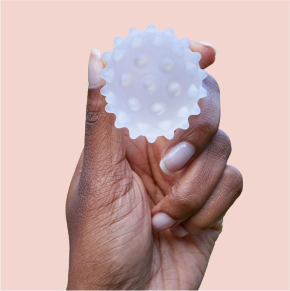
A clinical photograph of a Lactamo device, a specialized lactation aid designed for maternal health and breastfeeding support. The device is a small, handheld sphere, approximately 5 cm in diameter, constructed from a translucent, frosted medical-grade material. Its surface is characterized by multiple rounded, hollow protrusions or nodes distributed evenly across the sphere to provide varying levels of compression and tactile stimulation. The device is designed to be filled with a thermochromic gel, allowing for the delivery of hot or cold therapy in conjunction with breast massage and compression. It is used to address breastfeeding challenges such as engorgement, blocked ducts, and mastitis by facilitating milk flow through movement toward the nipple or axilla. The photograph shows the device held between fingers to demonstrate its scale and ergonomic design for self-application by postpartum patients.

A clinical photograph of a Lactamo device, a specialized lactation aid designed for maternal health and breastfeeding support. The device is a small, handheld sphere, approximately 5 cm in diameter, constructed from a translucent, frosted medical-grade material. Its surface is characterized by multiple rounded, hollow protrusions or nodes distributed evenly across the sphere to provide varying levels of compression and tactile stimulation. The device is designed to be filled with a thermochromic gel, allowing for the delivery of hot or cold therapy in conjunction with breast massage and compression. It is used to address breastfeeding challenges such as engorgement, blocked ducts, and mastitis by facilitating milk flow through movement toward the nipple or axilla. The photograph shows the device held between fingers to demonstrate its scale and ergonomic design for self-application by postpartum patients.

Searching Images

oxytocin prolactin hormone breastfeeding letdown reflex
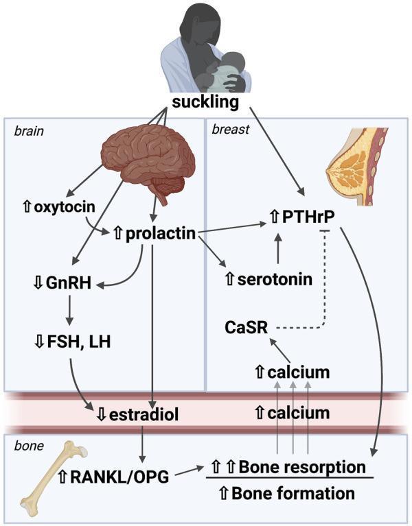
A pathophysiology diagram illustrating the brain-breast-bone axis and its regulation of mineral metabolism during lactation. The diagram begins with the 'suckling' stimulus, which triggers neural pathways to the brain and mammary gland. In the 'brain' section, suckling leads to increased levels of oxytocin and prolactin. Elevated prolactin inhibits Gonadotropic Releasing Hormone (GnRH), subsequently decreasing Follicle Stimulating Hormone (FSH) and Luteinizing Hormone (LH), which leads to reduced estradiol levels in the bloodstream. In the 'breast' section, suckling and prolactin increase Parathyroid Hormone-related Protein (PTHrP) and serotonin levels. The Calcium Sensing Receptor (CaSR) is shown providing negative feedback on PTHrP when mammary calcium levels are high. In the 'bone' section, the combined effect of low estradiol and high PTHrP increases the RANKL/OPG ratio, leading to stimulated bone resorption and bone formation (osteoclastic and osteocytic activity). This process liberates calcium into the circulation to be utilized by the mammary gland for milk production. The diagram effectively maps the hormonal cascade coordinating maternal skeletal turnover with neonatal nutritional demands.
A pathophysiology diagram illustrating the brain-breast-bone axis and its regulation of mineral metabolism during lactation. The diagram begins with the 'suckling' stimulus, which triggers neural pathways to the brain and mammary gland. In the 'brain' section, suckling leads to increased levels of oxytocin and prolactin. Elevated prolactin inhibits Gonadotropic Releasing Hormone (GnRH), subsequently decreasing Follicle Stimulating Hormone (FSH) and Luteinizing Hormone (LH), which leads to reduced estradiol levels in the bloodstream. In the 'breast' section, suckling and prolactin increase Parathyroid Hormone-related Protein (PTHrP) and serotonin levels. The Calcium Sensing Receptor (CaSR) is shown providing negative feedback on PTHrP when mammary calcium levels are high. In the 'bone' section, the combined effect of low estradiol and high PTHrP increases the RANKL/OPG ratio, leading to stimulated bone resorption and bone formation (osteoclastic and osteocytic activity). This process liberates calcium into the circulation to be utilized by the mammary gland for milk production. The diagram effectively maps the hormonal cascade coordinating maternal skeletal turnover with neonatal nutritional demands.

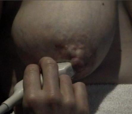
This clinical photograph displays a right lactating breast during the milk ejection reflex, alongside the application of a diagnostic ultrasound probe. The areola demonstrates significant physiological swelling and increased tension, resulting in a full, convex appearance. This visual change is indicative of the expansion of superficial milk ducts as oxytocin-induced myoepithelial contraction forces milk from the alveoli into the ductal system. A linear ultrasound transducer is positioned in direct contact with the inferior portion of the areola, stabilized by a hand to monitor ductal dilation and milk flow in real-time. The skin surface shows slight lobulation or prominence corresponding to the underlying distended ducts. This image illustrates the physical manifestations of the milk ejection reflex and the non-invasive clinical method of using ultrasound to study lactation physiology and breastfeeding dynamics.

This clinical photograph displays a right lactating breast during the milk ejection reflex, alongside the application of a diagnostic ultrasound probe. The areola demonstrates significant physiological swelling and increased tension, resulting in a full, convex appearance. This visual change is indicative of the expansion of superficial milk ducts as oxytocin-induced myoepithelial contraction forces milk from the alveoli into the ductal system. A linear ultrasound transducer is positioned in direct contact with the inferior portion of the areola, stabilized by a hand to monitor ductal dilation and milk flow in real-time. The skin surface shows slight lobulation or prominence corresponding to the underlying distended ducts. This image illustrates the physical manifestations of the milk ejection reflex and the non-invasive clinical method of using ultrasound to study lactation physiology and breastfeeding dynamics.

Multi-panel figure illustrating impaired milk ejection in cystathionine gamma-lyase deficient (Cth-/- or KO) mouse models. Panel A: Clinical photographs of surgically exposed abdominal mammary glands in wild-type (WT) and KO mice at lactating day 1 (L1) and day 14 (L14). Images compare milk ejection following vehicle (PBS) or oxytocin (+Oxt) application. In WT L1, oxytocin induces visible milk accumulation in larger ducts (Score 1), whereas KO L1 mice show a failure of milk ejection (Score 0). By L14, both genotypes exhibit Score 1. Panel B: Bar graph quantifying milk ejection scores (0 or 1), showing significant impairment specifically in L1 KO mice. Panel C: Western blot analysis of oxytocin receptor (Oxr) expression at 45 kDa, with Gapdh as a loading control at 35 kDa. The quantitative bar graph confirms significant upregulation of Oxr in both WT and KO mice during lactation compared to the virgin state. Panel D: Bar graph comparing serum oxytocin levels (pg/ml) between virgin and L1 stages, showing no significant difference between genotypes. This figure demonstrates that breastfeeding failure in Cth-/- models is due to peripheral oxytocin resistance rather than receptor deficiency or low circulating hormone levels.
Multi-panel figure illustrating impaired milk ejection in cystathionine gamma-lyase deficient (Cth-/- or KO) mouse models. Panel A: Clinical photographs of surgically exposed abdominal mammary glands in wild-type (WT) and KO mice at lactating day 1 (L1) and day 14 (L14). Images compare milk ejection following vehicle (PBS) or oxytocin (+Oxt) application. In WT L1, oxytocin induces visible milk accumulation in larger ducts (Score 1), whereas KO L1 mice show a failure of milk ejection (Score 0). By L14, both genotypes exhibit Score 1. Panel B: Bar graph quantifying milk ejection scores (0 or 1), showing significant impairment specifically in L1 KO mice. Panel C: Western blot analysis of oxytocin receptor (Oxr) expression at 45 kDa, with Gapdh as a loading control at 35 kDa. The quantitative bar graph confirms significant upregulation of Oxr in both WT and KO mice during lactation compared to the virgin state. Panel D: Bar graph comparing serum oxytocin levels (pg/ml) between virgin and L1 stages, showing no significant difference between genotypes. This figure demonstrates that breastfeeding failure in Cth-/- models is due to peripheral oxytocin resistance rather than receptor deficiency or low circulating hormone levels.

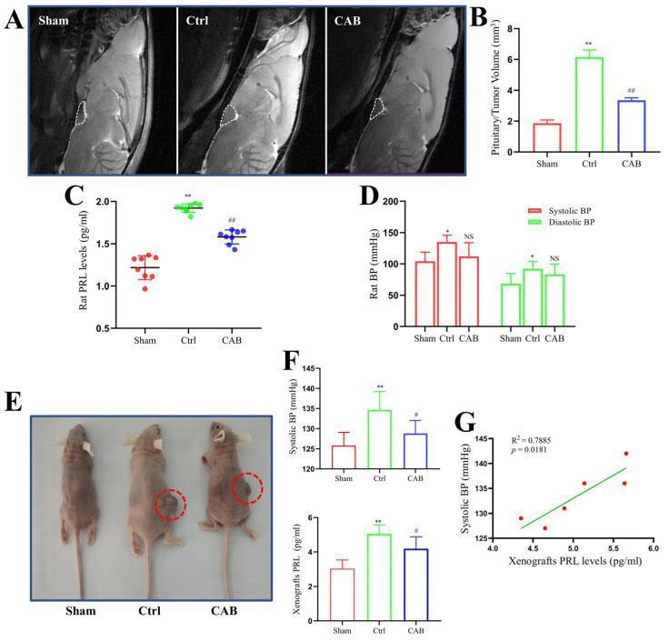
Educational figure illustrating blood pressure (BP) and hormone changes in rodent prolactinoma models across three experimental groups: Sham (control), Ctrl (untreated prolactinoma), and CAB (cabergoline-treated). Panel A presents sagittal T2-weighted MRI images of a rat in situ prolactinoma model, with white dashed lines delineating the pituitary/tumor region, showing significant enlargement in the Ctrl group compared to Sham, and reduction following CAB treatment. Panel B quantifies tumor volume (mm³), while Panel C shows corresponding serum prolactin (PRL) levels (pg/ml). Panel D provides a bar graph of systolic and diastolic blood pressure in rats. Panel E displays clinical photographs of a mouse xenograft model with tumors highlighted in red circles; the Ctrl group shows a prominent subcutaneous mass that appears reduced in the CAB group. Panels F and G correlate these findings with mouse systolic BP, serum PRL levels, and a regression analysis (R²=0.7885) showing a positive correlation between PRL and systolic BP. This figure demonstrates the efficacy of dopamine agonists in reducing tumor volume and hyperprolactinemia-associated hypertension.
Educational figure illustrating blood pressure (BP) and hormone changes in rodent prolactinoma models across three experimental groups: Sham (control), Ctrl (untreated prolactinoma), and CAB (cabergoline-treated). Panel A presents sagittal T2-weighted MRI images of a rat in situ prolactinoma model, with white dashed lines delineating the pituitary/tumor region, showing significant enlargement in the Ctrl group compared to Sham, and reduction following CAB treatment. Panel B quantifies tumor volume (mm³), while Panel C shows corresponding serum prolactin (PRL) levels (pg/ml). Panel D provides a bar graph of systolic and diastolic blood pressure in rats. Panel E displays clinical photographs of a mouse xenograft model with tumors highlighted in red circles; the Ctrl group shows a prominent subcutaneous mass that appears reduced in the CAB group. Panels F and G correlate these findings with mouse systolic BP, serum PRL levels, and a regression analysis (R²=0.7885) showing a positive correlation between PRL and systolic BP. This figure demonstrates the efficacy of dopamine agonists in reducing tumor volume and hyperprolactinemia-associated hypertension.

This medical illustration is a sagittal-view pathophysiology diagram of the human brain, detailing the oxytocinergic system and its interaction with the hypothalamic-pituitary-adrenal (HPA) axis. Key anatomical regions are labeled, including the prefrontal cortex (mPFC and vmPFC), anterior cingulate cortex, striatum, nucleus accumbens, amygdala, ventral tegmental area, and hippocampus. The diagram highlights the synthesis of oxytocin in the paraventricular nucleus (PVN) and supraoptic nucleus (SON) of the hypothalamus. Blue dashed lines represent central oxytocin projections terminating in various limbic and cortical regions, while solid blue arrows indicate projections to the pituitary gland and subsequent release into the peripheral bloodstream. Oxytocin receptor (OTR) expression is visually depicted by red highlighted areas and circles in target tissues. Additionally, the HPA axis is shown via the signaling of corticotropin-releasing hormone (CRH) to the pituitary and adrenocorticotropic hormone (ACTH) to the adrenal cortex, resulting in cortisol release. This educational visual explains the integration of neuropeptide signaling with neuroendocrine stress responses and social-motivational brain pathways.
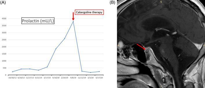
This medical illustration is a sagittal-view pathophysiology diagram of the human brain, detailing the oxytocinergic system and its interaction with the hypothalamic-pituitary-adrenal (HPA) axis. Key anatomical regions are labeled, including the prefrontal cortex (mPFC and vmPFC), anterior cingulate cortex, striatum, nucleus accumbens, amygdala, ventral tegmental area, and hippocampus. The diagram highlights the synthesis of oxytocin in the paraventricular nucleus (PVN) and supraoptic nucleus (SON) of the hypothalamus. Blue dashed lines represent central oxytocin projections terminating in various limbic and cortical regions, while solid blue arrows indicate projections to the pituitary gland and subsequent release into the peripheral bloodstream. Oxytocin receptor (OTR) expression is visually depicted by red highlighted areas and circles in target tissues. Additionally, the HPA axis is shown via the signaling of corticotropin-releasing hormone (CRH) to the pituitary and adrenocorticotropic hormone (ACTH) to the adrenal cortex, resulting in cortisol release. This educational visual explains the integration of neuropeptide signaling with neuroendocrine stress responses and social-motivational brain pathways.

This composite educational image illustrates the diagnosis and treatment response of a pituitary microadenoma. Panel A is a timeline graph displaying plasma prolactin levels (mU/L) over several years. It shows a significant longitudinal increase in prolactin, peaking at approximately 3800 mU/L, followed by a rapid precipitous decline to baseline levels reaching less than 500 mU/L immediately following the initiation of Cabergoline therapy, a dopamine agonist. Panel B is a sagittal T1-weighted magnetic resonance image (MRI) of the brain focusing on the sella turcica. A red arrow indicates a 5 mm hypointense lesion within the pituitary gland, which causes a subtle focal contour abnormality of the gland's superior profile. This finding is characteristic of a pituitary microadenoma. Together, these panels demonstrate the clinical correlation between hyperprolactinemia, neuroradiological evidence of a secretory tumor, and the high efficacy of medical management with dopamine agonists in normalizing hormone levels.

This composite educational image illustrates the diagnosis and treatment response of a pituitary microadenoma. Panel A is a timeline graph displaying plasma prolactin levels (mU/L) over several years. It shows a significant longitudinal increase in prolactin, peaking at approximately 3800 mU/L, followed by a rapid precipitous decline to baseline levels reaching less than 500 mU/L immediately following the initiation of Cabergoline therapy, a dopamine agonist. Panel B is a sagittal T1-weighted magnetic resonance image (MRI) of the brain focusing on the sella turcica. A red arrow indicates a 5 mm hypointense lesion within the pituitary gland, which causes a subtle focal contour abnormality of the gland's superior profile. This finding is characteristic of a pituitary microadenoma. Together, these panels demonstrate the clinical correlation between hyperprolactinemia, neuroradiological evidence of a secretory tumor, and the high efficacy of medical management with dopamine agonists in normalizing hormone levels.

Searching Images

breast milk storage guidelines expressed milk containers

This clinical photograph captures a close-up procedural demonstration of Antenatal Colostrum Expression (ACE). The image illustrates the 'C-hold' hand technique, where the breast tissue is compressed between the thumb and fingers, positioned approximately 2-3 cm behind the nipple at the edge of the areola. A second hand is shown holding a small oral syringe directly at the nipple tip to collect the expressed droplets of colostrum. The procedural focus is on the manual stimulation and compression of the lactiferous sinuses to facilitate milk removal. This visual serves as an educational tool for lactation consultants and expectant mothers, highlighting the correct hand placement and the use of a syringe for hygienic colostrum harvesting and storage during late pregnancy. The setting is likely an antenatal clinic or educational session designed to improve breastfeeding confidence and knowledge.

This clinical photograph captures a close-up procedural demonstration of Antenatal Colostrum Expression (ACE). The image illustrates the 'C-hold' hand technique, where the breast tissue is compressed between the thumb and fingers, positioned approximately 2-3 cm behind the nipple at the edge of the areola. A second hand is shown holding a small oral syringe directly at the nipple tip to collect the expressed droplets of colostrum. The procedural focus is on the manual stimulation and compression of the lactiferous sinuses to facilitate milk removal. This visual serves as an educational tool for lactation consultants and expectant mothers, highlighting the correct hand placement and the use of a syringe for hygienic colostrum harvesting and storage during late pregnancy. The setting is likely an antenatal clinic or educational session designed to improve breastfeeding confidence and knowledge.

This clinical photograph displays a medical-grade disinfection system used for neonatal care accessories, specifically for the steam sterilization of breast milk collection equipment. The image features a specialized microwave steam sterilization bag alongside a sterile collection cup. The bag is a flexible, translucent white pouch with yellow instructional graphics and a 'Hold here' safety zone. It displays a six-step illustrated guide for cleaning, adding water (60ml), and microwave heating times based on wattage. A tracking grid at the bottom is included to monitor the bag's 20-use lifecycle. Next to the bag is a cylindrical, transparent collection cup with a yellow screw-top lid and a red-bordered label for patient identification. This visual represents standard protocols for infection control in a Neonatal Intensive Care Unit (NICU) or home care setting, demonstrating the process of ensuring the microbiological safety of colostrum and breast milk storage containers through rapid steam treatment.

This clinical photograph displays a medical-grade disinfection system used for neonatal care accessories, specifically for the steam sterilization of breast milk collection equipment. The image features a specialized microwave steam sterilization bag alongside a sterile collection cup. The bag is a flexible, translucent white pouch with yellow instructional graphics and a 'Hold here' safety zone. It displays a six-step illustrated guide for cleaning, adding water (60ml), and microwave heating times based on wattage. A tracking grid at the bottom is included to monitor the bag's 20-use lifecycle. Next to the bag is a cylindrical, transparent collection cup with a yellow screw-top lid and a red-bordered label for patient identification. This visual represents standard protocols for infection control in a Neonatal Intensive Care Unit (NICU) or home care setting, demonstrating the process of ensuring the microbiological safety of colostrum and breast milk storage containers through rapid steam treatment.

A close-up clinical photograph demonstrating the Marmet technique for hand-expressing antenatal milk or colostrum. The image shows a person's hands positioned on a lactating breast. One hand supports the breast tissue, with the thumb and index finger placed approximately 2-3 cm behind the nipple at the edge of the areola, applying rhythmic compression to the underlying milk ducts. The areola appears physiologically hyperpigmented and slightly engorged, typical of late pregnancy or the early postpartum period. A small, clear, flip-top collection vial is held directly beneath the nipple to capture the expressed fluid. The vial contains a small volume of yellowish, viscous fluid, characteristic of colostrum. This visual serves as an educational guide for lactation support, specifically teaching manual expression and collection techniques for individuals in the late third trimester (antenatal milk expression) or early postpartum phase.
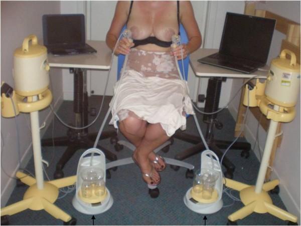
A close-up clinical photograph demonstrating the Marmet technique for hand-expressing antenatal milk or colostrum. The image shows a person's hands positioned on a lactating breast. One hand supports the breast tissue, with the thumb and index finger placed approximately 2-3 cm behind the nipple at the edge of the areola, applying rhythmic compression to the underlying milk ducts. The areola appears physiologically hyperpigmented and slightly engorged, typical of late pregnancy or the early postpartum period. A small, clear, flip-top collection vial is held directly beneath the nipple to capture the expressed fluid. The vial contains a small volume of yellowish, viscous fluid, characteristic of colostrum. This visual serves as an educational guide for lactation support, specifically teaching manual expression and collection techniques for individuals in the late third trimester (antenatal milk expression) or early postpartum phase.

This procedural photograph illustrates a clinical research setup for simultaneous, bilateral milk flow rate measurement using the Showmilk technique. A seated participant is positioned between two hospital-grade electric breast pumps connected to bilateral breast shields. The experimental apparatus utilizes clear collecting tubes that transport expressed milk from the shields down to purpose-built continuous weight measurement devices (Showmilks) positioned on the floor. These weighing devices feature circular bases with platforms housing multiple collection bottles for fractional sampling. The system is integrated with two pressure transducers to monitor vacuum levels. Data from the weight balances and transducers are transmitted via USB interfaces to two notebook computers for real-time visualization and recording of milk ejection patterns. This setup is designed to evaluate milk ejection frequency, flow rate peaks (g/s), and synchronicity between breasts during expression, providing objective data on the physiological mechanisms of lactation and the milk ejection reflex.

This procedural photograph illustrates a clinical research setup for simultaneous, bilateral milk flow rate measurement using the Showmilk technique. A seated participant is positioned between two hospital-grade electric breast pumps connected to bilateral breast shields. The experimental apparatus utilizes clear collecting tubes that transport expressed milk from the shields down to purpose-built continuous weight measurement devices (Showmilks) positioned on the floor. These weighing devices feature circular bases with platforms housing multiple collection bottles for fractional sampling. The system is integrated with two pressure transducers to monitor vacuum levels. Data from the weight balances and transducers are transmitted via USB interfaces to two notebook computers for real-time visualization and recording of milk ejection patterns. This setup is designed to evaluate milk ejection frequency, flow rate peaks (g/s), and synchronicity between breasts during expression, providing objective data on the physiological mechanisms of lactation and the milk ejection reflex.

Searching Images

infant tongue tie ankyloglossia breastfeeding difficulty
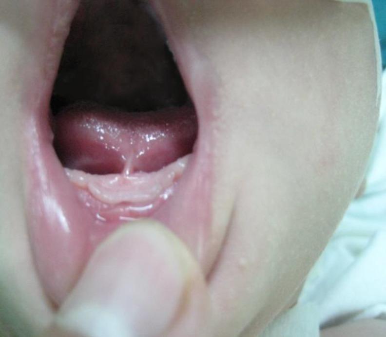
Clinical photograph of an infant's oral cavity illustrating ankyloglossia (tongue-tie). The image shows the ventral surface of the tongue elevated, revealing a short, restrictive lingual frenulum. This fibrous band of tissue attaches the midline of the underside of the tongue close to the mandibular alveolar ridge, limiting the tongue's range of motion. A slight heart-shaped notch or indentation is visible at the tip of the tongue where the frenulum pulls on the muscle. Surrounding oral structures include the lower gingiva, floor of the mouth, and the inner mucosa of the lower lip. This visual is diagnostic for identifying structural causes of breastfeeding difficulties, such as mastalgia in the mother or poor latch in the infant, often necessitating a frenotomy or frenuloplasty. The photograph is categorized under pediatric otolaryngology and intraoral imaging.
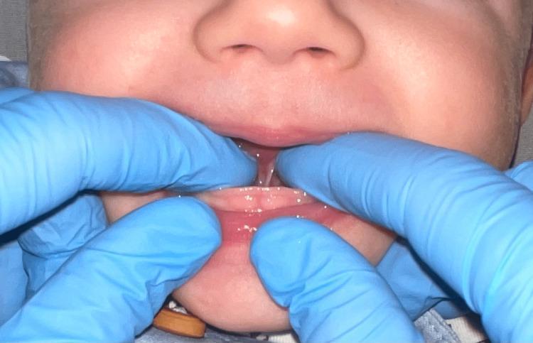
Clinical photograph of an infant's oral cavity illustrating ankyloglossia (tongue-tie). The image shows the ventral surface of the tongue elevated, revealing a short, restrictive lingual frenulum. This fibrous band of tissue attaches the midline of the underside of the tongue close to the mandibular alveolar ridge, limiting the tongue's range of motion. A slight heart-shaped notch or indentation is visible at the tip of the tongue where the frenulum pulls on the muscle. Surrounding oral structures include the lower gingiva, floor of the mouth, and the inner mucosa of the lower lip. This visual is diagnostic for identifying structural causes of breastfeeding difficulties, such as mastalgia in the mother or poor latch in the infant, often necessitating a frenotomy or frenuloplasty. The photograph is categorized under pediatric otolaryngology and intraoral imaging.

A clinical photograph of an infant's oral cavity during a physical examination for ankyloglossia (tongue-tie). Gloved fingers are shown retracting the lower lip and elevating the tongue to expose the sublingual region. The image demonstrates a prominent lingual frenulum, characterized as a thin, translucent membrane that appears short and restrictive. The frenulum attaches from the ventral surface of the tongue, near the anterior tip, down to the floor of the mouth in close proximity to the mandibular alveolar ridge. This anterior attachment and short length indicate a restricted range of motion for the tongue, characteristic of Coryllos grade 3 or similar classifications of ankyloglossia. This visual is intended for dental and pediatric education regarding the diagnostic appearance of symptomatic lingual frenula in breastfeeding infants.
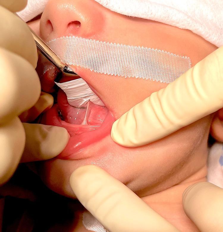
A clinical photograph of an infant's oral cavity during a physical examination for ankyloglossia (tongue-tie). Gloved fingers are shown retracting the lower lip and elevating the tongue to expose the sublingual region. The image demonstrates a prominent lingual frenulum, characterized as a thin, translucent membrane that appears short and restrictive. The frenulum attaches from the ventral surface of the tongue, near the anterior tip, down to the floor of the mouth in close proximity to the mandibular alveolar ridge. This anterior attachment and short length indicate a restricted range of motion for the tongue, characteristic of Coryllos grade 3 or similar classifications of ankyloglossia. This visual is intended for dental and pediatric education regarding the diagnostic appearance of symptomatic lingual frenula in breastfeeding infants.

This clinical photograph shows the oral cavity of an infant being examined for ankyloglossia (tongue-tie). The image demonstrates a restricted lingual frenulum, appearing as a tight, thin, membranous band of tissue connecting the ventral surface of the tongue to the floor of the mouth. The attachment is located close to the tongue tip, significantly limiting the tongue's range of motion and upward extension. During the examination, gloved fingers are used to retract the lips and stabilize the jaw, while a metallic grooved director or tongue elevator, assisted by white surgical gauze, lifts the tongue to expose the sublingual anatomy. A white adhesive bandage is visible on the upper lip, likely securing a medical device or providing stabilization during a pediatric procedure. This visual is characteristic of Coryllos grade 1 or 2 ankyloglossia, a condition often associated with breastfeeding difficulties. The image serves as an educational example for pediatric dentistry and otolaryngology regarding the diagnostic assessment of lingual restrictive tissues prior to frenotomy.
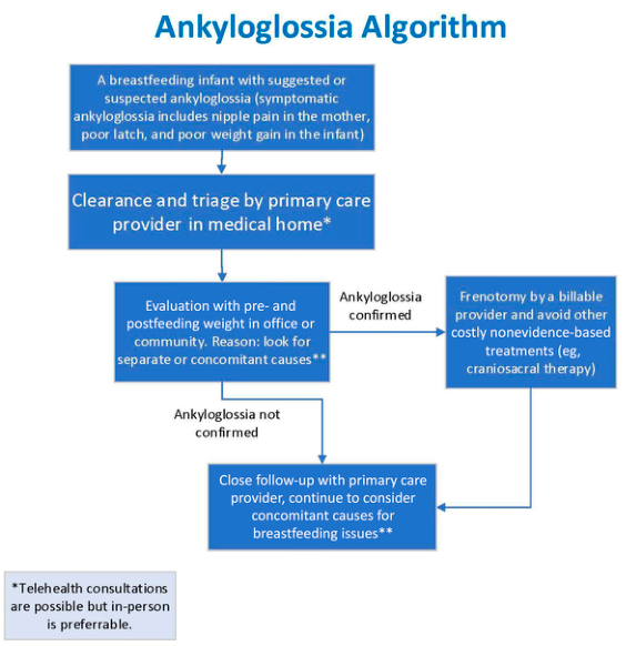
This clinical photograph shows the oral cavity of an infant being examined for ankyloglossia (tongue-tie). The image demonstrates a restricted lingual frenulum, appearing as a tight, thin, membranous band of tissue connecting the ventral surface of the tongue to the floor of the mouth. The attachment is located close to the tongue tip, significantly limiting the tongue's range of motion and upward extension. During the examination, gloved fingers are used to retract the lips and stabilize the jaw, while a metallic grooved director or tongue elevator, assisted by white surgical gauze, lifts the tongue to expose the sublingual anatomy. A white adhesive bandage is visible on the upper lip, likely securing a medical device or providing stabilization during a pediatric procedure. This visual is characteristic of Coryllos grade 1 or 2 ankyloglossia, a condition often associated with breastfeeding difficulties. The image serves as an educational example for pediatric dentistry and otolaryngology regarding the diagnostic assessment of lingual restrictive tissues prior to frenotomy.

Summary : This flowchart presents an algorithm for the evaluation and management of suspected ankyloglossia (tongue-tie) in breastfeeding infants, outlining steps from initial suspicion to confirmation, treatment, and follow-up.

flowchart:
# Nodes :
  • "A breastfeeding infant with suggested or suspected ankyloglossia (symptomatic ankyloglossia includes nipple pain in the mother, poor latch, and poor weight gain in the infant)" (rectangle)
  • "Clearance and triage by primary care provider in medical home*" (rectangle)
  • "Evaluation with pre- and postfeeding weight in office or community. Reason: look for separate or concomitant causes**" (rectangle)
  • "Ankyloglossia confirmed" (decision point, implied by branching)
  • "Frenotomy by a billable provider and avoid other costly nonevidence-based treatments (eg, craniosacral therapy)" (rectangle)
  • "Ankyloglossia not confirmed" (decision point, implied by branching)
  • "Close follow-up with primary care provider, continue to consider concomitant causes for breastfeeding issues**" (rectangle)
  • "*Telehealth consultations are possible but in-person is preferable." (footnote)
  • "**Concomitant causes for breastfeeding issues" (footnote)

# Connectors :
  • Downward arrows connect each step in sequence.
  • After "Evaluation with pre- and postfeeding weight...", the flow splits:
      – If "Ankyloglossia confirmed" → rightward arrow to "Frenotomy by a billable provider..." → downward arrow to "Close follow-up with primary care provider..."
      – If "Ankyloglossia not confirmed" → downward arrow to "Close follow-up with primary care provider..."
  • Footnotes are referenced by asterisks in relevant nodes.

# Layout :
  • Linear top-down flow with a decision split after evaluation.
  • Rightward branch for confirmed ankyloglossia, then rejoins main flow for follow-up.
  • Footnotes placed outside the main flow at the bottom left.

# Analysis :
  • The algorithm prioritizes initial evaluation and triage by a primary care provider, with a focus on ruling out other causes of breastfeeding issues.
  • Frenotomy is recommended only if ankyloglossia is confirmed, and non-evidence-based treatments are discouraged.
  • Close follow-up is emphasized regardless of confirmation, ensuring ongoing assessment for other potential causes.
  • Telehealth is mentioned as an option, but in-person evaluation is preferred.

Summary : This flowchart presents an algorithm for the evaluation and management of suspected ankyloglossia (tongue-tie) in breastfeeding infants, outlining steps from initial suspicion to confirmation, treatment, and follow-up. flowchart: # Nodes : • "A breastfeeding infant with suggested or suspected ankyloglossia (symptomatic ankyloglossia includes nipple pain in the mother, poor latch, and poor weight gain in the infant)" (rectangle) • "Clearance and triage by primary care provider in medical home*" (rectangle) • "Evaluation with pre- and postfeeding weight in office or community. Reason: look for separate or concomitant causes**" (rectangle) • "Ankyloglossia confirmed" (decision point, implied by branching) • "Frenotomy by a billable provider and avoid other costly nonevidence-based treatments (eg, craniosacral therapy)" (rectangle) • "Ankyloglossia not confirmed" (decision point, implied by branching) • "Close follow-up with primary care provider, continue to consider concomitant causes for breastfeeding issues**" (rectangle) • "*Telehealth consultations are possible but in-person is preferable." (footnote) • "**Concomitant causes for breastfeeding issues" (footnote) # Connectors : • Downward arrows connect each step in sequence. • After "Evaluation with pre- and postfeeding weight...", the flow splits: – If "Ankyloglossia confirmed" → rightward arrow to "Frenotomy by a billable provider..." → downward arrow to "Close follow-up with primary care provider..." – If "Ankyloglossia not confirmed" → downward arrow to "Close follow-up with primary care provider..." • Footnotes are referenced by asterisks in relevant nodes. # Layout : • Linear top-down flow with a decision split after evaluation. • Rightward branch for confirmed ankyloglossia, then rejoins main flow for follow-up. • Footnotes placed outside the main flow at the bottom left. # Analysis : • The algorithm prioritizes initial evaluation and triage by a primary care provider, with a focus on ruling out other causes of breastfeeding issues. • Frenotomy is recommended only if ankyloglossia is confirmed, and non-evidence-based treatments are discouraged. • Close follow-up is emphasized regardless of confirmation, ensuring ongoing assessment for other potential causes. • Telehealth is mentioned as an option, but in-person evaluation is preferred.

Here is a complete set of image-based quiz questions for a breastfeeding and breast milk quiz, organized by topic category. Each question references a real clinical image from the medical image library.

Image-Based Quiz Questions: Breastfeeding & Breast Milk


CATEGORY 1: Latch & Positioning


Q1. Dancer Hand Position

Dancer hand position - step-by-step breastfeeding technique
Question: The three-panel illustration above shows a specific hand technique used to support the breast and infant's head during breastfeeding. What is this technique called, and which anatomical landmarks guide correct finger placement?
Answer: This is the Dancer hand position. The thumb is placed near the infant's cheek, the fingers support the chin, and the palm cradles the breast underneath. The hand stabilizes both the breast and infant's occiput to facilitate an optimal latch.

Q2. Correct vs. Shallow Latch

Correct deep latch in breastfeeding infant
Question: This photograph shows an infant in a close belly-to-belly orientation with the mouth widely opened and deeply latched onto the breast. List three visual features that confirm this is a correct, deep latch.
Answer:
  1. Wide mouth opening covering a large portion of the areola (not just the nipple)
  2. Chin tucked firmly into the breast tissue
  3. Nose in light contact with the breast, cheeks rounded without dimpling, indicating effective vacuum

Q3. Identifying a Shallow/Poor Latch

Shallow breastfeeding latch - educational comparison
Question: The photograph above demonstrates a problematic latch. What specific visual findings confirm this is a shallow latch, and what two clinical consequences can this cause?
Answer: The infant's mouth shows a narrow angle of opening; significant areola is visible above the upper lip; very little areolar tissue is captured below the lower lip (infant is grasping the nipple only).
  • Consequence 1: Nipple trauma and maternal pain
  • Consequence 2: Inefficient milk transfer and poor infant weight gain

Q4. Hands-On Breastfeeding Support

Nurse supporting mother and infant during breastfeeding latch
Question: The nurse in this photograph is assisting a mother with breastfeeding positioning. What is the clinical term for this approach, and which two actions are the nurse performing with her hands?
Answer: This is the hands-on breastfeeding support technique. The nurse is:
  1. Supporting the infant's head and neck to maintain alignment
  2. Stabilizing the mother's breast to facilitate a proper latch

Q5. Nipple Shield Use

Infant breastfeeding with a nipple shield - gestalt positioning
Question: A white device is partially visible at the point of attachment in this image. What is this device, and in what two clinical situations is it commonly used?
Answer: This is a nipple shield. It is used in:
  1. Maternal nipple pain / nipple trauma where direct latch is too painful
  2. Preterm or weak-sucking infants who need assistance maintaining attachment

CATEGORY 2: Breast Anatomy & Milk Physiology


Q6. Lactating Breast Ductal Anatomy - Pre vs. Post Milk Ejection

Ultrasound of lactating breast pre and post milk ejection reflex
Question: These two ultrasound images compare the lactating breast before (a) and after (b) the milk ejection reflex. What structural change is visible in image (b), and what hormone drives this change?
Answer: In image (b), the milk ducts are significantly dilated/distended (ductectasia) compared to the narrower ducts in (a). Oxytocin drives myoepithelial cell contraction, forcing milk from the alveoli into the ductal system, causing ductal expansion. This also disproves the older concept of large milk "sinuses" - ducts are transport vessels, not storage reservoirs.

Q7. Normal Lactating Breast on Ultrasound

Ultrasound of lactating breast showing hyperechoic glandular tissue and dilated ducts
Question: This B-mode ultrasound of a lactating breast shows a specific pattern. Identify (a) the echogenicity of the glandular parenchyma, (b) what the dilated tubular structures represent, and (c) what the anechoic contents of these structures indicate.
Answer:
  • (a) Diffusely hyperechoic (bright) glandular tissue due to lactational changes
  • (b) Dilated lactiferous ducts (ductectasia)
  • (c) Anechoic to low-level echoes = accumulated milk or colostrum within the ducts

Q8. Milk Duct Branching Architecture

Ultrasound showing main duct and branch milk duct in lactating breast
Question: This ultrasound labels a "main duct" and a "branch milk duct" near the nipple. What are the measured diameters of each, and what does the branching pattern near the nipple tell us about normal lactating anatomy?
Answer: Main duct ~2.4 mm; branch milk duct ~1.7 mm. The branching pattern near the nipple demonstrates that the ductal system arborizes close to the surface - the smaller ducts drain glandular lobules and merge into a main collecting duct. Normal lactating anatomy does NOT feature large lactiferous sinuses; ducts converge near the nipple without prominent storage reservoirs.

Q9. Brain-Breast-Bone Axis During Lactation

Pathophysiology diagram of brain-breast-bone axis during lactation
Question: This pathophysiology diagram shows the brain-breast-bone hormonal cascade during breastfeeding. Trace the pathway: starting from suckling stimulus, what sequence of hormonal events eventually leads to maternal bone resorption?
Answer:
  1. Suckling stimulus → increased prolactin and oxytocin
  2. Elevated prolactin → inhibits GnRH → decreased FSH/LH → decreased estradiol
  3. Suckling + prolactin → increased PTHrP and serotonin from the breast
  4. Low estradiol + high PTHrP → increased RANKL/OPG ratio → bone resorption
  5. Calcium liberated from bone enters circulation → used for milk production

Q10. Milk Ejection Reflex - Clinical Appearance

Lactating breast during milk ejection reflex with ultrasound probe
Question: This photograph shows a lactating breast during the milk ejection reflex with an ultrasound probe applied. What visible physical change to the areola is shown, and what physiological mechanism causes this appearance?
Answer: The areola shows significant swelling and increased tension with a full, convex appearance. This is caused by oxytocin-induced myoepithelial contraction forcing milk from alveoli into the subareolar ductal system, causing superficial duct distension and areolar prominence. Ultrasound is used here to monitor this ductal dilation in real time.

CATEGORY 3: Colostrum & Milk Expression


Q11. Colostrum Expression - Marmet Technique

Marmet technique for hand expressing antenatal colostrum
Question: This photograph demonstrates the Marmet technique. (a) What is the correct finger placement for this technique? (b) What fluid is being expressed, and how can you identify it by appearance?
Answer:
  • (a) Thumb and index finger placed 2-3 cm behind the nipple at the edge of the areola, applying rhythmic compression to the underlying milk ducts
  • (b) Colostrum - visible as a yellowish, viscous fluid collected in the vial. Its golden-yellow color and thick consistency distinguish it from mature milk.

Q12. Antenatal Colostrum Expression (ACE) - C-Hold

C-hold technique for antenatal colostrum expression with syringe collection
Question: The image shows the "C-hold" technique for antenatal colostrum expression (ACE). (a) What device is being used to collect the colostrum? (b) Why is ACE recommended during late pregnancy?
Answer:
  • (a) An oral syringe held directly at the nipple tip for hygienic colostrum collection
  • (b) ACE is recommended to: (1) build maternal confidence in milk expression; (2) store colostrum for use if the newborn has feeding difficulties at birth; (3) particularly beneficial for mothers of infants at risk (e.g., diabetic mothers, planned caesarean, risk of hypoglycemia in neonate)

Q13. Breast Milk Components Infographic

Educational infographic of human breast milk components and neonatal development
Question: This infographic categorizes breast milk components into three panels. Name the three main component categories shown, and identify two specific bioactive factors listed under "Immune and Bioactive Factors."
Answer:
  • Three categories: (1) Immune and Bioactive Factors, (2) Macronutrients, (3) Micronutrients
  • Bioactive factors include: Lactoferrin, Lysozyme, sIgA (secretory immunoglobulin A), growth factors, immune cells
  • Macronutrients: Carbohydrates, Proteins, Lipids
  • Micronutrients: Minerals, Vitamins

Q14. Sterilization of Breast Milk Collection Equipment

Microwave steam sterilization bag for breast milk collection equipment in NICU
Question: The image shows equipment used in a NICU setting for breast milk collection hygiene. (a) What sterilization method is depicted? (b) The tracking grid on the bag monitors a specific lifecycle limit - what is it, and why is this important?
Answer:
  • (a) Microwave steam sterilization using a purpose-built sterilization bag (60 mL water added, microwave heated by wattage setting)
  • (b) The bag has a 20-use lifecycle limit. After 20 uses, the bag's integrity may be compromised and sterilization reliability cannot be guaranteed, posing infection risk in vulnerable NICU patients.

CATEGORY 4: Breastfeeding Complications


Q15. Four Breast/Nipple Pathologies Panel

Panel of four breast pathologies in lactating patients - engorgement, abscess, mastitis, nipple trauma
Question: This four-panel image shows distinct lactation-related breast conditions (A-D). Match each panel to its correct diagnosis:
  • Panel A: inverted nipple, taut/shiny skin
  • Panel B: periareolar erythema, necrotic nipple lesion, draining sinus
  • Panel C: skin wrinkling, nipple inversion, generalized inflammation
  • Panel D: raised hemorrhagic bleb on nipple (dark skin)
Answer:
  • Panel A = Breast engorgement with inverted nipple
  • Panel B = Breast abscess (periareolar, with nipple damage/draining sinus)
  • Panel C = Granulomatous mastitis (chronic inflammation, skin changes)
  • Panel D = Acute nipple trauma (hemorrhagic suction blister)

Q16. Areolar Hyperkeratosis - Candida Infection

Areolar hyperkeratosis with erythema and crusting in breastfeeding mother with candidiasis
Question: This image shows a breastfeeding mother's nipple-areola complex with diffuse redness, thickened/verrucous texture, scaling, and crusting. (a) What organism is responsible in the clinical context described? (b) What two conditions must be excluded in the differential diagnosis of this appearance?
Answer:
  • (a) Candida albicans (cutaneous candidiasis of the areola in a breastfeeding mother with recurrent yeast infections)
  • (b) Differentials to exclude: (1) Mammary Paget's disease (malignant, unilateral eczematous nipple change); (2) Contact dermatitis or eczema of the nipple-areola complex

Q17. Nipple Blisters from Vacuum Trauma

Fluid-filled blisters on nipple from breastfeeding vacuum trauma with nipple shield
Question: This photograph shows multiple small (2-4 mm), yellowish-white, fluid-filled vesicles on the nipple surface. (a) What is the mechanism causing these lesions? (b) What clinical device used during feeding was associated with their formation?
Answer:
  • (a) Suction-induced friction blisters caused by high intra-oral vacuum during breastfeeding - mechanical trauma from the pressure gradient during infant suckling
  • (b) Associated with use of a nipple shield, which can concentrate and amplify vacuum forces on the nipple surface

Q18. Lactation Simulation Model - Pathology Reference

Lactation simulation models showing various nipple morphologies and breastfeeding pathologies
Question: This clinical training model panel illustrates multiple lactation-related conditions and nipple morphologies. Name (a) four nipple morphologies shown and (b) three lactation pathologies depicted.
Answer:
  • (a) Nipple morphologies: Round, pinched, flat, bulbous
  • (b) Pathologies: Mastitis (erythema), plugged ducts (palpable nodules), milk blebs; also shown are surgical scars (augmentation/reduction) and axillary ectopic breast tissue

CATEGORY 5: Tongue-Tie (Ankyloglossia) & Breastfeeding


Q19. Identifying Ankyloglossia

Ankyloglossia - short lingual frenulum in infant with breastfeeding difficulty
Question: The image shows the ventral surface of an infant's tongue. (a) Identify the structure causing restriction. (b) What is the classic visual sign at the tongue tip? (c) Name two breastfeeding symptoms in the mother that suggest this condition.
Answer:
  • (a) A short, restrictive lingual frenulum attaching close to the mandibular alveolar ridge
  • (b) A heart-shaped notch/indentation at the tongue tip where the frenulum tethers the muscle
  • (c) Maternal symptoms: (1) Mastalgia/nipple pain and (2) poor infant latch/ineffective milk transfer

Q20. Ankyloglossia Management Algorithm

Flowchart for evaluation and management of suspected ankyloglossia in breastfeeding infants
Question: This flowchart presents the management algorithm for suspected ankyloglossia in a breastfeeding infant. (a) What initial step is required before frenotomy? (b) What intervention is recommended when ankyloglossia is confirmed? (c) What non-evidence-based treatment is explicitly discouraged in the algorithm?
Answer:
  • (a) Evaluation with pre- and post-feeding weight to look for other/concomitant causes of breastfeeding problems, with clearance and triage by a primary care provider
  • (b) Frenotomy by a billable provider
  • (c) Craniosacral therapy is explicitly listed as a costly, non-evidence-based treatment to avoid

CATEGORY 6: Milk Ejection Physiology & Expression Equipment


Q21. Bilateral Milk Flow Rate Measurement

Research setup for bilateral simultaneous breast milk flow measurement using Showmilk devices
Question: This research setup measures milk flow rate simultaneously from both breasts. (a) What device is shown for real-time milk weight measurement? (b) What two parameters are the pressure transducers monitoring? (c) What does synchronicity between breasts during expression tell us clinically?
Answer:
  • (a) Showmilk devices (purpose-built continuous weight measurement scales with collection bottles)
  • (b) Pressure transducers monitor vacuum levels on each breast shield
  • (c) Synchronous milk ejection between breasts indicates a coordinated oxytocin-driven response; asymmetry may suggest localized issues (blocked duct, mastitis on one side, or positional problems)

Q22. FTIR Spectroscopy for Breast Milk Purity Testing

FTIR spectroscopy analysis comparing human breast milk with cow milk samples
Question: This analytical chart shows FTIR spectroscopy used to evaluate breast milk purity. (a) What is the clinical/practical purpose of this test? (b) Identify two key spectral peaks and what macronutrients they represent.
Answer:
  • (a) To detect adulteration of donor human breast milk (e.g., with cow milk) and verify macronutrient composition - critically important for NICU donor milk safety
  • (b) Key peaks: 2,800-3,000 cm⁻¹ = lipid CH2 stretching (fat content); 1,650 cm⁻¹ (Amide I) = protein content; ~1,743 cm⁻¹ = lipid C=O stretching

Quick Reference Summary Table

#TopicImage TypeKey Teaching Point
1Dancer hand positionIllustrationTechnique for supporting breast + head
2Correct deep latchClinical photo3 signs of good attachment
3Shallow latchClinical photoNipple-only grasp causes pain + poor transfer
4Hands-on supportClinical photoNurse role in positioning
5Nipple shieldClinical photoIndications for use
6Pre/post milk ejection (US)UltrasoundDuctal dilation by oxytocin
7Normal lactating breast (US)UltrasoundHyperechoic tissue + dilated ducts
8Duct branchingUltrasoundNo large sinuses; ductal convergence near nipple
9Brain-breast-bone axisDiagramHormonal cascade from suckling to bone resorption
10Milk ejection reflexClinical photoAreolar swelling + US monitoring
11Marmet techniqueClinical photoFinger placement for hand expression
12ACE C-hold + syringeClinical photoAntenatal colostrum collection
13Milk componentsInfographicThree categories + key bioactive factors
14NICU sterilizationClinical photo20-use limit for steam bags
154-pathology panelClinical photoEngorgement vs. abscess vs. mastitis vs. trauma
16Areolar candidiasisClinical photoCandida vs. Paget's differential
17Vacuum blistersClinical photoNipple shield-related trauma mechanism
18Lactation simulation modelTraining modelNipple morphologies + common pathologies
19Ankyloglossia anatomyClinical photoFrenulum, heart-shaped tongue tip, symptoms
20Ankyloglossia algorithmFlowchartEvaluation → frenotomy; avoid craniosacral therapy
21Bilateral milk flowResearch photoOxytocin synchronicity measurement
22FTIR spectroscopyChartDonor milk purity + adulteration detection

These 22 image-based questions span the full scope of breastfeeding and breast milk education: latch technique, anatomy, physiology, milk composition, expression methods, complications, and specialized topics like tongue-tie and donor milk safety. Each is tied to a real clinical or educational image drawn from the medical image library.
This is a shared conversation. Sign in to Orris to start your own chat.