
Pain behind and deep under Achilles tendon . Give me possible structures that may be involved from physiotherapy perspective
Pain behind and deep under the Achilles tendon points to a specific anatomical region - the posterior heel/retromalleolar area. From a physiotherapy standpoint, the key structures include the retrocalcaneal bursa, Kager's fat pad, the deep surface of the Achilles tendon itself, and the calcaneal insertion. Let me pull the detailed anatomy and clinical context from the medical library.
Searching sources...
Reading File
Now let me search the library in parallel for the relevant anatomy and pathology:
Finding Sources
Finding Sources
Finding Sources
Searching the Web
structures involved posterior deep Achilles tendon pain physiotherapy anatomy retrocalcaneal
Now let me read the key textbook sections for detailed content:
Reading File
Reading File
Reading File
Finding Sources
Let me read a bit more of the Campbell's section and also get the enthesis/nerve supply data:
Reading File
Finding Sources
Finding Sources
Let me fetch the key anatomy images and get information about the enthesis organ:
Loading Image
Reading File
Excellent - I now have very rich, authoritative content. Let me compile the comprehensive answer.
Structures Involved in Pain Behind and Deep Under the Achilles Tendon - A Physiotherapy Perspective
Pain specifically described as behind and deep under the Achilles tendon localises to the posterior heel/retromalleolar region, which contains several distinct structures that physiotherapists must differentiate. This region is best understood through the concept of the Achilles Enthesis Organ - a collective of anatomically interrelated structures that all work together to reduce stress concentration at the calcaneal insertion.
The Achilles Enthesis Organ (The Master Concept)
The Achilles enthesis organ, as described in Rheumatology (Elsevier, 2022), comprises:
"The enthesis itself, the sesamoid and periosteal fibrocartilage, the bursa, and the fat pad collectively comprise the Achilles enthesis organ... Together, they all function in one way or another to reduce stress concentration at the enthesis itself."
Understanding this organ concept explains why these structures are so frequently co-affected. Here is a detailed breakdown:
1. Retrocalcaneal Bursa (Primary Suspect for Deep Pain)
This is the most clinically important structure for pain that is deep to and anterior to the Achilles tendon.
- Location: Between the anteroinferior wall of the Achilles tendon and the posterosuperior surface of the calcaneus - exactly "deep under" the tendon
- Structure: A saddle-shaped, synovial-lined space. Its anterior margin is fibrocartilage, posterior margin is the paratenon of the Achilles tendon, and the superior margin is adipose tissue
- Function: Reduces friction associated with tendon movement. During dorsiflexion, the Achilles tendon is compressed against the calcaneal tuberosity; the bursa allows smooth gliding
- Clinical features when inflamed: Deep posterior heel pain, fullness/swelling palpable medially and laterally to the tendon, pain worsened by ankle dorsiflexion (classic finding), morning stiffness on first rising
- Associations: Often co-occurs with Haglund deformity; linked to RA, seronegative spondyloarthropathies (ankylosing spondylitis, psoriatic arthritis), gout, as well as mechanical overuse in runners
(Campbell's Operative Orthopaedics 15th Ed 2026; Miller's Review of Orthopaedics 9th Ed; Rheumatology, Elsevier 2022)
2. Insertional Achilles Tendon (Deep Surface and Insertional Footprint)
The Achilles inserts broadly into the posterior calcaneal tuberosity over the lower half of the posterior calcaneal surface. The deep/anterior surface of the tendon at the insertion is the portion under compressive load.
- Insertional tendinopathy involves degeneration of tendon fibres at or within 2 cm of the calcaneal insertion
- Intratendinous calcification commonly develops with chronicity, creating focal compressive pain deep within the tendon
- The sesamoid fibrocartilage on the deep surface of the Achilles (adjacent to the retrocalcaneal bursa) and the periosteal fibrocartilage on the posterosuperior calcaneus are compressed against each other during dorsiflexion - the mechanical basis of deep insertional pain
- Key clinical distinction: Insertional tendinopathy pain is reproduced on the tendon itself at the heel bone, often with visible/palpable thickening; pain worsens with dorsiflexion and in closed-back shoes
(Campbell's Operative Orthopaedics 15th Ed 2026; Rheumatology, Elsevier 2022)
3. Kager's Fat Pad (Pre-Achilles Fat Pad)
This is often overlooked but clinically relevant from a physiotherapy standpoint.
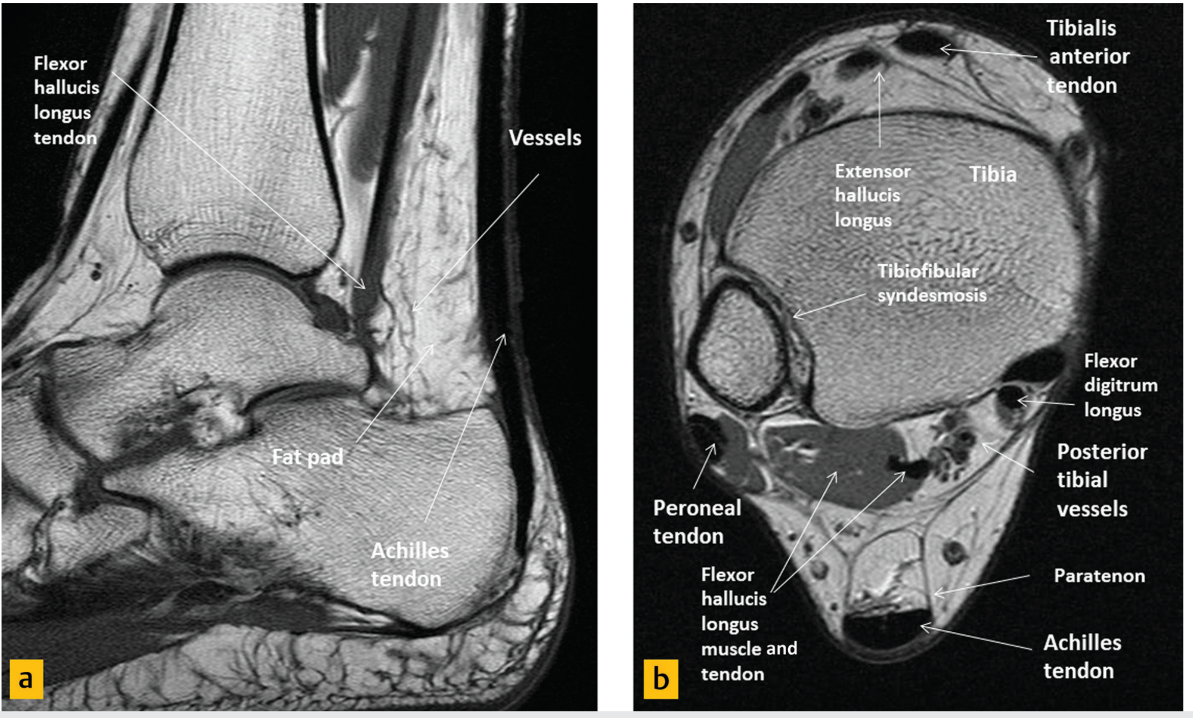
MRI of the hindfoot showing Kager's fat pad (triangular, anterior to Achilles tendon), alongside the FHL tendon - Imaging Anatomy Atlas Vol.3
- Location: A triangular space bounded by:
- Posterior: Achilles tendon
- Anterior: Flexor hallucis longus muscle and tendon
- Inferior: Superior cortex of the calcaneus
- Function: Highly vascular and sensory nerve-innervated (contains mechanoreceptors). During movement, the tip of Kager's fat pad slides in and out of the retrocalcaneal bursa to minimise pressure changes and promote boundary lubrication
- Pathology: In Achilles tendinopathy, oedema and fibrosis of Kager's fat pad is a recognised finding on MRI/ultrasound. This causes pain because it is richly innervated. Fat pad impingement can occur independently of tendon pathology
- Clinical note: A hardened or fibrotic Kager's fat pad may impair normal tendon gliding, perpetuating pain cycles even after the primary tendinopathy improves
(Imaging Anatomy Text and Atlas Vol.3; Rheumatology, Elsevier 2022)
4. Haglund Deformity (Posterosuperior Calcaneal Prominence)
While this is a bony structure, it is a key driver of soft tissue pain in this region.
- A prominent exostosis off the posterosuperior calcaneal tuberosity, located anterior to the Achilles tendon
- During dorsiflexion, the bony prominence repetitively abrades the deep surface of the Achilles tendon and compresses the retrocalcaneal bursa
- This leads to the classic triad: Haglund deformity + retrocalcaneal bursitis + insertional Achilles tendinopathy - often co-existing
- Physiotherapy relevance: Heel lifts reduce dorsiflexion range, taking mechanical stress off the tendon-bursa-bone interface; footwear modification (open-backed shoes) reduces compression
(Campbell's Operative Orthopaedics 15th Ed 2026; Miller's Review of Orthopaedics 9th Ed)
5. Calcaneal Bone / Insertional Footprint (Subtendinous Calcaneus)
- The posterosuperior calcaneal tuberosity where the Achilles inserts can develop:
- Calcaneal enthesophytes (insertional spurs) - bony outgrowths within the tendon at the insertion
- Bone marrow oedema (stress reaction) at the calcaneal tuberosity - visible on MRI as a signal change
- Calcaneal stress reactions cause deep, diffuse aching pain precisely "under" the tendon
- May be a primary source of pain independent of the overlying tendon
(General Anatomy and Musculoskeletal System, Thieme Atlas)
6. Flexor Hallucis Longus (FHL) Tendon
Sitting immediately anterior to Kager's fat pad in the posteromedial ankle:
- FHL runs in a groove under the talus (posterior talar process/os trigonum) and can develop tenosynovitis or stenosis
- Pain is often deep, posteromedial, and can mimic or co-exist with retrocalcaneal pathology
- Key differentiator: FHL pain is most prominent with toe/great toe motion, particularly in athletes (dancers, runners); a "trigger toe" phenomenon may be present
- Associated with posterior ankle impingement and os trigonum syndrome
(Rheumatology, Elsevier 2022 - Differential Diagnosis Table for Posterior Ankle Pain)
7. Superficial (Retro-Achilles / Pre-tendinous) Bursa
This is distinct from the retrocalcaneal bursa and sits posterior to (not deep under) the tendon, between the skin and the posterior Achilles tendon.
- An adventitious bursa - not always present anatomically
- Especially prone to friction from shoe heel counters
- Pain when wearing shoes is a hallmark; less painful on first arising than retrocalcaneal bursitis
- Relevant when differentiating purely deep pain (retrocalcaneal) from superficial posterior pain (retro-Achilles)
(Roberts and Hedges' Clinical Procedures in Emergency; Rheumatology, Elsevier 2022)
8. Os Trigonum / Posterior Talar Impingement
- The os trigonum is an accessory ossicle at the posterior lateral tubercle of the talus, present in ~5-14% of the population
- During forced plantarflexion, it becomes pinched between the tibia and calcaneus, causing posterior ankle impingement syndrome
- Pain is deep, posterior, and reproduced on full plantarflexion (contrast with retrocalcaneal bursitis which worsens on dorsiflexion)
- Often confused with Achilles pathology but requires very different physiotherapy management
(Rheumatology, Elsevier 2022 - Differential Diagnosis Table)
Summary Table - Key Differentiating Features
| Structure | Exact Location | Pain Provocation | Key Clinical Clue |
|---|---|---|---|
| Retrocalcaneal bursa | Deep to Achilles, anterior to tendon insertion | Dorsiflexion, palpation medial/lateral to tendon | Morning stiffness, consider systemic arthritis |
| Insertional Achilles tendon | At calcaneal footprint, deep surface | Dorsiflexion, direct palpation at bone | Calcification on X-ray, thickening |
| Kager's fat pad | Triangular space anterior to tendon | Tendon movement, compression | MRI oedema, often overlooked |
| Haglund deformity | Posterosuperior calcaneal tuberosity | Dorsiflexion, closed-back shoes | Visible bony lump, positive radiograph |
| Calcaneal enthesophyte/BMO | Within/at Achilles insertion on calcaneus | Loading, palpation | MRI confirms, X-ray spur |
| FHL tendon | Posteromedial, deep to fat pad | Great toe flexion, passive toe extension | Tendon pathology in runners/dancers |
| Retro-Achilles bursa | Posterior to tendon (superficial) | Shoe counter pressure | Pain only in shoes |
| Os trigonum | Posterior talus | Forced plantarflexion | Pain on plantarflexion, not dorsiflexion |
Physiotherapy Clinical Pearl
Campbell's Operative Orthopaedics makes a key point worth embedding in your assessment:
"In prolonged or severe cases, all aspects of insertional tendinopathy (retrocalcaneal bursitis, Achilles tendinopathy, and pretendinous bursitis) can exist concomitantly."
This means careful palpation - medial and lateral to the tendon for bursa, directly on the insertion for tendinopathy, and superficial for pretendinous bursa - is essential to identify the dominant pain generator before planning rehabilitation.
Sources: Campbell's Operative Orthopaedics 15th Ed 2026 | Rheumatology 2-Volume Set, Elsevier 2022 | Imaging Anatomy Text and Atlas Vol.3 | Miller's Review of Orthopaedics 9th Ed | General Anatomy and Musculoskeletal System, Thieme Atlas
give me proper reference to learn and read for my exam on Anatomy of Male and female pelvis with structures realted such as Muslces and ligament as well as nerves
For pelvic anatomy exam preparation, the gold-standard references are Gray's Anatomy, Clinically Oriented Anatomy (Moore), and the Thieme Atlas of Anatomy. Let me check what's actually available in the medical library so I can point you to specific chapters and page ranges you can read right now.
Searching sources...
Finding Sources
Good - I can see Gray's Anatomy for Students, Netter's Atlas, and the Thieme/Color atlases are all available. Let me now search all of them for pelvis-specific sections:
Finding Sources
Finding Sources
Finding Sources
Good. Now let me get the detailed TOC structure of the key pelvis chapters in Gray's and Thieme simultaneously:
Reading File
Reading File
Finding Sources
Reading File
Reading File
Now let me get the Thieme pelvic floor muscles section and the full Gray's TOC structure:
Finding Sources
Finding Sources
Finding Sources
Reading File
Reading File
Finding Sources
give me notes with reference
Reading all key sections in parallel:
Reading File
Reading File
Reading File
Reading File
Reading File
Reading File
Reading File
Reading File
Reading File
Reading File
Reading File
Reading File
Reading File
Reading File
Reading File
I now have comprehensive content from the textbooks. Let me fetch a couple of key anatomical images and then compile the full notes:
Loading Image
Loading Image
Loading Image
Loading Image
Loading Image
Here are your complete exam notes on the anatomy of the male and female pelvis, compiled directly from the textbooks in the library.
ANATOMY OF THE MALE AND FEMALE PELVIS
Exam Notes with Textbook References
Primary Sources:
- Gray's Anatomy for Students (Chapter 5: Pelvis and Perineum)
- General Anatomy and Musculoskeletal System - Thieme Atlas of Anatomy (Sections 1.20-1.22, 3.9-3.11)
PART 1 - OVERVIEW AND DIVISIONS
The pelvis and perineum are interrelated regions associated with the pelvic bones and terminal parts of the vertebral column. The pelvis is divided into two regions:
| Region | Also Called | Level | Contents |
|---|---|---|---|
| False (greater) pelvis | Major pelvis | Superior to linea terminalis | Part of abdominal cavity; lower abdominal viscera |
| True (lesser) pelvis | Minor pelvis | Inferior to linea terminalis | Pelvic cavity; urinary, GI, reproductive organs |
The perineum lies inferior to the pelvic floor (infra-levator). It contains the external genitalia and external openings of the genitourinary and GI systems.
(Gray's Anatomy for Students, p.497)
PART 2 - BONY PELVIS
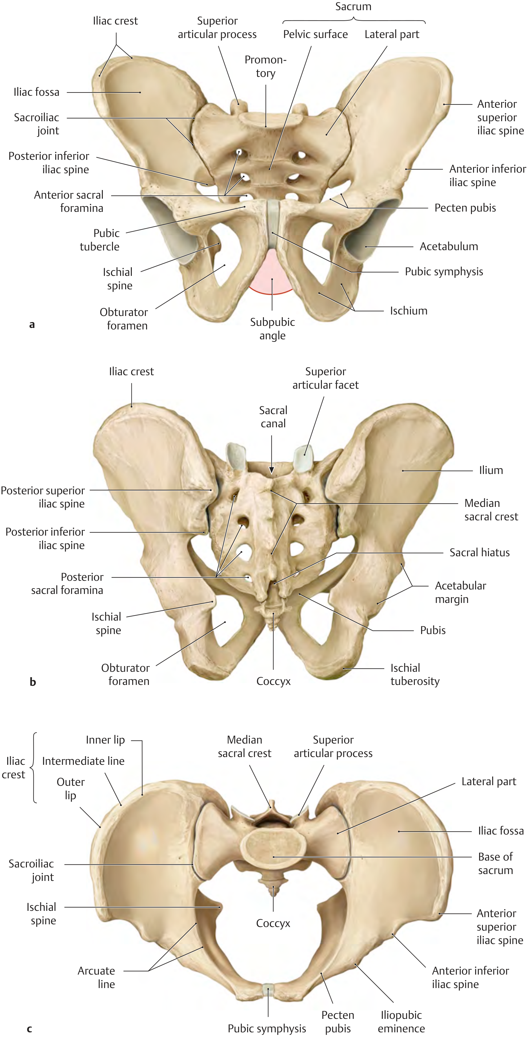
Bones
The pelvic skeleton consists of:
- Two pelvic (hip/coxal) bones - right and left
- Sacrum - articulates superiorly with L5 vertebra
- Coccyx - 4 fused coccygeal vertebrae
The Pelvic Bone (Hip Bone)
Each pelvic bone is formed by the fusion of three elements at the acetabulum (fuses between ages 16-18):
| Element | Position | Key Features |
|---|---|---|
| Ilium | Most superior | Iliac crest, ASIS, AIIS, iliac fossa, auricular surface, arcuate line |
| Pubis | Antero-inferior | Pubic crest, pubic tubercle, pecten pubis (pectineal line), superior and inferior rami |
| Ischium | Postero-inferior | Ischial spine, ischial tuberosity, greater and lesser sciatic notches |
Lateral surface: Bears the acetabulum (hip joint socket) and the obturator foramen (closed by obturator membrane except for obturator canal superiorly).
(Gray's Anatomy for Students, p.516-517)
Sacrum
- Formed by 5 fused sacral vertebrae
- Anterior edge of S1 body projects forward as the promontory (important obstetric landmark)
- 4 pairs of anterior sacral foramina (for anterior rami S1-S4)
- 4 pairs of posterior sacral foramina (for posterior rami S1-S4)
- Sacral canal is a continuation of the vertebral canal; ends as the sacral hiatus
(Gray's Anatomy for Students, p.520)
PART 3 - MALE vs. FEMALE PELVIS DIFFERENCES
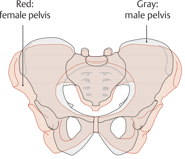
| Feature | Female | Male |
|---|---|---|
| Overall shape | Wider, broader, shallower | Narrower, deeper, taller |
| Pelvic inlet | Oval/round (wider) | Heart-shaped (narrower) |
| Subpubic angle | >80-90° (wide arch) | <70° (narrow arch) |
| Pelvic cavity | Cylindrical, roomier | Funnel-shaped, narrower |
| Sacrum | Shorter, wider, less curved | Longer, narrower, more curved |
| Ischial tuberosities | Farther apart | Closer together |
| Obturator foramen | Oval | Round |
| Obstetric conjugate | 11 cm (critical measurement) | N/A |
| Diagonal conjugate | 12.5-13 cm | N/A |
| AP diameter of outlet | 9 (+2) cm | N/A |
| Transverse diameter of inlet | 13.5 cm | N/A |
Clinical note: The obstetric conjugate (true conjugate = 11 cm) is the narrowest AP dimension of the pelvic inlet and the most important measurement for predicting successful vaginal delivery.
(Thieme Atlas of Anatomy, Section 1.21, p.161-162)
PART 4 - JOINTS OF THE PELVIS
1. Sacroiliac Joint
- Synovial joint (atypical - limited movement)
- Articular surfaces: ear-shaped (auricular) surfaces of ilium and sacrum
- Movements: Nutation (sacral promontory moves forward/down) and counter-nutation
- Stabilised by the strongest ligaments in the body (see Part 5)
- Clinical: Sacroiliac joint blockage from hard impact landing - tested by standing flexion test
2. Pubic Symphysis
- Secondary cartilaginous joint (symphysis)
- Connected by fibrocartilage disc
- Reinforced by superior and inferior (arcuate) pubic ligaments
- Slight movement possible, especially in pregnancy (relaxin effect)
3. Lumbosacral Joint
- Between L5 and S1
- Reinforced by iliolumbar ligaments
(Gray's Anatomy for Students, p.436-438; Thieme Atlas 1.22)
PART 5 - LIGAMENTS OF THE PELVIS
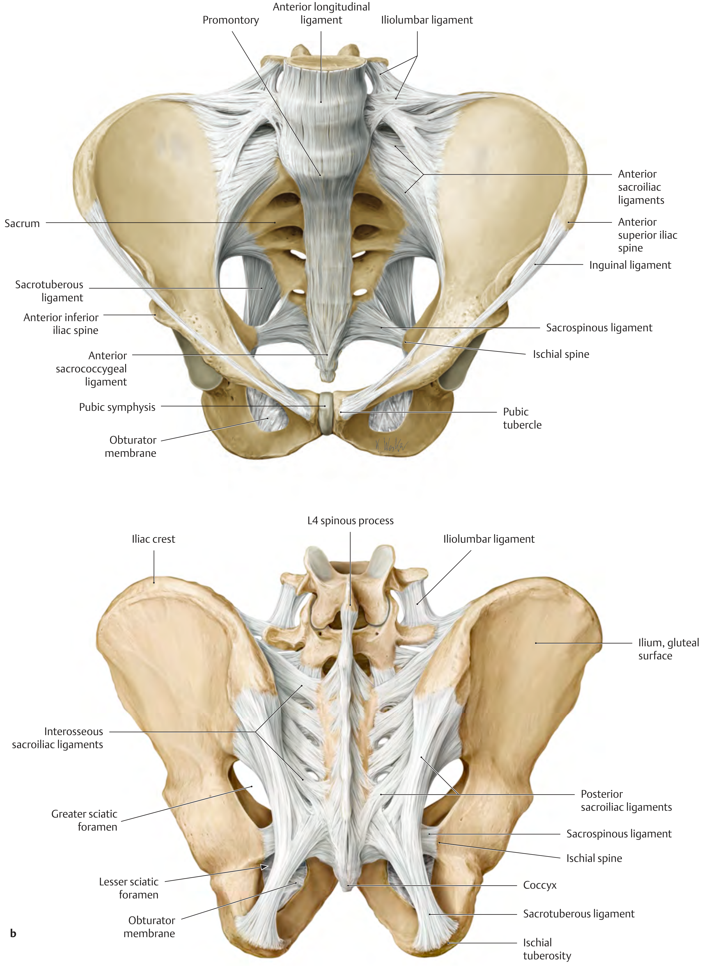
A. Ligaments of the Sacroiliac Joint
| Ligament | Location | Function |
|---|---|---|
| Anterior sacroiliac | Anterior surface of SI joint | Resists anterior displacement |
| Posterior sacroiliac | Posterior surface of SI joint | Resists posterior displacement |
| Interosseous sacroiliac | Deep, within the joint | Strongest; primary stabiliser |
| Iliolumbar | L4-L5 transverse processes to iliac crest | Stabilises lumbosacral junction |
B. Ligaments of the Pelvic Wall (Clinically Critical)
| Ligament | Attachment | What It Creates | Clinical Importance |
|---|---|---|---|
| Sacrospinous | Sacrum/coccyx → ischial spine | Greater and lesser sciatic foramina | Pudendal nerve loops around this |
| Sacrotuberous | Sacrum/PSIS/coccyx → ischial tuberosity | Greater and lesser sciatic foramina | Converts notches to foramina; stabilises sacrum |
Key concept: Both ligaments stabilise the sacrum by resisting upward tilting of the inferior sacrum. Together with the sciatic notches, they create the greater sciatic foramen (superior to sacrospinous ligament) and lesser sciatic foramen (between sacrospinous and sacrotuberous ligaments).
C. Ligaments Specific to the Pelvic Viscera (Women)
| Ligament | Between | Function |
|---|---|---|
| Broad ligament | Uterus → lateral pelvic walls | Peritoneal fold; contains uterine tube in its superior margin |
| Round ligament of uterus | Uterine fundus → labium majus (through inguinal canal) | Anteverts uterus |
| Cardinal (transverse cervical) ligament | Cervix/vaginal vault → lateral pelvic wall | Most important support of uterus |
| Uterosacral ligament | Cervix → posterolateral pelvic wall | Supports uterus; maintains anteversion |
| Pubocervical ligament | Cervix → posterior surface of pubic symphysis | Anterior support |
(Gray's Anatomy for Students, p.474; Thieme Atlas 1.21)
PART 6 - TRUE PELVIS: INLET, WALLS, FLOOR, OUTLET
Pelvic Inlet
Boundary (linea terminalis): Promontory → ala of sacrum → sacroiliac joint → arcuate line → pecten pubis → pubic crest → pubic symphysis
Pelvic Wall - Muscles
The walls of the true pelvis consist of bone + 2 ligaments + 2 muscles:
| Muscle | Origin | Insertion | Innervation | Function |
|---|---|---|---|---|
| Obturator internus | Internal surface of obturator membrane and surrounding bone | Greater trochanter of femur (via lesser sciatic foramen) | Nerve to obturator internus (L5, S1) | Lateral rotator of hip; forms lateral pelvic wall |
| Piriformis | Anterior surface of sacrum (S2-S4) | Greater trochanter of femur (via greater sciatic foramen) | Direct branches (S1, S2) | Lateral rotator of hip; forms posterolateral pelvic wall |
Pelvic Outlet
Diamond-shaped boundary:
- Anterior: Pubic symphysis → inferior border pubis → inferior pubic ramus → ischial ramus → ischial tuberosity (= pubic arch)
- Posterior: Ischial tuberosities → sacrotuberous ligaments → coccyx
(Gray's Anatomy for Students, p.524-528)
PART 7 - PELVIC FLOOR (PELVIC DIAPHRAGM)
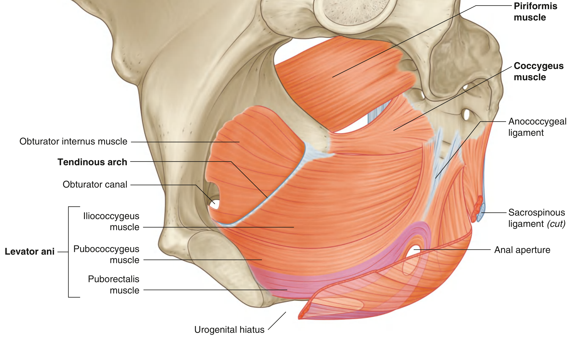
Structure of the Pelvic Floor - Three Levels (Thieme)
- Upper level: Pelvic diaphragm (levator ani + coccygeus + superior and inferior fasciae)
- Middle level: Deep urogenital muscles (deep perineal pouch)
- Lower level: Sphincters and erectile muscles
Muscles of the Pelvic Diaphragm
1. Levator Ani
| Part | Origin | Insertion | Key Function |
|---|---|---|---|
| Pubococcygeus | Posterior pubic body | Midline structures to coccyx | Main component; subdivides into puboprostaticus/pubovaginalis/puboanalis |
| Puborectalis | Pubic body | Forms a sling around rectum | Maintains anorectal angle (~90°); "pinch valve" for continence |
| Iliococcygeus | Tendinous arch (thickening of obturator internus fascia) | Anococcygeal raphe → coccyx | Supports pelvic floor |
Origin (overall): Line from posterior pubic body → tendinous arch (ATLA) in obturator internus fascia → ischial spine
Innervation: Direct branches from anterior ramus of S4 + inferior rectal branch of pudendal nerve (S2-S4)
Functions:
- Supports pelvic viscera
- Maintains anorectal angle; reinforces external anal sphincter
- In women: acts as vaginal sphincter
2. Coccygeus
| Details | |
|---|---|
| Origin | Ischial spine + pelvic surface of sacrospinous ligament |
| Insertion | Lateral margin of coccyx + adjacent sacrum |
| Innervation | Branches from anterior rami of S3 and S4 |
| Function | Completes the posterior pelvic floor; pulls coccyx forward after defecation |
Exam point: The urogenital hiatus is an anterior U-shaped gap in the levator ani through which the urethra (both sexes) and vagina (women) pass. The margins merge with the walls of these viscera. This is the anatomical weak point of the female pelvic floor.
(Gray's Anatomy for Students, p.529-530; Thieme Atlas 3.11)
Gender Difference in Levator Ani
In the female, there are connective tissue gaps between the muscular parts of the levator ani (making the female pelvic floor more susceptible to injury during childbirth). In the male, these gaps are minimal.
(Thieme Atlas 3.11, p.207)
PART 8 - FASCIA AND PERITONEAL RELATIONS
Pelvic Fascia
Pelvic fascia lines the pelvic walls, surrounds the bases of viscera, and forms sheaths around vessels and nerves.
In Women: Condensations form:
- Pubocervical ligament (anterior)
- Cardinal (transverse cervical) ligament (lateral) - most important uterine support
- Uterosacral ligament (posterior)
- Rectovaginal septum (separates vagina from rectum)
In Men:
- Prostatic fascia surrounds the prostate and contains the prostatic venous plexus
- Rectovesical septum separates prostate/bladder base from rectum
(Gray's Anatomy for Students, p.474)
Peritoneal Pouches (Important Exam Topics)
| Pouch | Location | Significance |
|---|---|---|
| Vesico-uterine pouch | Between bladder and uterus (women) | Shallow; anterior |
| Recto-uterine pouch (Pouch of Douglas) | Between uterus/vagina and rectum (women) | Deepest point of abdominopelvic cavity in supine position; site of fluid/pus collection |
| Rectovesical pouch | Between bladder and rectum (men) | Deepest peritoneal recess in men |
(Gray's Anatomy for Students, p.559-560)
PART 9 - NERVES OF THE PELVIS
A. Sacral Plexus
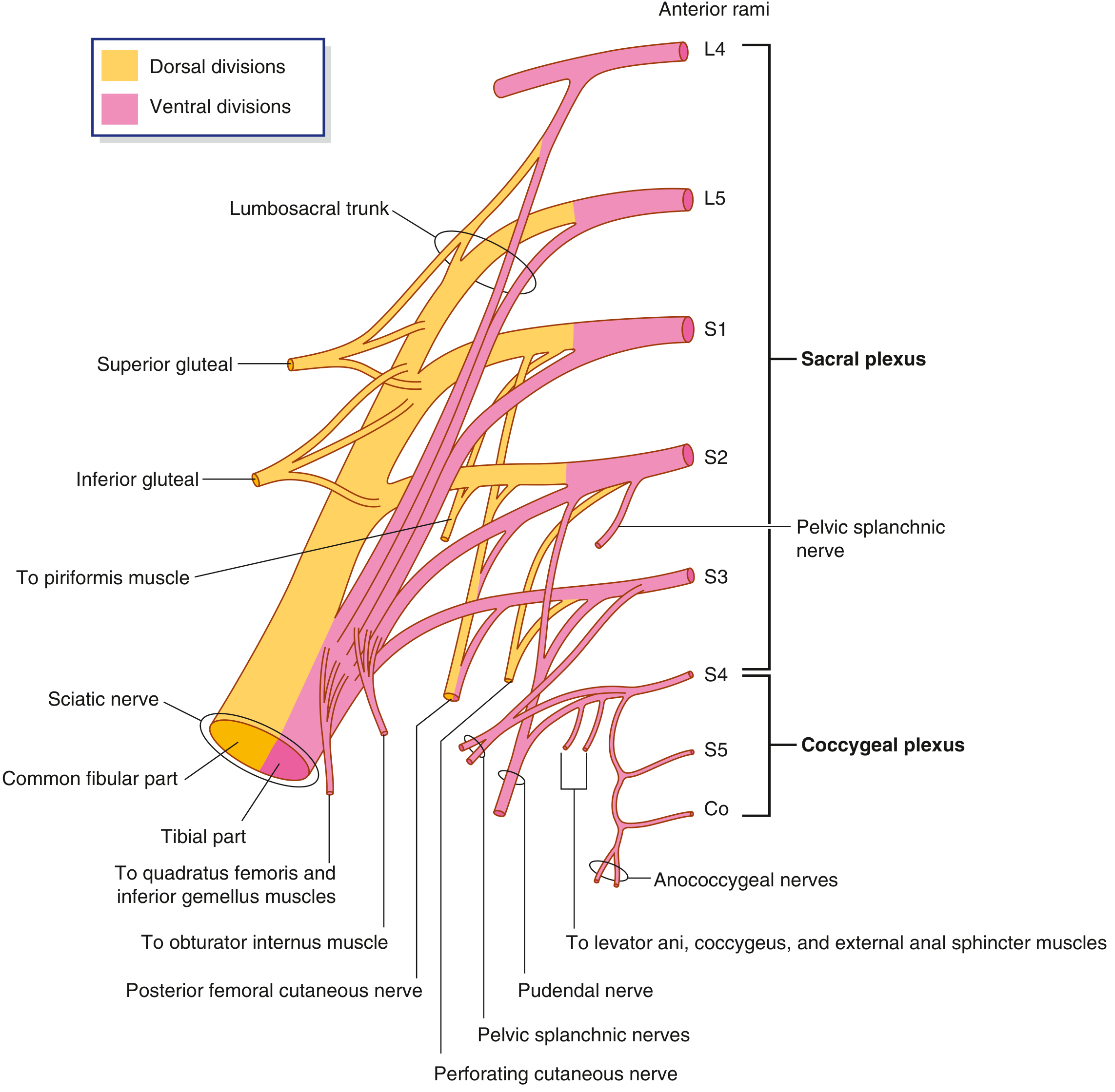
Formation: Anterior rami of L4-S4 (via lumbosacral trunk L4+L5 and sacral roots S1-S4)
Location: Anterior surface of piriformis muscle on the posterolateral pelvic wall
| Branch | Spinal Levels | Motor | Sensory |
|---|---|---|---|
| Sciatic nerve (largest nerve in body) | L4-S3 | All posterior thigh, all posterior leg, all foot muscles | Posterolateral foot/sole |
| Superior gluteal | L4-S1 | Gluteus medius, minimus, TFL | - |
| Inferior gluteal | L5-S2 | Gluteus maximus | - |
| Pudendal nerve | S2-S4 | All perineal muscles; external urethral + anal sphincters; levator ani | Most perineal skin; penis/clitoris |
| Pelvic splanchnic nerves | S2-S4 | Parasympathetic preganglionic to pelvic viscera (= nervi erigentes) | Visceral afferents |
| Nerve to obturator internus | L5-S2 | Obturator internus + superior gemellus | - |
| Nerve to quadratus femoris | L4-S1 | Quadratus femoris + inferior gemellus | - |
| Posterior femoral cutaneous | S1-S3 | - | Posterior thigh skin |
| Perforating cutaneous | S2-S3 | - | Gluteal skin |
Key route of pudendal nerve: Exits pelvic cavity via greater sciatic foramen → loops around ischial spine and sacrospinous ligament → re-enters via lesser sciatic foramen → enters the perineum via the pudendal canal (Alcock's canal). This is why the ischial spine is the landmark for pudendal nerve block.
(Gray's Anatomy for Students, Table 5.4, p.565-567)
B. Autonomic Nerves of the Pelvis
| Nerve | Type | Levels | Function |
|---|---|---|---|
| Pelvic splanchnic nerves | Parasympathetic (preganglionic) | S2-S4 | Erection; bladder contraction; rectal peristalsis |
| Sacral splanchnic nerves | Sympathetic (postganglionic) | L1-L2 via hypogastric plexus | Ejaculation; bladder neck contraction; vasoconstriction |
| Inferior hypogastric plexus | Mixed autonomic | - | Combines sympathetic + parasympathetic for pelvic viscera |
| Superior hypogastric plexus | Sympathetic | L1-L2 | Descends from abdomen to pelvis |
(Gray's Anatomy for Students, p.479, 488)
C. Obturator Nerve (from Lumbar Plexus - L2,3,4)
- Exits pelvis via obturator canal (obturator foramen)
- Motor: medial compartment thigh (adductors)
- Sensory: medial thigh
- Clinically: can be compressed by pelvic tumours/lymph nodes
PART 10 - SCIATIC FORAMINA AND COMMUNICATION ROUTES
| Foramen | Bounded By | Level relative to pelvic floor | Structures Passing Through |
|---|---|---|---|
| Greater sciatic foramen | Greater sciatic notch + sacrospinous ligament (superior border) | Above pelvic floor | Piriformis muscle; sciatic nerve; superior/inferior gluteal nerves + vessels; nerve to obturator internus; pudendal nerve (on exit only); posterior femoral cutaneous nerve |
| Lesser sciatic foramen | Lesser sciatic notch + sacrospinous + sacrotuberous ligaments | Below pelvic floor | Pudendal nerve + internal pudendal vessels (re-entering to perineum); nerve to obturator internus (tendon of obturator internus) |
(Gray's Anatomy for Students, p.527)
PART 11 - PELVIC SPACES AND SUBDIVISIONS
| Space/Level | Location | Contents |
|---|---|---|
| Peritoneal cavity of lesser pelvis | Upper level (supra-peritoneal) | Peritoneum-covered viscera |
| Subperitoneal space | Lower level (infra-peritoneal but supra-levator) | Viscera, vessels, nerves, fascia |
| Deep perineal space | Infra-levator | Deep perineal muscles (deep transverse perineal, external urethral sphincter) |
| Superficial perineal space | Infra-levator | Superficial perineal muscles, erectile tissue |
| Subcutaneous perineal space | Most superficial | Fatty tissue of perineum |
(Thieme Atlas 3.9, p.203)
SUMMARY - QUICK RECALL TABLE
| Topic | Key Points |
|---|---|
| Bones | 2 hip bones (ilium + ischium + pubis) + sacrum + coccyx |
| Joints | Sacroiliac (synovial), pubic symphysis (secondary cartilaginous), lumbosacral |
| Pelvic wall muscles | Obturator internus (medial wall), Piriformis (posterolateral wall) |
| Pelvic floor muscles | Levator ani (pubococcygeus, puborectalis, iliococcygeus) + Coccygeus |
| Levator ani innervation | S4 direct + pudendal nerve (S2-S4) |
| Critical ligaments | Sacrospinous (apex = ischial spine) + sacrotuberous (apex = ischial tuberosity) |
| Female fascial supports | Cardinal > uterosacral > pubocervical ligaments |
| Sacral plexus | L4-S4; formed on piriformis |
| Pudendal nerve | S2-S4; nerve of perineum; loops around ischial spine |
| Parasympathetics | S2-S4 pelvic splanchnics (erection, bladder, bowel) |
| Sympathetics | L1-L2 via hypogastric plexus (ejaculation, bladder neck) |
| Key pouches | Pouch of Douglas (women) = rectouterine; Rectovesical (men) |
RECOMMENDED READING CHAPTERS
| Book | Chapter / Section | Pages | What to Focus On |
|---|---|---|---|
| Gray's Anatomy for Students | Chapter 5: Pelvis and Perineum | 413-522 | Full chapter; especially Tables 5.2, 5.4 for muscles/nerves |
| Thieme Atlas of Anatomy - Vol 1 | Sections 1.20-1.22 (Bony pelvis, ligaments, measurements) | 158-167 | Diagrams of male/female differences, all ligaments labelled |
| Thieme Atlas of Anatomy - Vol 1 | Sections 3.9-3.11 (Pelvic floor structure and muscles) | 200-210 | Best diagrams of levator ani parts; pelvic floor levels |
| Color Atlas of Human Anatomy Vol. 2 | Pelvis section (Internal Organs) | Block 1 | Peritoneal relations; male/female comparative diagrams |
All four books are available in the library. The images directly above are from these textbooks.
This is a shared conversation. Sign in to Orris to start your own chat.