
describe the anatomy of anterior abdominal wall mention the measure to be taken to prevent inscisional hernia 10 mark main answer with proper diagram easy and flow charts with easy explanation for m.s main exam from berek and novak gynaecology exam
Reading File
Finding Sources
Finding Sources
Finding Sources
Reading File
Reading File
Finding Sources
Reading File
Reading File
Finding Sources
Reading File
Loading Image
Loading Image
Loading Image
I now have all the key information needed. Let me compose a comprehensive, exam-ready answer.
ANATOMY OF THE ANTERIOR ABDOMINAL WALL & PREVENTION OF INCISIONAL HERNIA
M.S. Main Exam Answer | 10 Marks
PART 1 - ANATOMY OF THE ANTERIOR ABDOMINAL WALL
LAYERS (Surface to Deep) - The KEY Flow Chart
SKIN
↓
SUBCUTANEOUS TISSUE (Superficial Fascia)
├── Fatty Layer (Fascia of Camper)
└── Membranous Layer (Fascia of Scarpa)
↓ (fuses below to form dartos in scrotum/labia majora)
MUSCLES & THEIR APONEUROSES
├── External Oblique Muscle / Aponeurosis
├── Internal Oblique Muscle / Aponeurosis
└── Transversus Abdominis Muscle / Aponeurosis
↓
TRANSVERSALIS FASCIA
↓
PREPERITONEAL FAT (Extra-peritoneal adipose tissue)
↓
PARIETAL PERITONEUM
DIAGRAM 1 - Cross-Section of Anterior Abdominal Wall (Above Umbilicus)
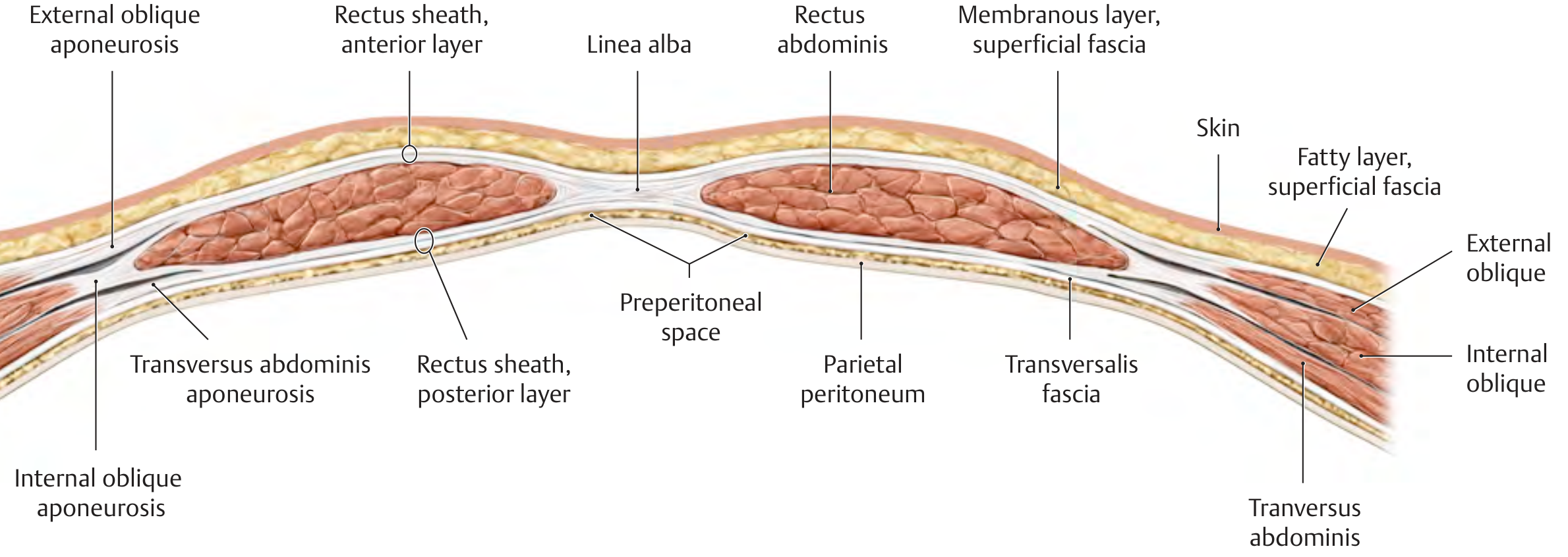
(THIEME Atlas of Anatomy - Cross-section showing all abdominal wall layers, linea alba, and rectus sheath)
A. SUPERFICIAL FASCIA
| Layer | Old Name | Contents |
|---|---|---|
| Fatty layer | Fascia of Camper | Fat, loose connective tissue |
| Membranous layer | Fascia of Scarpa | Dense membranous, important in surgery - closes as separate layer |
Exam Tip: Scarpa's fascia is clinically important - it forms a separate surgical layer; in wound closure it is sutured separately to reduce dead space and prevent seroma.
B. MUSCLES OF THE ANTERIOR ABDOMINAL WALL
FLAT MUSCLES (Lateral Group - 3 layers)
| Muscle | Origin | Insertion | Action | Nerve |
|---|---|---|---|---|
| External Oblique | Lower 8 ribs | Iliac crest, pubic tubercle, linea alba via aponeurosis | Compresses abdomen, flexion, rotation to opposite side | T7-T12 |
| Internal Oblique | Thoracolumbar fascia, iliac crest, inguinal ligament | Lower 3 ribs, linea alba, pubic crest | Compression, rotation to same side | T7-L1 |
| Transversus Abdominis | Lower 6 costal cartilages, thoracolumbar fascia, iliac crest | Linea alba, pubic crest via aponeurosis | Compresses abdomen | T7-L1 |
VERTICAL MUSCLES (Medial Group)
| Muscle | Origin | Insertion | Action |
|---|---|---|---|
| Rectus Abdominis | Pubic symphysis, pubic crest | Xiphoid process, costal cartilages 5-7 | Flexion of trunk, compresses abdomen |
| Pyramidalis | Pubic body | Linea alba | Tenses linea alba |
C. RECTUS SHEATH
This is the most important structure for the surgeon. It is formed by the aponeuroses of the 3 lateral muscles.
ABOVE ARCUATE LINE
┌─────────────────────────────┐
│ ANTERIOR LAYER │
│ External oblique aponeurosis │
│ + Ant half of int. oblique │
├─────────────────────────────┤
│ RECTUS ABDOMINIS MUSCLE │
├─────────────────────────────┤
│ POSTERIOR LAYER │
│ Post half of int. oblique │
│ + Transversus aponeurosis │
└─────────────────────────────┘
BELOW ARCUATE LINE
(3-5 cm below umbilicus)
┌─────────────────────────────┐
│ ANTERIOR LAYER │
│ All 3 aponeuroses pass in │
│ FRONT of rectus muscle │
├─────────────────────────────┤
│ RECTUS ABDOMINIS MUSCLE │
├─────────────────────────────┤
│ NO POSTERIOR LAYER │
│ Only transversalis fascia │
│ + parietal peritoneum │
└─────────────────────────────┘
Arcuate Line (of Douglas): 3-5 cm below umbilicus. Below this, posterior rectus sheath is ABSENT - making the lower abdomen a relatively weak zone.
D. LINEA ALBA & LINEA SEMILUNARIS
- Linea Alba: Fibrous band from xiphoid to pubic symphysis at the midline, formed by decussating fibers of all 3 aponeuroses. The site of midline (median) incisions in surgery.
- Linea Semilunaris: Lateral curved border of rectus abdominis - clinically relevant as the site of spigelian hernias.
E. BLOOD SUPPLY
SUPERIOR EPIGASTRIC (from internal thoracic)
↓ runs within rectus sheath
ANASTOMOSES
↑ runs within rectus sheath
INFERIOR EPIGASTRIC (from external iliac)
Other vessels: Deep circumflex iliac artery, lower posterior intercostal arteries (T7-T11), subcostal artery (T12).
F. NERVE SUPPLY
- T7-T11: Lower intercostal nerves (segmental supply)
- T12: Subcostal nerve
- L1: Iliohypogastric and ilio-inguinal nerves (lower abdominal wall)
All run between internal oblique and transversus abdominis before piercing to supply skin.
G. PERITONEAL FOLDS ON INTERNAL SURFACE (Posterior View)
5 folds converge toward umbilicus:
UMBILICUS
|
┌─────────────────────┼─────────────────────┐
│ │ │
Lateral fold Medial fold Median fold
(paired) (paired) (unpaired)
Contains Contains Contains
Inf. epigastric Obliterated umbilical Obliterated urachus
vessels artery
│ │ │
▼ ▼ ▼
Lateral Medial inguinal Supravesical
inguinal fossa fossa fossa
(Deep inguinal (Hesselbach's (above bladder)
ring - indirect triangle - direct
hernia site) hernia site)
DIAGRAM 2 - Internal Surface of Anterior Abdominal Wall
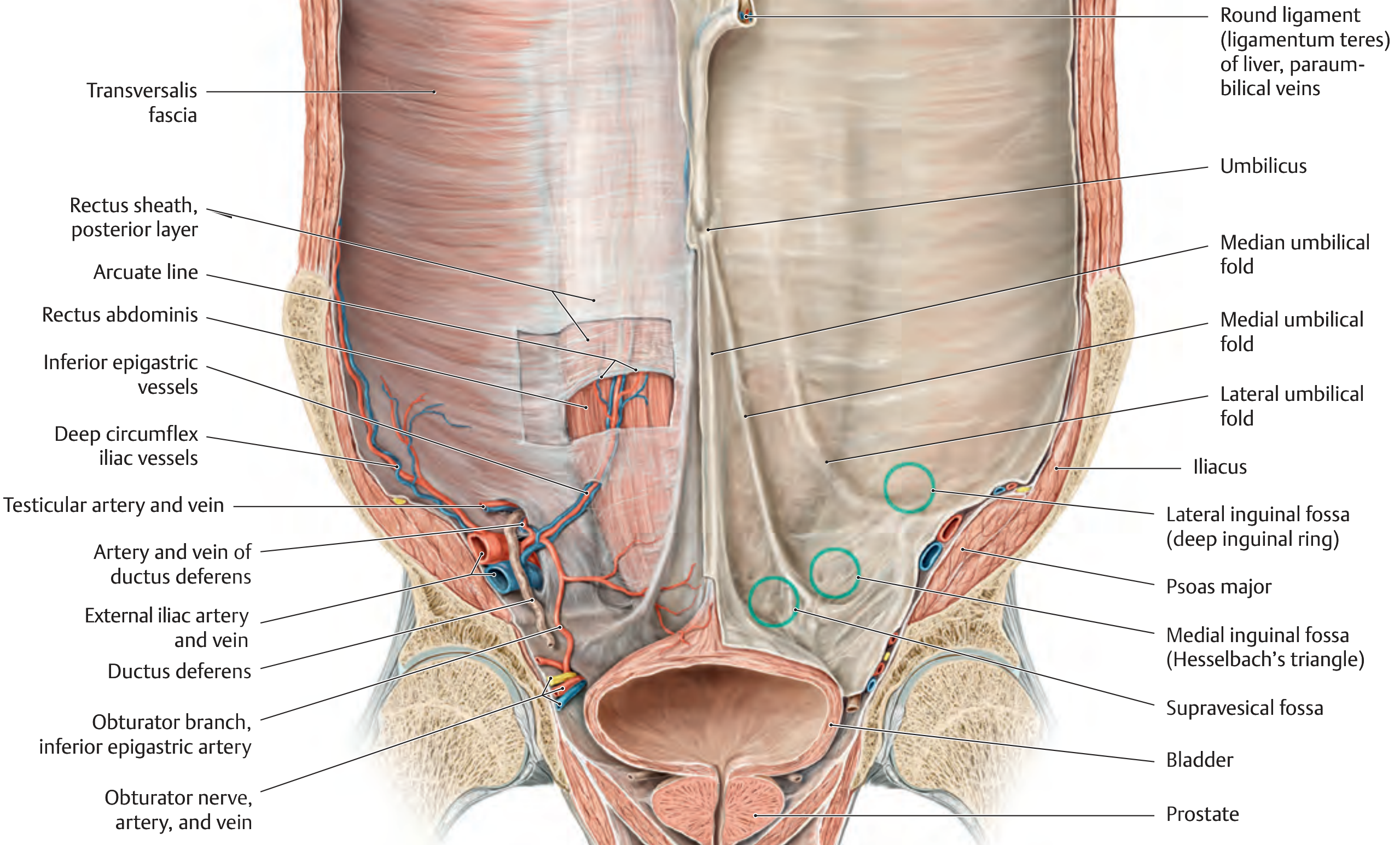
(THIEME Atlas - Internal view showing peritoneal folds, arcuate line, inferior epigastric vessels, Hesselbach's triangle)
H. WEAK SPOTS IN THE ANTERIOR ABDOMINAL WALL
| Weak Spot | Location | Hernia Type |
|---|---|---|
| Deep inguinal ring (lateral fossa) | Lateral to inf. epigastric vessels | Indirect inguinal hernia |
| Hesselbach's triangle (medial fossa) | Medial to inf. epigastric vessels | Direct inguinal hernia |
| Femoral ring | Below inguinal ligament, medial to femoral vein | Femoral hernia |
| Umbilicus | Defect in linea alba at umbilicus | Umbilical hernia |
| Linea alba | Epigastric region | Paraumbilical / epigastric hernia |
| Spigelian line | Lateral edge of rectus | Spigelian hernia |
| Previous incision sites | Any | Incisional hernia |
PART 2 - PREVENTION OF INCISIONAL HERNIA
DEFINITION
An incisional hernia is a protrusion of abdominal contents through a defect in the anterior abdominal wall at a previous surgical incision site.
RISK FACTORS (Flow Chart)
RISK FACTORS FOR INCISIONAL HERNIA
│
┌──────┴──────────────────┐
PATIENT SURGICAL
FACTORS FACTORS
│ │
┌──┴──┐ ┌───────┴──────┐
Obesity Smoking Wrong incision Poor closure
Malnutrition DM technique technique
Jaundice Steroids Wrong suture Infection
COPD Anaemia material Haematoma
Old age Cancer Drain through Tension
wound closure
PREVENTIVE MEASURES
A. PREOPERATIVE OPTIMIZATION
- Smoking cessation - reduces tissue hypoxia, improves wound healing
- Weight loss in obese patients - 5 kg reduction creates ~1 litre of extra intra-abdominal space (reduces tension)
- Nutritional optimization - protein supplementation in malnourished patients
- Control of DM - HbA1c < 7 before elective surgery
- Treat respiratory disease - reduces coughing strain on wound postoperatively
- Correction of anaemia and jaundice - jaundice impairs healing
B. CHOICE OF INCISION
| Incision | Hernia Risk | Notes |
|---|---|---|
| Transverse / Pfannenstiel | Low | Preferred in gynaecology - splits along Langer's lines |
| Midline (vertical) | Higher | Strong for access, but linea alba is relatively avascular |
| Paramedian | Low | Splits rectus sheath, healing reinforced by muscle |
Rule: Transverse incisions have lower hernia rates than vertical midline incisions because they align with the direction of tissue fibers.
C. WOUND CLOSURE TECHNIQUE (Most Important)
The "STITCH Trial" and subsequent evidence revolutionized closure technique:
TRADITIONAL (WRONG) MODERN EVIDENCE-BASED (CORRECT)
───────────────────── ──────────────────────────────
1 cm bites, 1 cm apart vs 5 mm bites, 5-8 mm from edge
Heavy non-absorbable vs 2/0 slowly absorbable suture
Suture:wound ratio < 4:1 vs Suture:wound ratio ≥ 4:1
Muscle included vs FASCIA ONLY - no muscle in bite
Key Rules for Closure:
- Sutures 5 mm apart and 5-8 mm back from wound edge
- Include fascia only in suture bites - do NOT incorporate muscle
- Suture length to wound length ratio must be ≥ 4:1 (prevents ischemia)
- Use 2/0 slowly absorbable suture (e.g., PDS, Monocryl) - NOT non-absorbable, NOT rapidly absorbable
- If continuous suture: do NOT pull too tight or place too close to fascial edge (cuts through)
- No evidence that interrupted is better than continuous suturing
D. DRAIN PLACEMENT
- Drains must ALWAYS exit through a separate stab incision - NEVER through the wound itself
- Drain through the wound prevents fascial apposition and directly causes hernia formation
E. PROPHYLACTIC MESH
PROPHYLACTIC MESH - Indications:
│
┌──────┴───────────┐
Obese patients Stoma formation
undergoing (parastomal hernia
bariatric surgery rate up to 50%
without mesh)
│
Substantially reduces
incisional hernia risk
- In high-risk patients (obese, bariatric surgery), prophylactic mesh placement substantially reduces incisional hernia risk
- Mesh also reduces parastomal hernia rate (which otherwise occurs in up to 50% of patients)
F. WOUND INFECTION PREVENTION
- Prophylactic antibiotics at induction
- Meticulous haemostasis - avoid haematoma
- Gentle tissue handling - avoid crush injury
- Avoid excessive wound tension
- Layered closure of Scarpa's fascia to eliminate dead space (prevents seroma)
SUMMARY FLOW CHART - PREVENTION
PREVENT INCISIONAL HERNIA
│
┌─────┴──────────────────────────┐
PREOPERATIVE INTRAOPERATIVE
│ │
Optimize patient: ┌───────┴──────────┐
- Stop smoking TECHNIQUE MATERIAL
- Lose weight - Transverse incision 2/0 slowly
- Treat DM/COPD - Fascia only in bites absorbable
- Nutrition - 5mm bites, 5-8mm (PDS/Monocryl)
- Correct anaemia from edge
- SL:WL ratio ≥ 4:1
- Drain NOT through wound
- Prophylactic mesh
in high-risk
│
POSTOPERATIVE
│
- Early mobilization
- Treat chest infection (prevents coughing strain)
- Abdominal binder in obese patients
- Avoid heavy lifting for 6 weeks
QUICK REVISION TABLE
| Factor | Recommendation |
|---|---|
| Suture material | 2/0 slowly absorbable (PDS) |
| Bite size | 5 mm apart, 5-8 mm from edge |
| SL:WL ratio | ≥ 4:1 |
| Tissue included | Fascia ONLY |
| Drain site | Separate stab incision |
| Best incision | Transverse / Pfannenstiel |
| High-risk prophylaxis | Prophylactic mesh |
| Preop | Stop smoking, lose weight, correct nutrition |
SOURCES
- General Anatomy and Musculoskeletal System - THIEME Atlas of Anatomy (Sections 3.7, 5.8)
- Bailey and Love's Short Practice of Surgery, 28th Edition - Chapter on Incisional Hernia, Prevention section (p. 2997-3019)
Exam Writing Tip for M.S.: In your main exam answer, always start with the layers table/flow chart, then go to muscles with a cross-section diagram, then rectus sheath (above vs below arcuate line is a favorite examiner question), then blood/nerve supply, then weak spots table. For prevention, the "5mm bites, 5-8mm back, SL:WL ratio 4:1, fascia only, 2/0 PDS" points are high-yield - examiners love these specific numbers. Prophylactic mesh in obese/bariatric patients is a modern high-yield addition.
describe anatomy of rectus sheath.write the different abdominal incisions used in obstetrics and gynaecology 10 mark answer from berek and novak with flow charts diagrams and proper explanation m.s obgyn main exam
Finding Sources
Finding Sources
Finding Sources
Finding Sources
Reading File
Finding Sources
Reading File
Finding Sources
Finding Sources
Finding Sources
Finding Sources
Reading File
Loading Image
Loading Image
Excellent - I now have all the key diagrams and textbook content from Berek & Novak's Gynecology and Gray's Anatomy. Let me now compose the comprehensive exam answer.
ANATOMY OF RECTUS SHEATH & ABDOMINAL INCISIONS IN OBG
M.S. OBG Main Exam Answer | 10 Marks
PART 1 - ANATOMY OF THE RECTUS SHEATH
INTRODUCTION
The rectus sheath is an aponeurotic fibrous compartment that encloses the rectus abdominis and pyramidalis muscles. It is formed by the interlacing aponeuroses of the three flat muscles of the lateral abdominal wall: the external oblique, internal oblique, and transversus abdominis.
(Berek & Novak's Gynecology, Fig. 5-18)
DIAGRAM 1 - Anterior View: Rectus Sheath & Abdominal Muscles (From Berek & Novak)
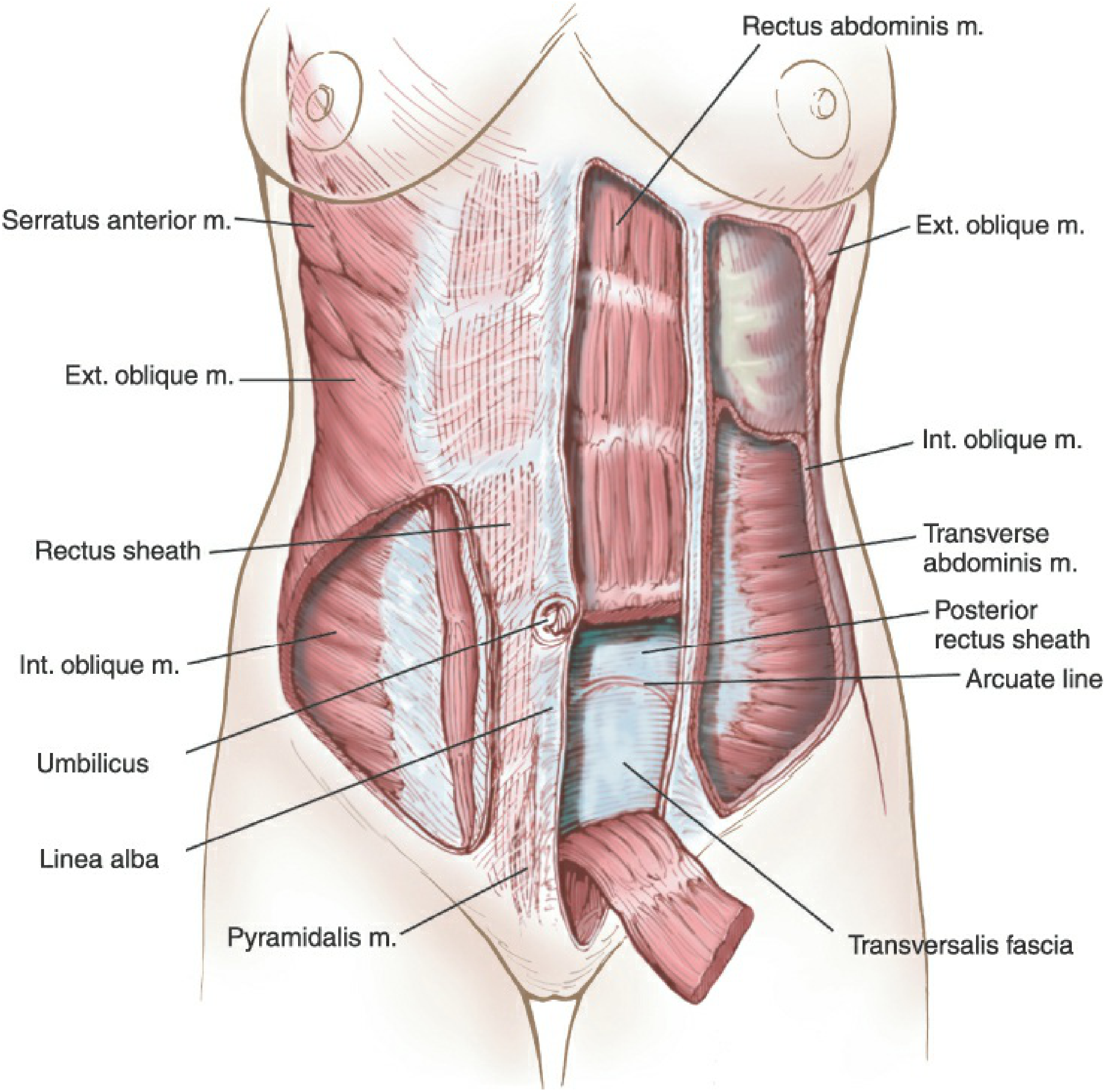
DIAGRAM 2 - Cross-Sections: Above and Below the Arcuate Line (Gray's Anatomy)
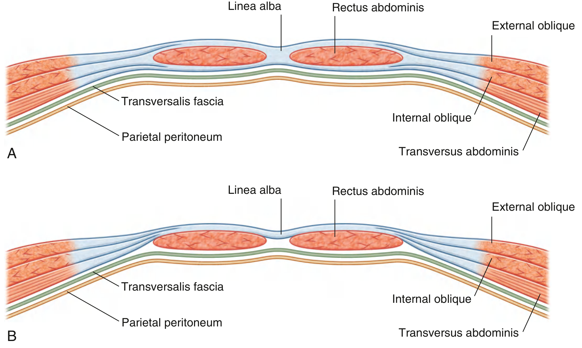
A. FORMATION - The Most Important Concept
The internal oblique aponeurosis SPLITS at the lateral margin of the rectus abdominis - this is the key to understanding the entire sheath.
THREE APONEUROSES CONTRIBUTING TO RECTUS SHEATH:
External Oblique Aponeurosis (never splits - always goes ANTERIOR)
+
Internal Oblique Aponeurosis (SPLITS into 2 lamellae at lateral margin of rectus)
+
Transversus Abdominis Aponeurosis (behavior CHANGES at arcuate line)
B. RECTUS SHEATH: ABOVE vs BELOW ARCUATE LINE
╔══════════════════════════════════╦══════════════════════════════════╗
║ ABOVE ARCUATE LINE ║ BELOW ARCUATE LINE ║
║ (Upper 3/4 of rectus) ║ (Lower 1/4 of rectus) ║
╠══════════════════════════════════╬══════════════════════════════════╣
║ ANTERIOR WALL: ║ ANTERIOR WALL: ║
║ • External oblique aponeurosis ║ ALL THREE pass anterior: ║
║ • Anterior lamella of internal ║ • External oblique aponeurosis ║
║ oblique aponeurosis ║ • Internal oblique aponeurosis ║
║ ║ • Transversus aponeurosis ║
╠══════════════════════════════════╬══════════════════════════════════╣
║ POSTERIOR WALL: ║ NO POSTERIOR WALL ║
║ • Posterior lamella of internal ║ • Only Transversalis fascia ║
║ oblique aponeurosis ║ • Preperitoneal fat ║
║ • Transversus aponeurosis ║ • Parietal peritoneum ║
╚══════════════════════════════════╩══════════════════════════════════╝
Key Exam Point (Berek & Novak): "The aponeurosis of the internal oblique muscle splits into anterior and posterior lamella above the arcuate line. Below this line, all three layers are anterior to the body of the rectus muscle."
C. THE ARCUATE LINE (Line of Douglas)
- Located midway between umbilicus and pubic symphysis (approximately 3-5 cm below umbilicus)
- Marks the point where the posterior rectus sheath ends
- Below the arcuate line, the rectus abdominis is in DIRECT contact with transversalis fascia
- Clinical importance: The inferior epigastric vessels enter the rectus sheath at the arcuate line - they can be damaged by lateral incisions or laparoscopic port insertion
D. BOUNDARIES & CONTENTS
| Feature | Detail |
|---|---|
| Medial boundary | Linea alba (where both sheaths fuse) |
| Lateral boundary | Linea semilunaris (spigelian line) |
| Contents | Rectus abdominis, pyramidalis, superior & inferior epigastric vessels, terminal branches T7-T12 |
| Tendinous intersections | 3 crossing lines that adhere to ANTERIOR sheath only (NOT posterior) |
E. LINEA ALBA & LINEA SEMILUNARIS
- Linea alba: Fibrous raphe at the midline from xiphoid to pubic symphysis formed by decussating fibers of all 3 aponeuroses. Site of midline laparotomy in gynecology.
- Linea semilunaris: Lateral border of the rectus sheath where aponeuroses leave the muscle - the site of Spigelian hernias.
F. BLOOD SUPPLY OF RECTUS SHEATH
SUPERIOR EPIGASTRIC ARTERY
(from internal thoracic artery)
|
↓ travels inside rectus sheath, BEHIND rectus muscle
ANASTOMOSES at umbilical level
↑ travels inside rectus sheath, entering at arcuate line
INFERIOR EPIGASTRIC ARTERY
(from external iliac artery)
Surgical significance (Berek & Novak): The inferior epigastric artery is "vulnerable to damage by abdominal incisions in which the rectus muscle is completely or partially transected, during placement of lateral laparoscopic ports, or by excessive lateral traction on the rectus."
G. NERVE SUPPLY
- T7-T11: Lower intercostal nerves
- T12: Subcostal nerve
- L1: Iliohypogastric and ilio-inguinal nerves (lower wall)
- Injury to iliohypogastric/ilio-inguinal nerves occurs in ~4% of Pfannenstiel incisions (Berek & Novak)
H. TENDINOUS INTERSECTIONS
- 3 fibrous bands cross the rectus abdominis transversely
- Located at: xiphoid, umbilicus, midway between the two
- Fused to the anterior rectus sheath only - NOT the posterior
- Divide the rectus into muscular segments
- These intersections explain why blood/pus in a rectus sheath hematoma is compartmentalized
PART 2 - ABDOMINAL INCISIONS IN OBG
CLASSIFICATION FLOW CHART
ABDOMINAL INCISIONS IN OBG
│
┌───────┴────────────────────────┐
VERTICAL TRANSVERSE
│ │
┌──┴──────┐ ┌───────┴──────────────┐
Midline Paramedian Pfannenstiel Maylard
(Median) (most common) Cherney
│
┌────┴────┐
Modified Joel-Cohen
Pfannenstiel (Misgav
Ladach)
1. VERTICAL (MIDLINE / MEDIAN) INCISION
Position: From pubic symphysis to umbilicus (or above as needed)
SKIN → SUBCUTANEOUS FAT → LINEA ALBA → TRANSVERSALIS FASCIA → PERITONEUM
| Feature | Detail |
|---|---|
| Where | Through the linea alba - avascular midline fibrous band |
| Wound access | Excellent - can extend upward easily |
| Blood loss | Minimal (linea alba is relatively avascular) |
| Hernia risk | Higher (avascular linea alba heals slowly) |
| Nerve damage | Minimal |
| Closure | Linea alba sutured in single layer |
Indications in OBG:
- Emergency laparotomy (ruptured ectopic, massive hemorrhage)
- Radical hysterectomy / pelvic exenteration
- Ovarian cancer staging and debulking
- Where maximum exposure is required
- When significant bowel involvement is anticipated
Advantages: Rapid entry, easily extended, minimal blood loss, suitable for any pelvic procedure
Disadvantages: More incisional hernias, cosmetically poor, slower healing, greater postoperative pain
2. PFANNENSTIEL INCISION (Most Common in OBG)
Named after: Hermann Pfannenstiel (1862-1909)
SKIN (curved transverse, 2-3 cm above pubic symphysis)
↓
SUBCUTANEOUS FAT
↓
ANTERIOR RECTUS SHEATH (incised transversely - "smile" shape)
↓
SHEATH REFLECTED UP AND DOWN (freed from rectus muscle)
↓
RECTUS MUSCLES SEPARATED IN MIDLINE (not cut - split vertically)
↓
TRANSVERSALIS FASCIA + PERITONEUM (opened vertically)
Key anatomy: The skin incision is transverse but the peritoneum is opened vertically - giving two different planes of entry.
| Feature | Detail |
|---|---|
| Skin incision | Curved, 10-15 cm, 2-3 cm above pubic symphysis |
| Fascial incision | Transverse |
| Peritoneal entry | Vertical (midline) |
| Muscles | Retracted - NOT cut |
| Cosmesis | Excellent (hidden by pubic hair line) |
| Hernia risk | Low |
| Access | Limited - cannot extend for wider field |
Indications:
- Cesarean section (most common indication)
- Total abdominal hysterectomy (elective)
- Myomectomy
- Pelvic lymph node dissection (limited)
- Any elective lower abdominal gynecologic surgery
Advantages: Excellent cosmesis, low hernia rate, strong healing, muscles not cut (minimal pain), aligned with Langer's lines
Disadvantages: Limited access, cannot extend easily, risk of hematoma in sheath, ilio-inguinal nerve injury (~4%)
3. MAYLARD INCISION
SKIN (transverse, same level as Pfannenstiel)
↓
ANTERIOR RECTUS SHEATH (incised transversely)
↓
RECTUS ABDOMINIS MUSCLES CUT TRANSVERSELY (bilateral)
↓ ← Inferior epigastric vessels LIGATED before cutting
PERITONEUM (opened transversely)
| Feature | Detail |
|---|---|
| Difference from Pfannenstiel | Rectus muscles are CUT through |
| Inferior epigastric vessels | Must be ligated before muscle division |
| Access | Wider than Pfannenstiel |
| Weakness | Post-op muscle weakness, more pain |
Indications:
- Radical hysterectomy (Wertheim's)
- Pelvic lymph node dissection
- Extensive pelvic surgery requiring wider access
- When Pfannenstiel gives insufficient exposure
4. CHERNEY INCISION
SKIN (transverse - same as Pfannenstiel)
↓
ANTERIOR RECTUS SHEATH (incised transversely)
↓
RECTUS MUSCLES DETACHED AT THEIR INSERTION (pubic symphysis)
↓ ← Retracted UPWARD (not cut across)
PERITONEUM (opened vertically)
| Feature | Detail |
|---|---|
| Key difference | Tendinous insertion of rectus is DIVIDED at pubic symphysis |
| Muscles | Not cut - simply detached and reflected upward |
| Access to retropubic space | Excellent (space of Retzius) |
| Inferior epigastric vessels | Preserved |
Indications:
- Need for access to retropubic space (space of Retzius)
- Burch colposuspension
- Radical cystectomy
- Pelvic floor reconstruction
5. JOEL-COHEN INCISION (Misgav-Ladach / Modified for LSCS)
STRAIGHT transverse skin incision
(3 cm above pubic symphysis - slightly higher than Pfannenstiel)
↓
SUBCUTANEOUS FAT opened bluntly by fingers
↓
FASCIA incised in CENTER, then extended by INDEX FINGERS
↓
RECTUS MUSCLES separated by BLUNT FINGER DISSECTION
↓
PERITONEUM opened BLUNTLY (no sharp dissection)
| Feature | Detail |
|---|---|
| Technique | Mostly blunt dissection - minimal sharp cutting |
| Speed | Faster than Pfannenstiel |
| Blood loss | Less (blunt technique = less vessel injury) |
| Use | Modified Misgav-Ladach caesarean section |
Advantages: Faster, less blood loss, less analgesic requirement, shorter operation time
Disadvantages: Limited exposure compared to Pfannenstiel
COMPARISON TABLE - QUICK REVISION
| Incision | Skin | Fascia | Muscles | Peritoneum | Best Use |
|---|---|---|---|---|---|
| Midline | Vertical | Linea alba | Separated | Vertical | Emergency, cancer |
| Pfannenstiel | Transverse | Transverse | Separated (not cut) | Vertical | LSCS, TAH |
| Maylard | Transverse | Transverse | CUT transversely | Transverse | Radical Hyst |
| Cherney | Transverse | Transverse | Detached at insertion | Vertical | Retzius space |
| Joel-Cohen | Transverse | Blunt | Blunt | Blunt | Quick LSCS |
FLOW CHART: HOW TO CHOOSE THE INCISION
NEED FOR ABDOMINAL SURGERY IN OBG
│
┌──────┴──────────────────────────┐
EMERGENCY ELECTIVE
│ │
Midline ┌───────┴───────────────┐
(fastest, Limited Wide access
maximum Access needed?
access) needed? │
│ Maylard/
Pfannenstiel Cherney
(or Joel-Cohen (or Midline)
for LSCS)
NERVE AT RISK IN EACH INCISION
| Incision | Nerve at Risk | Consequence |
|---|---|---|
| Pfannenstiel | Ilio-inguinal (L1), Iliohypogastric (L1) | Groin/thigh numbness, chronic pain (~4%) |
| Maylard | Femoral branch of genitofemoral nerve | Lateral thigh numbness |
| Midline | Minimal nerve risk | - |
CLINICAL PEARL - BEREK & NOVAK
"The incisions least likely to result in damage to the integrity and innervation of the abdominal wall muscles include a midline incision through the linea alba and a transverse incision through the recti muscle fibers that respects the integrity of its innervation."
SUMMARY DIAGRAM - INCISION LEVELS ON ABDOMEN
XIPHOID
│
│
───────┼──────── T7
│
═══════╪══════ SUBCOSTAL (liver, gallbladder)
│
│
UMBILICUS
│
═══════╪══════ JOEL-COHEN (3 cm above symphysis)
═══════╪══════ PFANNENSTIEL / MAYLARD / CHERNEY (2-3 cm above symphysis)
│
─────PUBIC─────
SYMPHYSIS
◄──────────────────────────►
MIDLINE INCISION (from symphysis to wherever needed - can cross all levels)
SOURCES
- Berek & Novak's Gynecology - Chapter 5: Anatomy of the Abdominal Wall (Rectus Sheath, pp. 188-192)
- Gray's Anatomy for Students - Chapter 4: Rectus Sheath (p. 344-345, Fig. 4.33)
- General Anatomy and Musculoskeletal System - THIEME Atlas, Section 3.7
M.S. Exam Strategy: Always draw the cross-section comparing ABOVE vs BELOW arcuate line - examiners specifically ask this. For incisions, draw a labeled anterior abdomen diagram showing incision sites at their correct levels. The Pfannenstiel steps (transverse skin → transverse fascia → vertical peritoneum) are frequently asked in short notes. Maylard vs Cherney distinction (cut muscle vs detach insertion) is a high-yield differentiating point.
describe the anatomy and importance of lower uterine segment 10 mark answer with proper flowcharts diagram easy explanation for m.s main exam from williams
Finding Sources
Finding Sources
Your credits are exhausted. You can purchase a top-up or subscribe to a plan to continue using Orris. If you're already subscribed, your daily credits will reset tomorrow.
This is a shared conversation. Sign in to Orris to start your own chat.