

Question 1
MS Annual 2018 - Paper II: Orthopaedics (Principles of General Surgery)
Question 1 (35 marks): Emergency Management of a Polytrauma Patient
Definition
Initial Approach: The "Golden Hour" - ATLS Protocol
PRIMARY SURVEY (Life-threatening conditions - identify and treat simultaneously)
- Assess airway patency - look for blood, vomit, foreign body, laryngeal fracture
- In unconscious patients: jaw thrust (not head-tilt), suction, oropharyngeal airway
- Definitive airway (endotracheal intubation) if GCS ≤8, respiratory distress, or airway burns
- Cervical spine immobilization (hard collar + manual in-line stabilization) until C-spine cleared
- Surgical airway (cricothyrotomy) if intubation fails
- Expose the chest; assess rate, depth, symmetry, and work of breathing
- Immediately treat life-threatening chest injuries:
- Tension pneumothorax: needle decompression (2nd ICS, MCL) → chest drain
- Open pneumothorax: three-sided occlusive dressing → chest drain
- Massive haemothorax: large-bore chest drain; autotransfusion if available
- Flail chest: O2, analgesia, positive-pressure ventilation if paradoxical movement causes respiratory failure
- Pulse oximetry and supplemental O2 to all patients
- Assess: pulse rate, blood pressure, capillary refill, skin colour
- External haemorrhage: direct pressure, tourniquet for limb bleeding
- Two large-bore (≥16G) IV cannulae; send blood for cross-match, FBC, coagulation, metabolic panel
- Fluid resuscitation: balanced crystalloid (Ringer's lactate), then blood products
- Massive Transfusion Protocol (MTP): 1:1:1 ratio of packed RBCs : FFP : platelets
- Avoid trauma-induced coagulopathy (TAC) - correct hypothermia, acidosis, coagulopathy (the "lethal triad")
- Identify internal bleeding: FAST ultrasound to check pleural, pericardial, peritoneal spaces
- Five spaces blood can accumulate: (1) pleural, (2) intra-abdominal, (3) pelvic/retroperitoneal, (4) soft tissues around long-bone fractures, (5) external wounds
- Pelvic binder for open-book pelvic fractures
- Emergency laparotomy if FAST shows free fluid in hypotensive patient
- Glasgow Coma Scale (GCS) - see Question 3b
- Pupillary size and reaction (unequal pupils suggest herniation)
- Blood glucose - treat hypoglycaemia
- GCS ≤8 = severe TBI; requires urgent neurosurgical assessment
- Fully undress the patient (log-roll for posterior examination)
- Prevent hypothermia: warm blankets, warm IV fluids, warm environment
SECONDARY SURVEY (Head-to-toe examination after primary survey and initial resuscitation)
- Systematic examination: head, neck, chest, abdomen, pelvis, extremities, back
- Detailed history: AMPLE (Allergies, Medications, Past illness, Last meal, Events)
- Imaging: chest X-ray, pelvic X-ray, FAST; CT head/cervical/thoracoabdominal as indicated
DAMAGE CONTROL ORTHOPAEDICS (DCO)
- In haemodynamically unstable patients, avoid early definitive fracture fixation
- Temporary external fixation for long-bone fractures
- Definitive fixation (intramedullary nailing) only when patient is physiologically stable
- Femoral fracture stabilization within 24 hours is generally safe once stabilized
Monitoring
- Urinary catheter (monitor urine output ≥0.5 mL/kg/hr in adults)
- Nasogastric tube (decompress stomach, prevent aspiration)
- Continuous ECG, SpO2, ETCO2
- Repeat primary survey after any intervention
Question 2 (35 marks): Ideal Below-Knee Amputation Stump - Precautions in Children, Elderly, and Diabetics
Characteristics of an Ideal BKA Stump
- Ideal residual tibial length: 12-15 cm from the tibial tuberosity (or 50-55% of tibial length)
- Too short: poor prosthetic control and socket suspension
- Too long: inadequate soft tissue coverage, poor blood supply distally
- Cylindrical or gently tapered - NOT bulbous or conical
- Avoids bony prominences at the end of the stump
- Long posterior myocutaneous flap (fish-mouth incision): brings well-vascularized gastrocnemius-soleus muscle and posterior skin over the cut tibia
- The scar should be non-adherent, non-tender, well-healed
- Scar should lie posteriorly - not on the end-bearing surface or anteriorly (where the prosthesis socket applies pressure)
- Tibia cut transversely; anterior border bevelled at 45° to prevent pressure under prosthesis
- Fibula cut 1-2 cm shorter than tibia
- Bone ends smooth (no sharp spurs)
- Myodesis (muscle to bone) or myoplasty (muscle to muscle flaps) for good padding and muscle balance
- Prevents muscle retraction, stump atrophy, and neuroma formation
- Major nerves (sural, saphenous, peroneal) divided under traction - retracts and avoids painful neuroma at weight-bearing area
- Major vessels ligated individually and securely
- Healthy, mobile, non-adherent
- Equal tension closure - no dog-ears, no excess tension
- Rigid dressing (plaster cast) or pneumatic post-amputation mobility (PPAM) aid to control oedema and shape the stump
Special Precautions
Children
- Avoid disarticulation through a growth plate - prefer amputation through bone rather than disarticulation at a joint when possible, as a bony overgrowth (appositional bone formation at the cut end) is a well-recognized complication in children
- Bony overgrowth: tibia and fibula continue to grow but the skin/soft tissue does not grow as fast, leading to a spike that may perforate the skin; revision surgery is often needed
- Longer stumps are preferred to preserve growth potential, but this must be balanced against overgrowth risk
- Consider epiphysiodesis at the fibular distal physis if fibular overgrowth becomes problematic
- Prosthetic fitting is adjusted frequently as the child grows
- Preserve as much length and growth potential as possible
Elderly
- High incidence of vascular disease (atherosclerosis, PVD): ensure adequate blood supply for healing
- Assess with ankle-brachial index (ABI), Doppler flowmetry, transcutaneous PO2
- ABI <0.5 or TcPO2 <20 mmHg predicts poor healing
- Skin is thin and fragile: handle tissues gently, avoid excessive retraction
- Osteoporosis: bone more brittle; careful cutting technique
- Poor wound healing: nutritional assessment (albumin >3.5 g/dL) pre-op
- Comorbidities: cardiac and pulmonary optimization before surgery
- Prefer regional anaesthesia (spinal) over general if possible
- Early mobilization is critical - prolonged bed rest causes rapid deterioration
- Higher level amputation (AKA) may be needed if BKA healing is doubtful
Diabetic Patients
- Assess vascularity carefully: diabetics have macrovascular (atherosclerosis) and microvascular disease
- TcPO2, toe pressure, angiography if needed; consider revascularization before amputation if feasible
- Infection: common in diabetics; debride all necrotic/infected tissue; bone biopsy to exclude osteomyelitis
- Pre-op IV antibiotics; wound cultures
- Neuropathy: loss of protective sensation means pressure necrosis can occur post-op without patient awareness
- Poor wound healing: hyperglycaemia impairs neutrophil function, collagen synthesis, and angiogenesis
- Tight perioperative glycaemic control (target BGL 140-180 mg/dL intraoperatively)
- Leave wound open or use delayed primary closure if there is any evidence of infection or contamination
- Scar must be posterior and non-adherent to avoid pressure areas
- Regular podiatric and stump review post-op
- Frequent prosthetic socket adjustments as stump volume changes
Question 3: Short Notes
3a. Glasgow Coma Scale (GCS) - 10 marks
| Component | Response | Score |
|---|---|---|
| Eye Opening (E) | Spontaneous | 4 |
| To voice/verbal command | 3 | |
| To pain | 2 | |
| None | 1 | |
| Verbal Response (V) | Oriented | 5 |
| Confused | 4 | |
| Inappropriate words | 3 | |
| Incomprehensible sounds | 2 | |
| None | 1 | |
| Motor Response (M) | Obeys commands | 6 |
| Localises pain | 5 | |
| Withdraws from pain | 4 | |
| Abnormal flexion (decorticate) | 3 | |
| Abnormal extension (decerebrate) | 2 | |
| None | 1 |
- 13-15: Mild brain injury
- 9-12: Moderate brain injury
- ≤8: Severe brain injury - intubation required
- Always record the breakdown (e.g., E3V4M5 = 12), not just the total
- Record the best response elicited from any limb
- Painful stimulus: sternal rub, supraorbital pressure, or trapezius squeeze
- Used in the D (Disability) step of ATLS primary survey
- Serial GCS monitoring is essential - a fall of ≥2 points is clinically significant
- Intubated/aphasic patients: record verbal as "T" (e.g., 10T)
3b. Flail Chest - 10 marks
- Paradoxical movement impairs effective ventilation
- Underlying pulmonary contusion is almost always present and is the main cause of hypoxia
- Pain from multiple rib fractures causes splinting, atelectasis, and retained secretions
- Pendulluft (movement of air between the two lungs) worsens ventilation-perfusion mismatch
- Paradoxical chest wall movement on inspection
- Severe pain, tachypnea, hypoxia (SpO2 <90%)
- Crepitus at fracture sites
- Diagnosis confirmed on CXR or CT chest
- Oxygen - high-flow O2 to maintain SpO2 >95%
- Analgesia - essential to allow adequate ventilation
- Thoracic epidural anaesthesia (TEA) is the gold standard for pain control
- IV opioids, intercostal nerve blocks, paravertebral blocks
- Positive Pressure Ventilation (PPV) - indicated if:
- PaO2 <60 mmHg on O2
- PaCO2 >50 mmHg
- Respiratory rate >35 or <8/min
- GCS <8 (unable to protect airway)
- This provides "internal pneumatic splinting" of the flail segment
- Surgical rib fixation (ORIF) - considered in severe cases where weaning from ventilation fails; evidence supports reduced ICU stay and ventilator days
- Treat associated injuries: haemopneumothorax (chest drain), pulmonary contusion (careful fluid balance)
3c. Fat Embolism - 10 marks
- Most common: traumatic fractures of marrow-containing long bones (femur, tibia); risk increases with number of fractures
- Other causes: orthopaedic procedures (reaming/nailing), liposuction, fatty liver trauma, severe burns, pancreatitis
- Mechanical: fat globules embolize to pulmonary capillaries causing obstruction and RV strain
- Biochemical (more important): lipase action on neutral fat releases free fatty acids (FFA) that cause diffuse vasculitis with capillary leakage in the lungs, brain, and skin
- Respiratory distress - dyspnoea, tachypnoea, hypoxaemia (PaO2 <60 mmHg), bilateral infiltrates on CXR
- Neurological signs - confusion, agitation, drowsiness, coma (cerebral fat embolism)
- Petechial rash - distributed over the head, neck, anterior chest, and axillae (only in 20-50% of cases; absence does not exclude diagnosis)
- Early stabilization of long-bone fractures (within 24 hours in stable patients)
- Prophylactic corticosteroids are controversial but some evidence supports benefit if given early
- Primarily supportive - the mainstay of treatment
- High-flow O2; mechanical ventilatory support (invasive or non-invasive) if ARDS develops
- Careful fluid balance to avoid fluid overload in injured lungs
- IV corticosteroids (methylprednisolone 1.5 mg/kg every 8 hours) - may reduce severity
- Albumin infusion (binds FFA)
- No proven benefit for heparin, ethanol, or dextran
- With meticulous supportive care, most patients survive; however, severe FES with widespread CT opacities and ARDS carries high mortality
DCO vs ETC
DCO vs ETC in Polytrauma
Background: The Core Question
The Physiological Basis: "Double Hit" Theory
- Amplify the cytokine response beyond a critical threshold
- Precipitate SIRS → MODS (multiple organ dysfunction syndrome)
- Cause ARDS, acute renal failure, coagulopathy
Early Total Care (ETC)
Definition
Rationale (historical - 1980s-90s evidence)
- Bone et al. (1989) showed that early femoral nailing (within 24 h) reduced pulmonary complications (ARDS, fat embolism syndrome, pneumonia) and ICU/hospital stay compared to delayed fixation
- Early fracture stabilization eliminates the ongoing source of fat embolism, pain, and blood loss
- Reduces DVT and PE risk (fracture haematoma = prothrombotic)
- Easier nursing care and early mobilization
When ETC is Appropriate - The "Fit" Patient
| Parameter | ETC Safe If: |
|---|---|
| Haemodynamics | Stable (responds to resuscitation) |
| Lactate | <2.5 mmol/L |
| Base deficit | <6 mEq/L |
| Coagulation | PT/APTT near normal |
| Temperature | >35°C |
| ISS | <40 (relative) |
| Head injury | Absent or mild (GCS >8) |
| Chest injury | Absent or mild |
Procedure
- Primary intramedullary nailing of femur + tibia
- ORIF of other major fractures
- All in one sitting or within same day
Damage Control Orthopaedics (DCO)
Definition
Concept (derived from Damage Control Surgery - DCS)
Three Stages of DCO
| Stage | Phase | Action |
|---|---|---|
| Stage 1 | Acute (first few hours) | Temporary external fixation of long-bone fractures; haemorrhage control; ATLS resuscitation |
| Stage 2 | ICU recovery (24-72 h) | Physiological restoration: correct coagulopathy, hypothermia, acidosis ("lethal triad"); normalize lactate, inflammatory markers |
| Stage 3 | Definitive (once stable) | Conversion to IMN or ORIF when patient is physiologically optimized |
Conversion Timing
- External fixator → IMN for femur: safe within 3 weeks (if pin sites clean)
- External fixator → IMN for tibia: safe within 7-10 days (if pin sites clean)
- If pin sites are infected: remove fixator, allow skin healing, then stage the nailing separately
When DCO is Indicated - The "Borderline" or "Unstable" Patient
- Haemodynamic instability despite resuscitation
- Lactate >2.5 mmol/L / Base deficit >6 mEq/L
- Coagulopathy (INR >1.5, fibrinogen <1.5 g/L)
- Hypothermia <35°C
- Severe traumatic brain injury (GCS ≤8) - aggressive reaming raises ICP
- Severe thoracic injury (bilateral lung contusions, PaO2/FiO2 <200)
- ISS >40
- Damage control laparotomy already performed
- ISS 20-40 with chest injury
- Moderate TBI
- Bilateral femoral fractures
- Polytrauma + coagulopathy being corrected
- Resuscitated but not yet "fit"
Head-to-Head Comparison
| Feature | ETC | DCO |
|---|---|---|
| Timing of definitive fixation | Within 24 h of injury | Staged; temporary fixation first, definitive later |
| Patient selection | Stable, low ISS | Unstable/borderline, high ISS |
| First procedure | Definitive IMN | External fixation (Ex-Fix) |
| Surgical time (first op) | Longer | Short (30-60 min) |
| Blood loss (first op) | More | Minimal |
| Risk of second hit | Present (if patient marginal) | Avoided by staging |
| ARDS/MODS risk | Higher in wrong patient | Lower |
| Fat embolism | Prevented by early stabilization | Partially prevented by Ex-Fix |
| DVT/PE risk | Reduced by early fixation | Slightly higher during Ex-Fix phase |
| Pin site infection | Not applicable | 5-10%; must monitor |
| Hospital stay | Shorter (if patient tolerates) | Longer (due to staged procedures) |
| Nursing care | Easier post-IMN | Ex-Fix bulky but patient mobile |
| Cost | One OR visit | Two OR visits |
| Evidence | Bone et al. 1989; Nahm et al. 2011 | Pape et al. 2002; Rotondo et al. |
Current Consensus: "Safe Definitive Surgery" / "Early Appropriate Care (EAC)"
- Stable patients → ETC (IMN within 24 h)
- Unstable/borderline patients → DCO
- Borderline patients who are optimized within 6-8 hours of resuscitation → may proceed to ETC
- Stable → ETC
- Borderline → DCO (or ETC with caution if rapidly optimized)
- Unstable → DCO mandatory
- In extremis → DCO + abbreviated life-saving surgery only
Key Takeaways for Exams
- ETC = fix it all now (within 24 h); appropriate for stable patients; reduces ARDS/FES/pneumonia
- DCO = stabilize temporarily (Ex-Fix) → resuscitate → definitive fixation later; avoids second hit
- The "second hit" = major surgery on a primed inflammatory state → MODS
- DCO does NOT mean no surgery - it means rapid temporary stabilization in the OR
- Convert femoral Ex-Fix to IMN within 3 weeks; tibial within 7-10 days
- Modern approach = Early Appropriate Care (EAC): patient-specific decision based on physiology, not a fixed protocol
Gas gangrene
gas gangrene clostridial myonecrosis clinical appearance
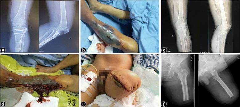
This composite of six images (a-f) documents the clinical progression and management of gas gangrene in a lower limb. Panes (a), (c), and (f) are radiological studies: (a) shows a tibial plateau fracture with internal fixation; (c) demonstrates diffuse subcutaneous emphysema and gas tracking within the soft tissues of the lower leg; and (f) shows the postoperative femoral stump after mid-thigh amputation. Clinical photographs (b), (d), and (e) illustrate the macroscopic pathology. Pane (b) shows the lower limb with severe ecchymosis, darkened necrotic skin, and tense serosanguinous bullae (tension blisters). Pane (d) captures the surgical decompression, revealing extensively necrotic, non-viable musculature with a brownish-red, 'cooked' appearance and thin, malodorous secretions. Pane (e) shows the post-amputation stump left open for drainage. Collectively, these images illustrate the diagnostic triad of clostridial myonecrosis: rapid soft tissue destruction, subcutaneous gas on X-ray, and characteristic muscular necrosis, emphasizing the necessity of emergency debridement or amputation in orthopedic infections.
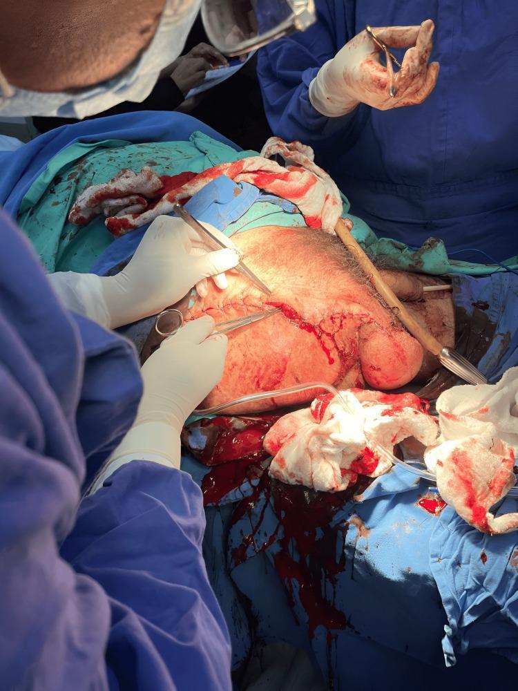
Clinical photograph of an intraoperative field during a right hip disarticulation, a major surgical procedure performed for the management of extensive tissue necrosis and gas gangrene. The image captures surgeons in sterile blue gowns and gloves utilizing instruments, including forceps and a scalpel, to manage the surgical site. The anatomy shown includes the right groin, hip region, and pelvic landmarks. A large surgical incision is visible across the upper thigh area, with active bleeding and significant accumulation of blood-soaked gauze and drapes indicating a complex, high-stakes intervention. The exposed skin and subcutaneous tissues exhibit signs of trauma and compromised vascularity. This visual serves as a clinical illustration of radical surgical debridement and amputation required to arrest life-threatening soft tissue infections like clostridial myonecrosis in patients with underlying comorbidities such as diabetes mellitus. Key educational features include surgical field management, identification of major anatomical planes during disarticulation, and the visual appearance of necrotizing infection progression.
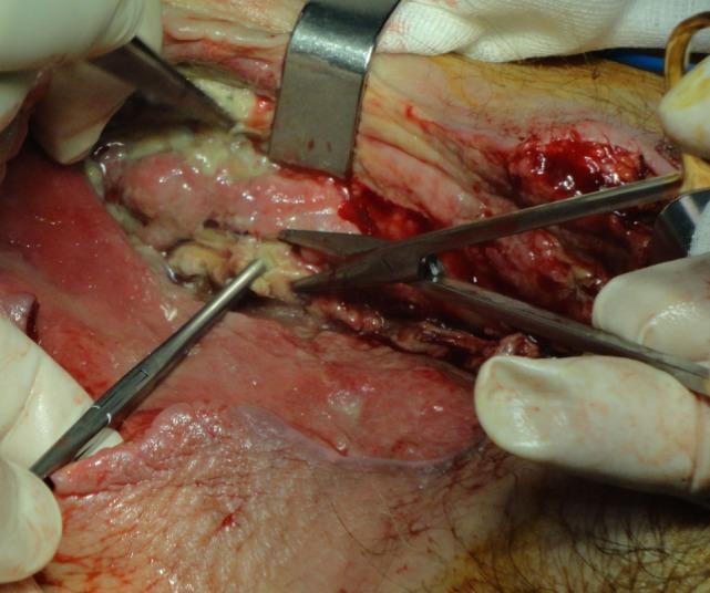
This clinical photograph shows an intraoperative view of an aggressive surgical debridement of necrotizing fasciitis (specifically gas gangrene) in the proximal right thigh. The surgical field reveals deep tissue planes with significant pathology. Visible features include areas of pale, yellowish-white necrotic tissue and purulent material juxtaposed with erythematous, inflamed soft tissue. The texture of the affected area is irregular and disrupted, showing signs of tissue liquefaction and liquefactive necrosis. Surgical instruments, including forceps and scissors, are actively being used by gloved hands to excise devitalized muscle and fascia, a procedure known as necrosectomy. A metal retractor is positioned to maintain visualization of the deep wound. The image demonstrates the hallmark clinical management of necrotizing soft tissue infections, emphasizing the need to extend debridement until viable, healthy tissue margins are reached. This content is relevant for surgical training and emergency medicine, illustrating the gross appearance of clostridial myonecrosis and the necessary surgical intervention to prevent systemic sepsis.
Gas Gangrene (Clostridial Myonecrosis)
Definition
Aetiology
Causative Organisms
| Organism | Notes |
|---|---|
| Clostridium perfringens (= C. welchii) | Most common (80-90%); commonest cause |
| C. oedematiens (C. novyi) | Severe, rapidly fatal |
| C. septicum | Associated with colonic malignancy (spontaneous gas gangrene) |
| C. bifermentans, C. sporogenes | Less common |
| Non-clostridial organisms | Coliforms, Bacteroides, streptococci; found in 60-85% of cases as co-infectors |
- Anaerobic, spore-forming, Gram-positive rods
- Found in soil, faeces, and mucous membranes
- Spores are resistant to chemical skin sterilization
- Grow best in necrotic, ischaemic, low-oxygen tissue
Types of Clostridial Infection (Campbell's Classification)
| Type | Description |
|---|---|
| Type 1 | Clostridial contamination only - positive culture, NO clinical signs |
| Type 2 | Clostridial cellulitis - gas in soft tissue, foul smell, NO systemic toxicity, muscle spared |
| Type 3 | Gas gangrene (clostridial myonecrosis) - true muscle necrosis + systemic sepsis |
Predisposing Factors / Conditions
- Traumatic wounds: crush injuries, missile/gunshot wounds, open fractures (especially with soil contamination)
- Closed fractures (also possible due to ischaemic muscle)
- Primary closure of a contaminated wound (creates anaerobic microenvironment)
- Surgery (especially bowel surgery, amputation in vascular disease)
- Immunosuppression (diabetes, malignancy, steroids)
- Chronic oedema / peripheral vascular disease
- Penetrating foreign body (especially farm or soil-contaminated)
- Aerobic co-infection (consumes remaining O2, creating anaerobic conditions)
- Intramuscular injections in drug users
Pathophysiology
Mechanism of Toxin Action
| Toxin | Action |
|---|---|
| Alpha-toxin (lecithinase/phospholipase C) | Most important - destroys RBCs, WBCs, platelets, fibroblasts, and muscle cells; causes massive haemolysis |
| Theta-toxin | Myocardial suppression, haemolysis, cardiotoxicity |
| Kappa-toxin (collagenase) | Destroys connective tissue and blood vessels |
| Mu-toxin (hyaluronidase) | Spreads infection through tissue planes |
| Nu-toxin (DNase) | Tissue liquefaction |
Gas Production
- Bacterial fermentation produces hydrogen sulphide (H2S) and CO2 (not just hydrogen)
- Gas spreads along muscle planes and fascial spaces
- Creates the classic crepitus and radiographic gas shadows
Vicious Cycle
Clinical Features
Incubation Period
- Typically < 24 hours (range: 1 hour to 6 weeks)
- Onset is sudden and dramatic - the rapidity of progression is a hallmark
Symptoms (in order of appearance)
- Rapid, severe, and disproportionate to the wound appearance
- Increases relentlessly - this is the cardinal warning sign
- Wound oedema and tense swelling of the limb
- Serosanguineous (thin, watery, "dishwater") discharge - not frank pus
- Characteristic sickly-sweet/mousy odour (very distinctive)
- Skin changes: initially pale/white → bronzed/brown → blue-black
- Haemorrhagic bullae and blebs (contain dark, non-clotting fluid)
- Crepitus on palpation (gas in tissues) - present within 24 hours
- Muscle colour: pale/grey, "cooked meat" appearance, does not bleed or contract when cut
- Fever (often low-grade paradoxically)
- Tachycardia (out of proportion to fever)
- Anxiety and altered mental status - patients often appear apprehensive/toxic early
- Tachypnoea
- Hypotension → septic shock
- Haemolysis → haemoglobinuria → acute kidney injury
- ARDS
- Jaundice
- Death (within hours in untreated cases)
Investigations
Bedside / Emergency
| Investigation | Finding |
|---|---|
| Plain X-ray | Gas in soft tissues along muscle planes (feathery pattern); early and reliable sign |
| CT scan | More sensitive for gas; defines extent; useful for trunk/abdominal involvement |
| Gram stain of exudate | Large Gram-positive rods WITHOUT neutrophils (toxins kill WBCs) - pathognomonic |
| Peripheral blood smear | Haemolysis, fragmented RBCs |
| Urine | Haemoglobinuria (port-wine urine) |
Laboratory
- FBC: haemolytic anaemia, thrombocytopaenia, leukocytosis
- Metabolic panel: metabolic acidosis, raised creatinine (AKI), elevated LFTs (jaundice)
- Coagulation: DIC in severe cases
- Blood cultures: anaerobic cultures (takes 48-72 h; do NOT delay treatment)
- Culture medium requires anaerobic environment + sodium thioglycolate (reducing agent)
Key Point: Diagnosis is CLINICAL - do NOT wait for cultures before acting.
Differential Diagnosis
| Condition | Distinguishing Features |
|---|---|
| Clostridial cellulitis (Type 2) | Gas in subcutaneous tissue only; muscle NOT involved; less systemic toxicity; slower progression |
| Necrotising fasciitis | Fascial plane involvement; mixed organisms; gas less prominent; "dishwater" fluid; skin initially normal |
| Non-clostridial myositis | Caused by anaerobic streptococci (Peptostreptococcus); delayed onset (days); gram-positive cocci; muscle may recover |
| Crepitant cellulitis | E. coli, Klebsiella, Bacteroides; subcutaneous gas; no systemic toxaemia |
| Compartment syndrome | No gas; no skin changes; no systemic toxaemia |
Treatment
Principle: Time is muscle - Time is life. Do not delay surgery.
1. Resuscitation (simultaneous with surgery prep)
- ICU admission with continuous monitoring
- 2 large-bore IV access; aggressive fluid resuscitation
- Blood products for haemolysis and coagulopathy
- Endotracheal intubation if shocked or obtunded
- Urinary catheter; monitor urine output hourly
- Blood cultures before antibiotics (but do not delay antibiotics)
2. Antibiotics (IV - immediate)
- Penicillin G (high dose: 10-24 MU/day in divided doses) - kills clostridia
- Clindamycin (600-900 mg IV 8-hourly) - inhibits toxin synthesis (protein synthesis inhibitor); acts even on non-dividing organisms; synergistic with penicillin
- Add 3rd generation cephalosporin (cefotaxime/ceftriaxone) for Gram-negatives
- Crushing/farm contamination: Penicillin + Cephalosporin + Aminoglycoside
- Clindamycin-resistant C. perfringens: Vancomycin
3. Surgical Treatment (THE MAINSTAY - EMERGENCY)
- Operate immediately - every hour of delay worsens outcome
- Wide, radical surgical debridement of ALL necrotic tissue
- Muscle that does not bleed or contract to electrical stimulation = dead = must be removed
- Wounds left WIDE OPEN - never close primarily
- Pack lightly with saline-soaked gauze
- Return to OR every 24-48 hours for repeat inspection and debridement until infection controlled
- Tissue with questionable viability: leave and reassess at next debridement
- Indicated when infection is extensive, limb is non-salvageable, or patient in septic shock
- Life-saving and should not be delayed in established gas gangrene with systemic toxicity
- Amputation stump left OPEN - never closed
- Disarticulation (joint-level amputation) may be needed to get clear of infected tissue
4. Hyperbaric Oxygen (HBO)
- Adjunct - not a substitute for surgery
- Mechanism: high-pressure O2 creates hostile environment for anaerobes; inhibits alpha-toxin
- Given at 2-2.5 atmospheres, 3 sessions/day for acute infection
- Evidence suggests helps stabilize limb amputations secondary to clostridial infection
- Only if immediately available and does not delay surgery
- Controversy remains about evidence quality
Prevention
- Thorough wound debridement of all open/contaminated wounds - remove all devitalized tissue
- Never close contaminated wounds primarily - leave open or use delayed primary closure
- Repeat debridement every 24-48 h for high-risk wounds
- IV prophylactic penicillin for contaminated crush injuries and open fractures
- Tetanus prophylaxis (different organism but same contaminated wound mechanism)
- Early recognition of at-risk wounds (crush, soil contamination, vascular ischaemia)
Prognosis
- Without treatment: 100% fatal
- With optimal treatment: mortality 5-30% for extremity infections
- Trunk/abdominal/spontaneous gas gangrene: mortality up to 50-70%
- Poor prognostic factors: diabetes, cirrhosis, leucopenia, delay in diagnosis/treatment, truncal involvement
Comparison: Gas Gangrene vs Necrotising Fasciitis
| Feature | Gas Gangrene | Necrotising Fasciitis |
|---|---|---|
| Layer involved | Muscle (myonecrosis) | Fascia (fasciitis) |
| Organisms | Primarily Clostridium (anaerobic) | Mixed (Type 1) or Group A Strep (Type 2) |
| Gas in tissues | Prominent | Less prominent |
| Onset | Very rapid (<24 h) | Rapid (24-72 h) |
| Skin appearance | Brown/black, bullae | Red → dusky → necrotic |
| "Dishwater" fluid | Yes | Yes |
| Muscle necrosis | Yes (hallmark) | Variable (late) |
| Antibiotic | Pen G + Clindamycin | Pip-tazo + Clindamycin ± Carbapenems |
| Surgery | Radical debridement ± amputation | Radical fasciotomy + debridement |
Continue